Week 9 — Cell-Free Systems
Part A: General and Lecture- Specific
1. Explain the main advantages of cell-free protein synthesis over traditional in vivo methods, specifically in terms of flexibility and control over experimental variables. Name at least two cases where cell-free expression is more beneficial than cell production.
- Flexibility & Control: Unlike living cells, CFPS is an “open” system. You can directly manipulate the reaction environment—adjusting pH, redox potential, or adding non-natural amino acids—without worrying about maintaining cell viability.
- Speed: It bypasses the time-consuming steps of transformation, cell culture, and scale-up, allowing for results in hours rather than days.
- Beneficial Cases:
- Toxic Proteins: Expressing proteins that would kill a living host cell (e.g., certain antimicrobial peptides).
- Rapid Prototyping: Testing many different genetic designs quickly in a high-throughput format.
2. Describe the main components of a cell-free expression system and explain the role of each component.
- Cell Extract: Provides the molecular “machinery,” including ribosomes, tRNAs, and initiation/elongation factors.
- Energy Source: Molecules like phosphoenolpyruvate (PEP) or creatine phosphate used to regenerate ATP and GTP.
- Amino Acids: The essential building blocks for synthesizing the protein chain.
- DNA Template: The genetic instructions (plasmid or linear PCR product) for the target protein.
- Buffer/Salts: Maintains optimal pH and ionic strength (especially Mg2+ and K+) required for ribosomal function.
3. Why is energy provision regeneration critical in cell-free systems? Describe a method you could use to ensure continuous ATP supply in your cell-free experiment.
- Criticality: Energy is consumed rapidly during transcription and translation. Without a regeneration system, the reaction stops once the initial ATP pool is depleted, leading to very low yields.
- Method: Use an enzymatic substrate system, such as the Creatine Phosphate/Creatine Kinase system, which transfers a phosphate group back to ADP to regenerate ATP in situ.
4. Compare prokaryotic versus eukaryotic cell-free expression systems. Choose a protein to produce in each system and explain why.
- Prokaryotic (e.g., E. coli): High yield, fast, and inexpensive. However, it lacks complex post-translational modifications (PTMs).
- Protein: GFP (Green Fluorescent Protein) for simple reporting or biosensing where complex folding isn’t required.
- Eukaryotic (e.g., CHO or Wheat Germ): Lower yield but capable of complex folding and PTMs like glycosylation.
- Protein: Human Insulin, which requires specific disulfide bond formation and folding pathways not present in bacteria.
5. How would you design a cell-free experiment to optimize the expression of a membrane protein? Discuss the challenges and how you would address them in your setup.
- Challenges: Membrane proteins are hydrophobic and often aggregate or misfold when synthesized in an aqueous cell-free mix without a lipid environment.
- Optimization Strategy: Integrate synthetic lipid bilayers
6. Imagine you observe a low yield of your target protein in a cell-free system. Describe three possible reasons for this and suggest a troubleshooting strategy for each.
- Template Degradation: - Reason: Presence of RNases or DNases in the extract.
- Strategy: Use high-purity DNA and supplement the reaction with RNase inhibitors.
- Energy Depletion: - Reason: Fast consumption of ATP/GTP.
- Strategy: Increase the concentration of the energy buffer or use a dialysis-based continuous-exchange system.
- Codon Bias: - Reason: The DNA sequence uses codons that are rare in the organism the extract was made from.
- Strategy: Use a codon-optimized gene sequence or supplement the reaction with a mixture of rare tRNAs.
Homework question from Kate Adamala
1. Design an example of a useful synthetic minimal cell as follows:
- Pick a function and describe it.
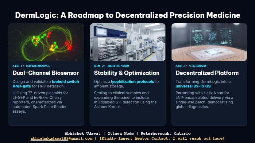
- Function: DermLogic is a dual-channel AND-gate biosensor designed for point-of-care HPV detection and therapeutic antisense RNA delivery.
- Input/Output: The input is extracellular HPV L1 and E6/E7 RNA sequences; the output is a dual-fluorescence signal (GFP/mCherry) and the synthesis of therapeutic antisense RNA.
- Could this function be realized by cell-free Tx/Tl alone, without encapsulation?
- Yes, the core logic functions in a bulk cell-free mix. However, encapsulation is required for the “therapeutic patch” vision to protect the RNA payload from degradation and to concentrate the reagents for faster kinetics.
- Could this function be realized by genetically modified natural cell?
- It is difficult. Natural cells have complex innate immune responses (like interferon pathways) that might interfere with or degrade synthetic RNA logic circuits and antisense outputs.
- Describe the desired outcome of your synthetic cell operation.
- A low-cost, decentralized tool that stratifies HPV risk and simultaneously produces a customized therapeutic response without a cold chain.
2. Design all components that would need to be part of your synthetic cell.
- What would be the membrane made of?
- A robust, shelf-stable lipid bilayer composed of POPC and Cholesterol, or potentially a polymeric/hybrid vesicle for better stability during lyophilization.
- What would you encapsulate inside?
- BL21 DE3 Lysate, the pDermLogic-v1 plasmid, T7 RNA Polymerase, amino acids, and an energy regeneration system (PEP/ATP).
- Which organism your Tx/Tl system will come from?
- Bacterial (E. coli) is preferred. It is highly efficient for T7-driven transcription and the toehold switches are optimized for bacterial ribosomes.
- How will your synthetic cell communicate with the environment?
- Through expressed membrane pores like alpha-hemolysin (aHL). These allow the viral RNA “triggers” to enter the cell and the fluorescent/therapeutic outputs to be detected or released.
3. Experimental details
- List all lipids and genes.
- Lipids: POPC, Cholesterol.
- Genes:
sfGFP(Channel A reporter),mCherry(Channel B reporter), and a custom antisense RNA sequence targeting the HPV E6/E7 junction.
- How will you measure the function of your system?
- Using a dual-channel fluorescence readout (Spark Plate Reader) to monitor the AND-gate logic, and denaturing PAGE gel electrophoresis to verify the production of the ~21 nt antisense RNA.
Homework question from Peter Nguyen
- Pitch: “Bio-Sensing Textiles: A garment that changes color upon detecting hazardous environmental pathogens.”
- How it works: Freeze-dried cell-free systems containing a specific RNA-based biosensor (riboswitch) are embedded into fabric fibers. Upon exposure to a specific pathogen and moisture (sweat or atmospheric water), the system rehydrates, triggers the sensor, and expresses a chromoprotein that visibly stains the fabric.
- Societal Challenge: This addresses the need for passive, wearable safety monitoring for healthcare workers or soldiers in environments with invisible biological threats.
- Addressing Limitations: The “one-time use” nature is addressed by making the sensor a disposable patch integrated into the garment; activation is solved by leveraging inherent moisture or the user’s perspiration as the rehydration trigger.
Homework question from Ally Huang
- Background Information: Microgravity and cosmic radiation cause significant muscle atrophy and DNA damage in astronauts. Monitoring real-time protein expression in space is difficult due to bulky equipment. BioBits® allows for rapid diagnostic tests on the ISS with a minimal footprint.
- Target: Myostatin (MSTN) protein levels, which are key regulators of muscle growth and indicators of muscle wasting.
- Relation to Space Biology: Tracking Myostatin levels allows researchers to quantify the rate of muscle degradation in microgravity. By using a cell-free biosensor, we can monitor these levels in real-time without needing to return samples to Earth.
- Hypothesis/Research Goal: Hypothesis: BioBits® can be engineered to produce a fluorescent signal proportional to the concentration of Myostatin mRNA. The goal is to create a “just-add-sample” diagnostic kit for astronauts to monitor their physical health during long-duration missions.
- Experimental Plan: We will test astronaut saliva samples. Control: Rehydrated BioBits® with a known concentration of MSTN DNA. Experimental: BioBits® rehydrated with the astronaut’s sample. Data will be collected using the P51 Molecular Fluorescence Viewer to observe the intensity of the green fluorescence.
Part B: Final Project Integration — DermLogic
Aim 1: Cell-Free Logic Validation
The core of my final project, DermLogic, relies on the E. coli BL21 DE3 cell-free system to execute a complex molecular AND-gate. This week’s focus on cell-free systems directly informs my strategy for:
- Signal Processing: Using dual-channel toehold switches (L1-GFP and E6/E7-mCherry) to stratify HPV risk.
- On-Demand Therapeutics: Leveraging the “open” nature of cell-free systems to synthesize ~21 nt antisense RNA molecules immediately upon pathogen detection.
Lyophilization Strategy
Following the principles of BioBits®, DermLogic is designed to be a shelf-stable, “just-add-water” (or sample) diagnostic. My validation plan for Aim 1 includes:
- Cryoprotectant Optimization: Testing a mix of 100mM Trehalose and 0.1% BSA to ensure the T7 RNA Polymerase and ribosomes remain functional after freeze-drying.
- Stability Testing: Comparing the fluorescence kinetics of fresh vs. rehydrated pellets using the Spark Plate Reader to calculate signal retention.
Key Component: By encapsulating this reaction in a POPC/Cholesterol lipid bilayer with alpha-hemolysin pores, the system transforms from a bulk reaction into a “synthetic cell” capable of localized therapeutic delivery.