Week 10 Lab: Mass Spectrometry
Lab 10
Overview
In this lab, we used several types of mass spectrometry techniques at the Waters Immerse Lab in Cambridge to analyze proteins and large biomolecular complexes. The experiments focused on measuring protein molecular weight, protein folding and structure, peptide sequencing, and oligomeric assembly states.
The primary protein analyzed throughout the lab was enhanced Green Fluorescent Protein (eGFP), which was studied using multiple workflows on the Waters Xevo G3 QTof and Waters BioAccord LC-MS systems. We also analyzed Keyhole Limpet Hemocyanin (KLH) using charge detection mass spectrometry (CDMS).
Part I — Intact Protein Molecular Weight Measurement
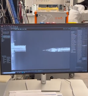
In the first experiment, intact eGFP was analyzed on the Waters Xevo G3 QTof LC-MS system under denaturing conditions. The protein sample was first buffer exchanged into ammonium acetate using spin columns and then injected into the LC-MS system.
The liquid chromatography system separated the sample before electrospray ionization and mass analysis. Because the LC solvents and acidic conditions denatured the protein, eGFP unfolded during analysis and produced a broad charge state distribution.
Using the measured mass-to-charge (m/z) values and charge states, we calculated the molecular weight of eGFP and compared it to the predicted molecular weight from the amino acid sequence.
This experiment demonstrated how LC-MS can accurately determine intact protein mass and confirm protein identity.
Part II — Native vs. Denatured Protein Structure
In the second experiment, eGFP was analyzed in both native and denatured forms using direct infusion mass spectrometry on the Waters Xevo G3 QTof.
The native protein remained folded in ammonium acetate buffer and produced lower charge states because fewer protonation sites were exposed. After adding formic acid, the protein unfolded and produced broader, higher charge state distributions.
Comparing the native and denatured spectra showed how protein structure affects electrospray ionization behavior and charge state formation.
This experiment demonstrated that mass spectrometry can provide information not only about molecular weight, but also about higher-order protein structure and folding state.
Part III — Peptide Mapping and Protein Sequencing
In the third experiment, eGFP was digested with trypsin to generate smaller peptide fragments. Trypsin cleaves proteins after lysine (K) and arginine (R) residues, producing predictable peptide fragments.
The peptide mixture was analyzed using the Waters BioAccord LC-MS system. Liquid chromatography separated the peptides, and tandem mass spectrometry fragmented selected peptide ions to determine amino acid sequences.
Using peptide masses and fragmentation spectra, we generated a peptide map of eGFP and confirmed the primary amino acid sequence of the protein.
This experiment demonstrated how LC-MS/MS can identify proteins, determine peptide sequences, and measure sequence coverage.
Part IV — Charge Detection Mass Spectrometry of KLH
In the final experiment, Keyhole Limpet Hemocyanin (KLH) was analyzed using the Waters Xevo CDMS system.
KLH forms extremely large oligomeric protein complexes in the megadalton mass range, which are difficult to analyze using conventional mass spectrometry because their charge states overlap heavily.
Charge detection mass spectrometry (CDMS) directly measures both the charge and mass of individual ions, allowing accurate mass measurements of very large protein assemblies.
Using CDMS, we identified multiple oligomeric states of KLH, including decamers and larger multidecameric complexes.
This experiment demonstrated how advanced mass spectrometry methods can analyze extremely large biomolecular assemblies that are inaccessible using standard LC-MS techniques.
Conclusion
Overall, this lab introduced several important applications of mass spectrometry in modern biochemistry and biotechnology.
Using the Waters Xevo G3 QTof, Waters BioAccord, and Waters Xevo CDMS systems, we measured:
- Intact protein molecular weight
- Protein folding and structure
- Peptide sequences
- Oligomeric assembly states
These experiments demonstrated how mass spectrometry can provide structural, molecular, and sequence-level information about biological molecules with high sensitivity and precision.