Week 09 HW: cell free systems
- Advantages of cell-free systems Cell-free protein synthesis (CFPS) offers a highly flexible and controllable environment compared to in vivo expression systems. Because there are no living cells, experimental conditions such as pH, ionic strength, redox environment, DNA concentration, cofactors, and additives can be directly tuned without affecting cell viability. This enables rapid optimization and prototyping of genetic constructs.
Additionally, CFPS is significantly faster, allowing protein production within hours instead of requiring cell growth, transformation, and induction steps.
Cell-free systems are particularly advantageous in cases such as:
- Toxic proteins: proteins that would inhibit or kill host cells can be produced safely
- Membrane proteins: can be expressed with detergents, liposomes, or nanodiscs to improve folding and functionality
 *Generated by ChatGBT)
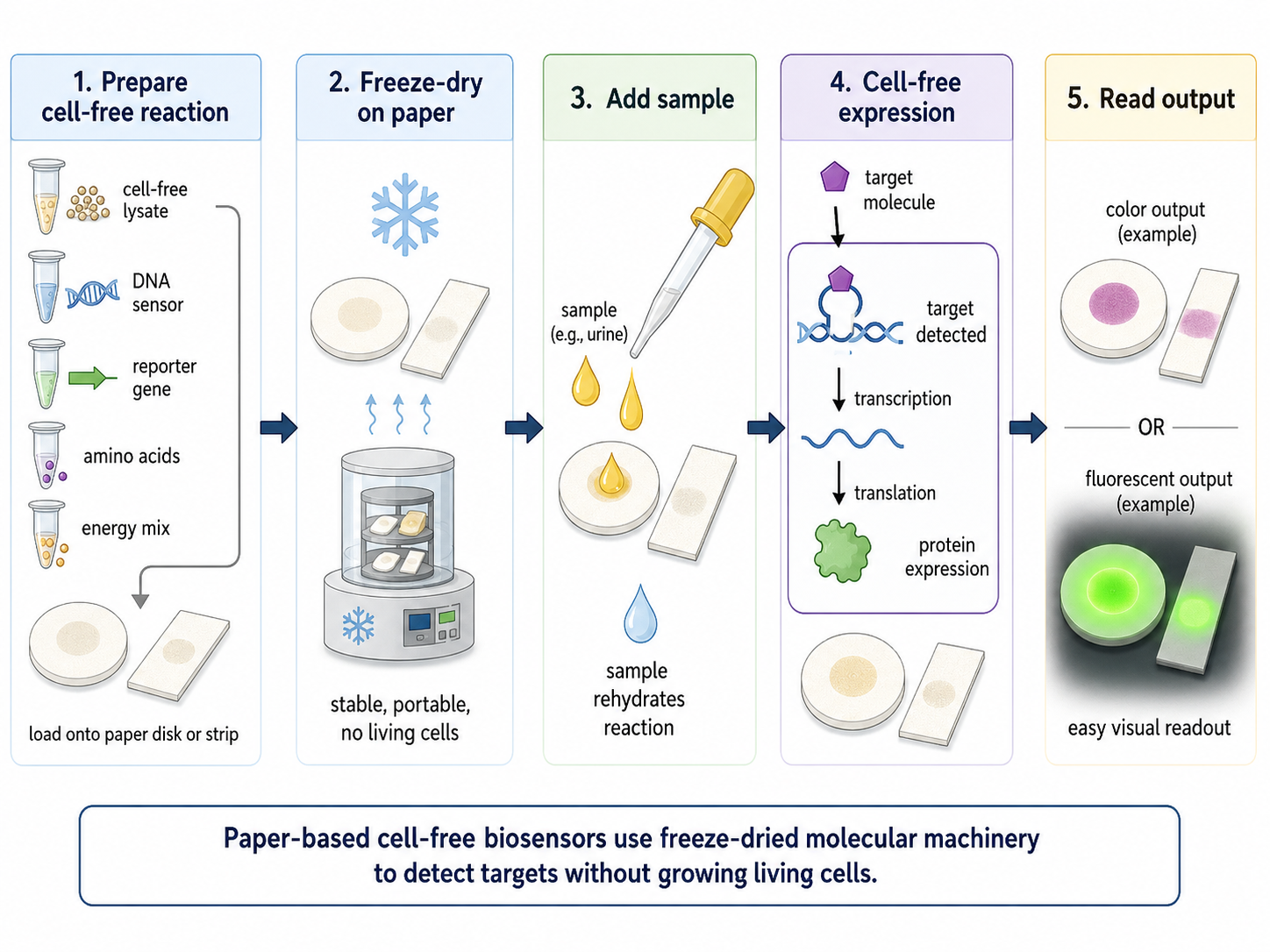
A real-world example is freeze-dried paper-based diagnostics, where cell-free reactions are dried onto paper and then reactivated by adding a liquid sample. Instead of growing engineered bacteria, the paper contains the transcription–translation machinery needed to make a reporter protein when a target molecule is detected. This is useful for low-resource testing because it avoids maintaining living genetically modified cells and can be made portable.
This is one interesting Example project
- Components of a cell-free system
A typical cell-free expression system includes:
Cell extract / TX-TL machinery
- Provides ribosomes, tRNAs, enzymes, and factors required for transcription and translation
- DNA or mRNA template - Encodes the protein of interest
- Amino acids Building blocks for protein synthesis
- Nucleotides (ATP, GTP, CTP, UTP) - Required for transcription and energy transfer
- Energy regeneration system - Maintains ATP/GTP supply during the reaction
- Buffer + cofactors (Mg²⁺, K⁺, etc.) - Maintain optimal biochemical conditions
- Optional additives (chaperones, lipids, detergents)- Help folding or membrane protein insertion
- Why energy regeneration is critical
ATP and GTP are consumed during:
- transcription
- tRNA charging
- ribosomal translation
- Without regeneration, the reaction stops quickly.
Solution: Use an energy regeneration system such as: phosphoenolpyruvate (PEP) + pyruvate kinase or creatine phosphate + creatine kinase. These systems continuously regenerate ATP, allowing sustained protein production.
Prokaryotic vs eukaryotic systems
For a prokaryotic CFPS system, I would express GFP or sfGFP because it is a small, well-characterized reporter protein that folds efficiently in bacterial systems and gives an easy fluorescent readout.
For a eukaryotic CFPS system, I would express a human receptor fragment or a glycosylated protein, because eukaryotic systems are better suited for proteins that require complex folding, disulfide bonds, or post-translational modifications. For example, a mammalian lysate would be more appropriate for testing a human membrane receptor than an E. coli lysate.
| Feature | Prokaryotic CFPS | Eukaryotic CFPS |
|---|---|---|
| Speed | Fast | Slower |
| Yield | High | Lower |
| Complexity | Simple | Complex |
| PTMs | Limited | Full (glycosylation, etc.) |
- Designing a membrane protein experiment
Challenges:
- Poor solubility
- Misfolding
- Aggregation Approach:
- Add detergents or liposomes to mimic membranes
- Include chaperones
- Optimize Mg²⁺, temperature, and energy system
Membrane proteins are challenging because their hydrophobic regions normally need a lipid membrane to fold correctly. In a cell-free experiment, I would express the same DNA template under different membrane-like conditions, such as no additive, mild detergent, liposomes, nanodiscs, or membrane vesicles.
I would compare total protein yield using a tag such as GFP or His-tag, but also test function, because a membrane protein can be expressed but still misfolded. Key variables to optimize would include DNA concentration, magnesium/potassium concentration, temperature, incubation time, and lipid or detergent concentration.
Main challenges include aggregation, incorrect folding, and additives interfering with the cell-free reaction. I would address these by testing membrane mimics, using lower temperatures for slower folding, and including a soluble reporter control such as GFP to check that the cell-free system still works.
- Troubleshooting low protein yield Low yield could have several causes:
1. Poor DNA template design
The promoter, ribosome binding site, or coding sequence may not work well.
Troubleshooting: Check the sequence, use the correct promoter such as T7, test another RBS/UTR, and compare with a GFP control.
2. Reaction conditions are not optimized
Salt, magnesium, DNA concentration, temperature, or energy mix may be suboptimal.
Troubleshooting: Run a small optimization matrix testing DNA amount, Mg²⁺/K⁺ levels, temperature, and incubation time.
3. The protein is unstable or misfolded
The target protein may aggregate, degrade, or require cofactors/chaperones.
Troubleshooting: Lower the temperature, add folding helpers, cofactors, detergents, liposomes, or nanodiscs, and check the product by SDS-PAGE or fluorescence.
Homework question from Kate Adamala
Design of a useful synthetic minimal cell: I ask myself would it be possible to construct my final project idea completly as syntetic cell? Light-controlled bacterial cellulose patterning
1. Pick a function and describe it
The function of this synthetic minimal cell would be to translate a light pattern into a chemical signal that controls bacterial cellulose production in a nearby engineered bacterial cellulose culture.
Instead of asking the synthetic cell to produce cellulose itself, It would problebly be more feasible to design a light-responsive signaling protocell. When exposed to blue light, the synthetic cell would produce or release a small molecule signal, such as AHL, which could activate a genetic circuit in engineered Komagataeibacter rhaeticus. This would connect the minimal cell homework to my final project logic:
light → genetic regulation → changed cellulose production → patterned material structure.
My final project already proposes using optogenetic control to spatially regulate bacterial cellulose production in K. rhaeticus, with the long-term goal of creating patterned differences in cellulose density, thickness, and structure during growth. In the original final project, the proposed logic is:
Light → Opto-T7RNAP → PT7 → sRNA → ↓ UGPase → ↓ cellulose.
For the synthetic minimal cell version, I would slightly modify this into a communication system:
Light → synthetic minimal cell → AHL signal → engineered K. rhaeticus → sRNA → ↓ UGPase → ↓ cellulose.
This keeps the same material goal, but makes the synthetic cell act as a programmable signaling layer.
a) What would the synthetic cell do?
The synthetic cell would sit in or near a bacterial cellulose growth system and respond to projected blue light.
Input:
A spatial blue-light pattern, for example stripes, dots, gradients, or moiré-like projected patterns.
Internal operation:
Inside the synthetic cell, a cell-free transcription/translation system would express a light-controlled signaling module. When illuminated, the system would produce or release AHL.
Output:
A diffusible AHL signal that activates a receiver circuit in engineered K. rhaeticus. The bacteria would then express an sRNA targeting UGPase, reducing UDP-glucose supply and therefore locally reducing cellulose biosynthesis.
Material output:
Regions exposed to light would produce less cellulose, while dark regions would produce more cellulose. After growth and drying, this could create a bacterial cellulose sheet with spatial differences in thickness, density, flexibility, or optical structure.
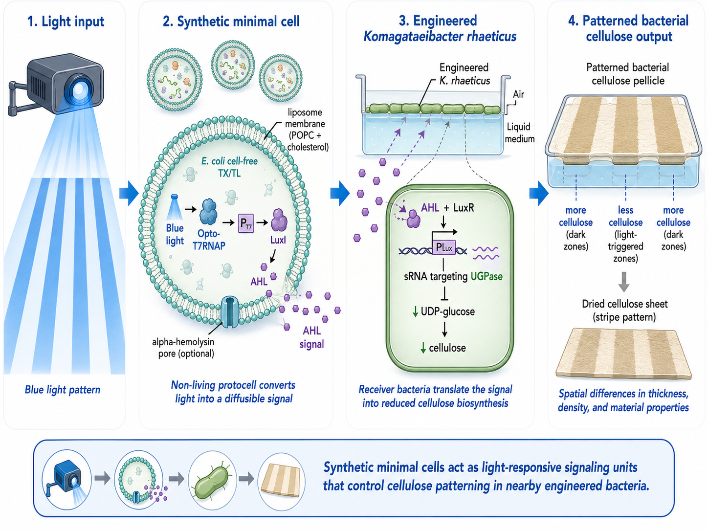 Illustration generated by ChatGBT
b) Could this be done by cell-free Tx/Tl alone, without encapsulation?
Partly, but not as well.
A bulk cell-free Tx/Tl reaction could express a reporter, enzyme, or signaling molecule in response to DNA-programmed logic. However, without encapsulation, the reaction would not behave like a cell-like unit. The components would diffuse freely, and the spatial boundary between “on” and “off” regions would be poorly defined.
For my project, encapsulation is useful because the synthetic cell acts as a localized microreactor. The membrane keeps the Tx/Tl machinery, DNA, enzymes, and cofactors together, while allowing selected small molecules to move in or out. This is important if the goal is spatial patterning rather than only bulk expression.
So, cell-free Tx/Tl alone could demonstrate the molecular logic, but encapsulation is needed to make it a synthetic minimal cell with compartmentalized behavior.
c) Could this function be realized by a genetically modified natural cell
Yes. In fact, my final project mainly proposes a genetically modified natural cell: engineered Komagataeibacter rhaeticus.
A natural-cell version could contain the full circuit directly inside K. rhaeticus:
Light → Opto-T7RNAP → PT7 → UGPase-targeting sRNA → reduced cellulose production.
This is probably the most direct route for making a real bacterial cellulose material, because K. rhaeticus naturally produces bacterial cellulose at the air–liquid interface. However, the synthetic minimal cell version is interesting because it separates the sensing/signaling layer from the cellulose-producing organism. This could make the system more modular and safer to test: the minimal cell does not grow, divide, or evolve like a natural genetically modified organism.
d) Desired outcome of the synthetic cell operation
The desired outcome is a bacterial cellulose pellicle whose material structure is patterned by light.
After projecting a light pattern during growth, the material would show local differences in:
- cellulose thickness
- density
- transparency
- mechanical stiffness or flexibility
- possibly layered moiré-like visual or structural effects
This connects to the long-term goal of my final project: moving bacterial cellulose from passive sheet growth toward programmable biofabrication, where light becomes a design interface for controlling material formation during growth.
2. Components of the synthetic minimal cell
a) Membrane
The membrane would be a phospholipid liposome, because liposomes are commonly used as compartments for synthetic cell prototypes and can encapsulate cell-free Tx/Tl reactions. A possible simple membrane composition would be:
- POPC as the main phospholipid
- cholesterol to improve membrane stability
- a small fraction of fluorescent lipid for microscopy tracking, for example Rhodamine-PE or NBD-PE
The membrane should be semi-permeable. Small molecules such as nutrients, ions, and possibly AHL should be able to exchange with the environment, while large components such as ribosomes, DNA, enzymes, and Tx/Tl machinery remain trapped inside.
b) Encapsulated inside
The inside of the synthetic cell would contain:
- bacterial cell-free Tx/Tl system
- DNA templates encoding the light-responsive circuit
- amino acids
- NTPs
- energy regeneration system
- salts and magnesium
- cofactors
- AHL-producing enzyme module or AHL-release module
- optional fluorescent reporter for debugging
c) Tx/Tl system
I would use an E. coli-based Tx/Tl system. Bacterial Tx/Tl is appropriate because the circuit does not require mammalian post-translational modifications. A mammalian system would only be needed if I wanted to use mammalian regulatory systems such as Tet-ON or mammalian promoters. For this project, bacterial expression is enough.
E. coli Tx/Tl is also commonly used for synthetic cell prototypes, including liposome-encapsulated systems expressing reporters and membrane proteins such as alpha-hemolysin and MscL.
d) Communicate
The synthetic cell would communicate chemically with engineered K. rhaeticus through AHL.
AHL is useful because it is a small quorum-sensing molecule and can diffuse between compartments more easily than large biomolecules such as proteins or RNA. This matters because the synthetic cell membrane should not need to release large genetic components.
To improve exchange, I could include a membrane pore or channel. A possible gene is:
- hla from Staphylococcus aureus, encoding alpha-hemolysin, a pore-forming protein
- alternatively mscL from E. coli, encoding the mechanosensitive channel of large conductance
Alpha-hemolysin is often used in synthetic-cell work because it can form pores in lipid membranes and allow small-molecule exchange. It has also been expressed using cell-free systems and inserted into phospholipid membranes.
In the receiver bacteria, K. rhaeticus would contain a LuxR/pLux-based AHL receiver system. This is close to the original cellulose-control work by Florea et al., where bacterial cellulose production was externally controlled using a genetic toolkit in K. rhaeticus.
Synthetic minimal cell genes
Light-control / expression layer
- Split T7 RNA polymerase system, Opto-T7RNAP
- nMagHigh1 / pMag light-induced dimerization domains
- T7 promoter, PT7
- optional reporter: sfGFP or mCherry
Communication output
Option A: AHL production
- luxI: AHL synthase gene, produces AHL signal
Option B: AHL release / permeability support
- hla: alpha-hemolysin pore from Staphylococcus aureus
- or mscL: mechanosensitive channel from E. coli
Engineered K. rhaeticus receiver genes
- luxR: AHL-responsive transcription factor
- pLux promoter: activated by AHL-LuxR
- sRNA targeting UGPase mRNA
- optional Hfq-binding region to support sRNA function
- target pathway: UGPase / UDP-glucose pathway controlling cellulose precursor supply
In the receiver cell, the output logic would be:
AHL → LuxR/pLux → sRNA → lower UGPase → less UDP-glucose → reduced cellulose production.
3. Experimental setup
I would grow engineered K. rhaeticus in a shallow bacterial cellulose culture system. Synthetic minimal cells would be added into or near the growth interface. A blue-light projector or LED mask would expose defined regions of the culture.
I would test simple light patterns first:
- full light
- full dark
- stripes
- dots
- gradient
- offset stripe layers for moiré-like effects
Controls
Important controls would include:
- no synthetic cells
- synthetic cells without DNA
- synthetic cells without light
- full-light positive control
- K. rhaeticus receiver without LuxR/pLux
- reporter-only version before connecting to cellulose regulation
Measurements
I would measure function at two levels.
1. Molecular / circuit function
- fluorescence reporter output from synthetic cells
- AHL response using a reporter strain or pLux-GFP receiver
- microscopy to confirm synthetic cell localization
- time-course fluorescence under light and dark conditions
2. Material output
After cellulose growth, I would measure:
- pellicle thickness
- dry weight
- transparency / optical density
- image contrast between light and dark regions
- mechanical properties such as tensile strength or flexibility
- microscopy of cellulose structure
Success would mean that the projected light pattern is converted into a measurable spatial difference in the bacterial cellulose material.
This synthetic minimal cell would act as a non-living, light-responsive signaling unit for programmable bacterial cellulose fabrication. The minimal cell would not produce cellulose directly. Instead, it would sense blue light and communicate with engineered K. rhaeticus through AHL. The natural bacteria would remain responsible for cellulose production, while the synthetic cell would provide spatial control.
This design is useful because it separates sensing, signaling, and material production into modular layers. It could be tested first with fluorescent reporters and later connected to cellulose regulation. The final goal would be a bacterial cellulose sheet whose density and structure are patterned by light during growth.
Homework question from Peter Nguyen
Summary
Based on my idea 1 for my final project I would develop a Bacterial cellulose cosmetic skinmask that would sense the “health” of the customers skin. Facemasks are populair single use product, however they are “dumb” providing a singulair batch of substances without telling you anything about what your skin acctually needs.
BC is already a compelling cosmetic substrate because it holds a lot of water, conforms well to skin, and has been tested as a moisturizing sheet mask material. In one evaluation, instead of putting living engineered cells on the face, a safer “synthetic biology” route is to embed freeze-dried cell-free gene expression (TX-TL) into the BC sheet as small patterned “sensor dots.” These cell-free circuits stay inactive when dry, then turn on when the mask hydrates during wear; outputs can be colorimetric (visible) or optical.
How will the idea work,
Because freeze-dried cell-free circuits activate upon rehydration, a conventional pre-hydrated sheet mask would trigger prematurely during storage. A practical design might be a dry-stored BC mask (or a separate paper sensor tab) that is activated only at time of use by releasing fluid.
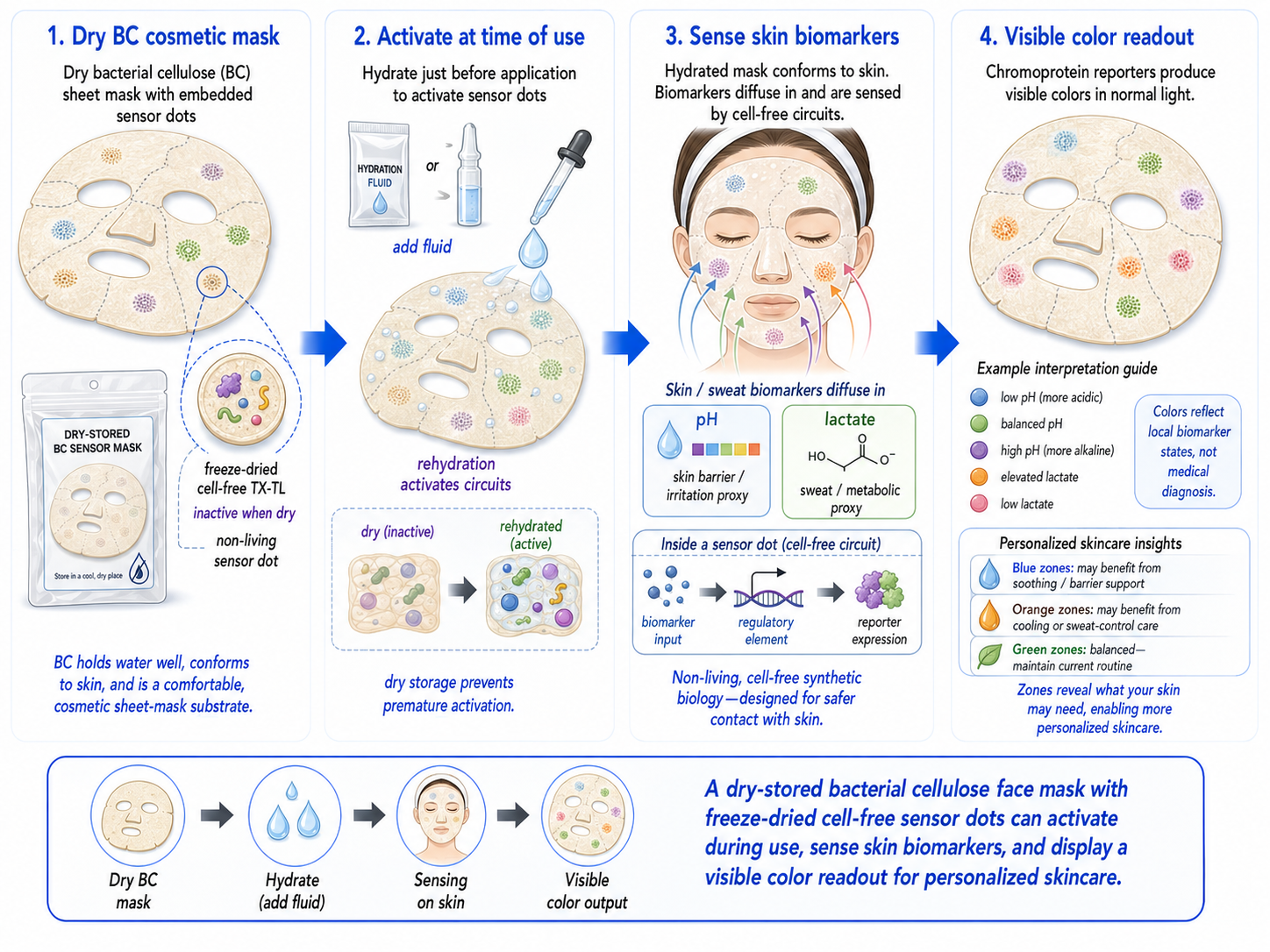 Illustration generated by ChatGBT
System:
- Input (skin/sweat biomarker): pH (skin barrier/irritation proxy), lactate (sweat/metabolic proxy).
- Sensing layer (cell-free circuit): a biomarker-responsive regulatory element controls whether a reporter is expressed.
- Output (visible color): express a chromoprotein (strong color under normal light) so the mask visibly shifts color in specific zones without any instrument; chromoproteins are attractive for “naked-eye” readouts.
Market need
The advantage of this concept is that facemask is already concidered as single use products so the one time use limitation of freeze dried system is becoming a desirable feature.
Limitation of cell-free reactions
A main limitation of freeze-dried cell-free systems is that they are usually one-time-use and activate when water is added. In this project, I would turn that limitation into part of the product design. Cosmetic sheet masks are already single-use products, so the fact that the TX-TL sensor dots only work once actually matches the use case.
The biggest challenge is premature activation. A normal pre-hydrated sheet mask would activate the freeze-dried TX-TL reactions during storage, before the customer uses it. To avoid this, the BC mask would be stored dry, with the freeze-dried sensor dots inactive. The hydration liquid could be kept in a separate sachet or breakable reservoir and released only at the time of use.
To improve stability, the cell-free sensor dots could be freeze-dried with stabilizers such as trehalose or sucrose, then sealed in moisture-barrier packaging. The mask would need to be protected from humidity, heat, and light during storage. Each sensor dot could also be patterned as a small protected region inside the BC sheet, so the reaction components stay localized.
The system would be designed as a single-use readout: hydrate the mask, apply it to the skin, allow biomarkers such as pH or lactate to diffuse into the sensor dots, then read the color change. After use, the mask would be discarded like a conventional cosmetic mask, but with the added value that it gives local information about the skin condition.
References — Project
Brown, D. M. et al. (2025). Semiautomated Production of Cell-Free Biosensors. ACS Synthetic Biology.
https://pubmed.ncbi.nlm.nih.gov/40073441/Amnuaikit, T. et al. (2011). Effects of a cellulose mask synthesized by a bacterium on facial skin characteristics and user satisfaction.
https://pmc.ncbi.nlm.nih.gov/articles/PMC3417877/Nguyen, P. Q. et al. (2021). Wearable materials with embedded synthetic biology sensors for biomolecule detection. Nature Biotechnology.
https://www.nature.com/articles/s41587-021-00950-3Pardee, K. et al. (2014). Paper-Based Synthetic Gene Networks. Cell.
https://pubmed.ncbi.nlm.nih.gov/25417167/Ba, F. et al. Chromoproteins: visible tools for advancing synthetic biology.
https://pubmed.ncbi.nlm.nih.gov/41309430/
Homework question from Ally Huang

1. Background
My proposal is to develop a freeze-dried BioBits paper-based diagnostic for astronaut urine monitoring. The system would work like “smart toilet paper”: it rehydrates on contact with urine and produces a visible or fluorescent signal if an infection marker is present. This addresses the need for low-resource, non-invasive health monitoring in space, where medical infrastructure is limited. UTIs are relevant because immune changes in microgravity may increase infection risk. The project combines synthetic biology, paper-based diagnostics, and cell-free systems for autonomous health monitoring.
2. Molecular / genetic target
Bacterial 16S rRNA sequence specific to Escherichia coli as a biomarker for urinary tract infection.
3. Relation to space biology challenge
Astronauts can experience immune dysregulation and altered microbial behavior in microgravity, which may increase infection risk. UTIs are relevant during long missions because hygiene is constrained and medical support is limited. Detecting E. coli 16S rRNA in urine would provide a direct molecular indicator of a common UTI-causing bacterium. A paper-based cell-free diagnostic could enable rapid, on-site detection without complex lab equipment, supporting earlier intervention and reducing health risks during extended space travel.
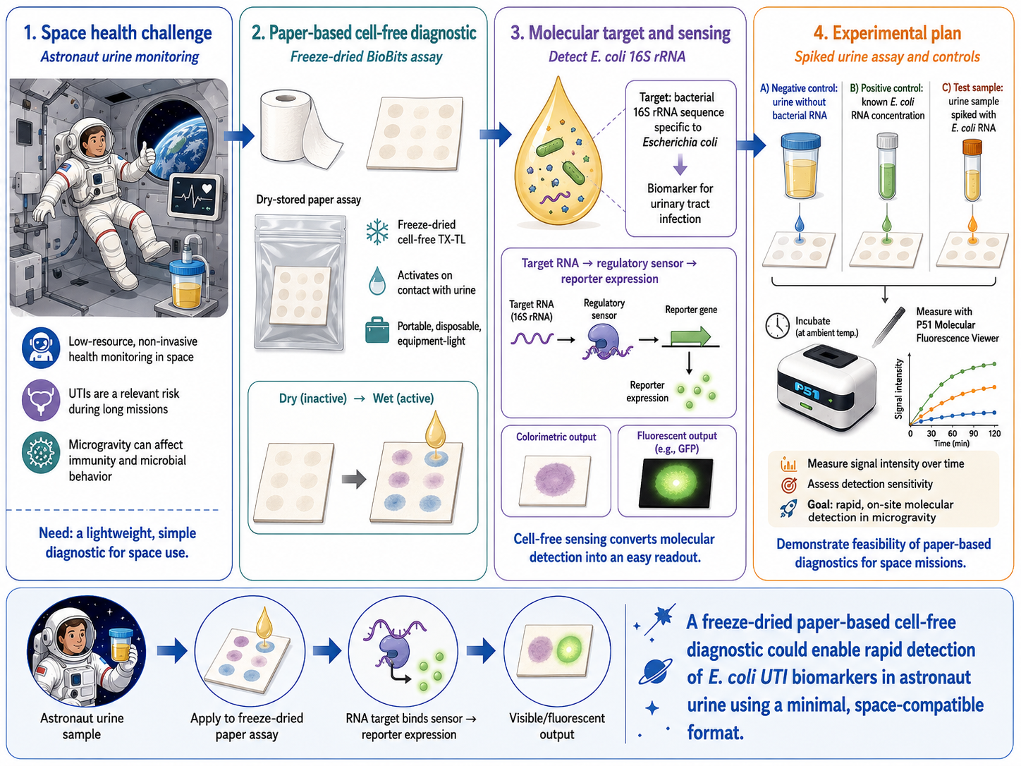
Illustration generated by ChatGPT
4. Hypothesis / research goal
I hypothesize that a freeze-dried BioBits cell-free system embedded in paper can detect E. coli RNA in urine and produce a measurable colorimetric or fluorescent output after rehydration. The assay would contain a DNA construct designed to respond to the target RNA sequence and trigger reporter expression, such as GFP. Because freeze-dried cell-free systems can remain inactive during storage and activate with simple hydration, they are well suited for space applications. The goal is to test whether molecular detection and signal generation can occur reliably in a lightweight, disposable, equipment-light format suitable for microgravity environments.
5. Experimental plan
Urine samples spiked with E. coli RNA will be applied to freeze-dried BioBits paper assays. Controls will include urine without bacterial RNA as a negative control and samples with known RNA concentrations as positive controls. After rehydration, the assays will be incubated and analyzed for color change or fluorescence using the P51 Molecular Fluorescence Viewer. Data collected will include signal intensity over time and detection sensitivity. This will test whether paper-based cell-free diagnostics can detect UTI biomarkers in a simple space-compatible format.
References
Pardee, K. et al. (2014). Paper-Based Synthetic Gene Networks. Cell, 159(4), 940–954.
https://doi.org/10.1016/j.cell.2014.10.004Saengsawang, N. et al. (2023). Validation of quantitative loop-mediated isothermal amplification assay using a fluorescent distance-based paper device for detection of Escherichia coli in urine. Scientific Reports, 13, 18781.
https://www.nature.com/articles/s41598-023-46001-6