Week 09 – Cell-Free Systems
Part A: General and Lecturer-Specific Questions
Q.1. One of the main advantages of cell-free protein synthesis over traditional in vivo methods is the flexibility and level of control over the system. In normal in vivo systems, cells are still trying to survive, grow, divide, regulate metabolism, and maintain their own functions. Because of that, researchers are working with the cell’s own priorities and limitations.
In a cell-free setup, that limitation becomes much smaller because the system is open. Researchers can directly adjust variables like ion concentrations, energy sources, cofactors, temperature, chaperones, membrane components, and even parts of the genetic code itself without worrying about keeping a cell alive. I think this is where the engineering side of synthetic biology becomes very clear because instead of only observing biology, we can start designing and tuning the system for a specific purpose.
Cell-free expression is also useful in situations where living cells usually struggle. One example is toxic proteins because these proteins can harm or kill host cells during production. Another example is membrane proteins, which are often difficult because they can misfold or aggregate inside living cells. In cell-free systems, membrane-like environments such as liposomes, detergents, or nanodiscs can be added directly during synthesis to help improve folding and function.
Q.2. The most important component is the cell extract or lysate because it contains the core machinery needed for protein synthesis, including ribosomes, tRNAs, translation factors, enzymes, and other cellular components.
The system also needs a DNA or mRNA template containing the gene of interest. This acts as the instruction set that tells the system which protein should be produced.
Amino acids are another essential component because they serve as the building blocks used to assemble the protein.
An energy source is also required because protein synthesis consumes a large amount of ATP and GTP. Energy regeneration systems help maintain the reaction so protein production can continue for a longer period of time.
Salts and buffers are important because they maintain the correct chemical environment. Ribosomes and enzymes are sensitive to factors such as pH and ion concentrations, particularly magnesium levels, so these conditions need to remain stable.
Additional components can also be added depending on the goal of the experiment. For example, liposomes or detergents can help support membrane protein production, chaperones can assist with protein folding, cofactors can improve protein activity, and non-natural amino acids can introduce new properties into the final protein.
Q.3. Energy regeneration is critical because protein synthesis requires a continuous energy supply. Processes such as transcription, translation, and amino acid charging continuously consume ATP and GTP during the reaction. If ATP becomes depleted, protein production will stop even if all the other components are still present.
Unlike living cells, cell-free systems do not naturally sustain long-term metabolic activity unless the reaction is intentionally designed to support it. That is why energy regeneration becomes an important part of the system rather than just an additional component.
One commonly used strategy is phosphoenolpyruvate (PEP), which acts as a phosphate donor and regenerates ATP from ADP to keep the reaction running. Other systems may also use energy substrates such as glucose or maltodextrin because enzymes already present in the extract can process them gradually, allowing a more sustained energy supply during longer reactions.
Q.4. Prokaryotic systems, especially E. coli-based systems, are usually faster, cheaper, and easier to optimize. They can produce high protein yields relatively quickly, which makes them useful for rapid prototyping and for proteins that do not require complex processing.
I would use an E. coli system to produce GFP because it generally folds well in bacterial systems and does not require complicated post-translational modifications. It is also easy to monitor since fluorescence can quickly show whether the protein is being expressed successfully.
Eukaryotic systems are more complex, but they are often better for proteins that need proper folding, disulfide bond formation, chaperone support, or post-translational modifications such as glycosylation.
For a eukaryotic system, I would produce erythropoietin (EPO) because it is a human glycoprotein that depends on proper glycosylation for normal biological function. Since bacterial systems cannot perform the same type of processing as mammalian systems, a eukaryotic environment would be more suitable.
Overall, I would choose the system based on what the protein actually needs rather than automatically choosing the more complex option.
Q.5. Membrane proteins are difficult mainly because of their hydrophobic regions. Once they are outside a membrane environment, they can misfold or aggregate pretty easily.
If I were designing a cell-free experiment for a membrane protein, I would make sure the membrane-like environment is already present during protein synthesis instead of trying to add it later.
I would probably test different conditions using:
• liposomes • detergents • nanodiscs • synthetic membrane systems
Nanodiscs seem especially interesting because they provide a stable lipid bilayer environment while still remaining relatively controlled experimentally.
One challenge would definitely be aggregation, so I would optimize factors such as:
• temperature • magnesium concentration • detergent concentration • translation conditions
Another issue would be proper insertion and folding of the protein into the membrane environment. Chaperones or slower translation conditions could help give the protein more time to fold correctly.
Q.6. It could be poor DNA quality or an inefficient template design. I would first check the DNA integrity and then test different construct designs or template concentrations.
Or it could be issues with the lysate itself. Since the lysate contains the machinery needed for protein production, including ribosomes, enzymes, and translation factors, poor extract quality or non-optimal reaction conditions could affect protein yield. I would probably test a fresh lysate preparation and optimize factors such as magnesium or salt concentrations.
A third possibility is protein aggregation or incorrect folding, especially for membrane proteins or larger proteins. In that case, I would try lower temperatures, add chaperones, or include membrane mimetics such as nanodiscs or liposomes to improve protein stability and folding.
Part B: Homework question from Kate Adamala
- A synthetic minimal cell would function as a stress sensor for biological soil crust environments. Since biocrust organisms experience environmental stress from drought and increasing salinity, the system would detect osmotic stress conditions before the soil ecosystem becomes severely damaged.
Input: High salt concentration / osmotic stress signal
Output of SMC: GFP fluorescence signal
The idea is that when osmotic stress increases, the synthetic cell activates a stress-responsive system and produces a measurable fluorescent signal.
B. Partially yes. Cell-free Tx/Tl could generate the sensing reaction, but encapsulation would help organize the system into a defined compartment and better mimic environmental sensing conditions.
C. Yes. A bacterial cell could be engineered to detect osmotic stress. However, natural cells introduce their own metabolism and stress responses, which may affect the signal. Using a synthetic minimal cell would provide more direct control over the system.
- Experimental details
Lipids
• POPC • Cholesterol
Genetic components
• proU promoter (osmotic-responsive promoter system) • GFP reporter gene • hla gene encoding α-hemolysin membrane pores
I would expose the synthetic minimal cells to different salt concentrations and measure GFP fluorescence.
Conditions: • low salt concentration (control) • moderate salt concentration • high salt concentration
If the system works correctly, GFP expression should increase when osmotic conditions activate the sensing system. I would compare the fluorescence intensity across the different conditions to determine whether the synthetic cell responds to environmental changes.
Measurements: • fluorescence microscopy • plate-reader fluorescence measurements
References
Adamala et al. (2017) – Engineering genetic circuit interactions within and between synthetic minimal cells Adamala, K. P., Martin-Alarcon, D. A., Guthrie-Honea, K. R., & Boyden, E. S. Nature Chemistry, 9(5), 431–439. Supports: synthetic minimal cells, liposome encapsulation, genetic circuits.
Mellies et al. (1994) – The Escherichia coli proU promoter element and its contribution to osmotically signaled transcription activation Mellies, J., Bremer, E., & Villarejo, M. Journal of Bacteriology, 176(12), 3638–3645. Supports: osmotic-response system (proU promoter) used in your sensing design.
Hilburger et al. (2019) – Controlling Secretion in Artificial Cells with a Membrane AND Gate Hilburger, C. E., Jacobs, M. L., Lewis, K. R., Peruzzi, J. A., & Kamat, N. P. ACS Synthetic Biology, 8(6), 1224–1230. Supports: α-hemolysin pores and membrane communication in artificial cells.
Shin & Noireaux (2012) – An E. coli cell-free expression toolbox: Application to synthetic gene circuits and artificial cells Shin, J., & Noireaux, V. ACS Synthetic Biology, 1(1), 29–41. Supports: bacterial cell-free transcription/translation systems.
Guo et al. (2017) – Insights on osmotic tolerance mechanisms in Escherichia coli gained from omics studies Guo, Y., Winkler, J., & Kao, K. C. Biotechnology for Biofuels, 10, 38. Supports: osmotic stress biology and bacterial responses to salt changes.
Homework question from Peter Nguyen
I trust that a wearable bracelet or skin patch with freeze-dried cell-free biosensors built into the material that activate with moisture and detect environmental heavy metal exposure through a visible color change.
The bracelet or patch would contain small freeze-dried cell-free reaction spots embedded inside a hydrogel or flexible wearable material. Sweat or environmental moisture would naturally reactivate the biological reactions. Heavy metals from polluted air particles, contaminated water, or environmental exposure could then interact with the sensing system. Once activated, the biosensors could detect heavy metals such as lead, mercury, or cadmium and produce a visible color response.
Heavy metal exposure can happen gradually and people may not realize they are being exposed until symptoms start appearing later on. Long-term exposure can also contribute to oxidative damage and broader health effects. A wearable biosensing material could help provide easier real-time monitoring for people who work in polluted or higher-risk environments.
One limitation is that freeze-dried systems need hydration to become active. In this design, sweat or environmental moisture would naturally activate the system only when the bracelet or patch is being worn. Another limitation is that many cell-free reactions are still single-use. To help with this, the material could contain replaceable sensing layers or small compartments that activate gradually instead of all at once. Protective hydrogels or polymer coatings could also help improve stability and reduce damage from heat, oxidation, or UV exposure.
References
Pardee, K., et al. (2014). Paper-based synthetic gene networks. Cell, 159(4), 940–954. DOI:10.1016/j.cell.2014.10.004
Carlson, E. D., et al. (2012). Cell-Free Protein Synthesis: Applications Come of Age. Biotechnology Advances, 30(5), 1185–1194. DOI:10.1016/j.biotechadv.2011.09.016
Shin, J., & Noireaux, V. (2012). An E. coli cell-free expression toolbox: Application to synthetic gene circuits and artificial cells. ACS Synthetic Biology, 1(1), 29–41. DOI:10.1021/sb200016s
Heikenfeld, J., et al. (2018). Wearable sensors: modalities, challenges, and prospects. Lab on a Chip, 18(2), 217–248. DOI:10.1039/C7LC00914C
Homework question from Ally Huang
Background information Long-duration space missions will likely depend on portable biotechnology systems such as freeze-dried cell-free reactions for diagnostics and on-demand protein production. One major challenge in space is exposure to solar and cosmic radiation, which can damage DNA and interfere with biological function. NASA has linked deep-space radiation exposure to increased risks of cancer, nervous system damage, degenerative diseases, and acute radiation sickness. Since BioBits systems rely on DNA templates to produce proteins, I became curious about whether DNA used in these systems would still function properly after radiation exposure. Since cosmic radiation is difficult to reproduce in a classroom setting, UV-B exposure will be used as an accessible model of DNA damage.
Molecular or genetic target UV-B-exposed DNA templates encoding GFP and RFP fluorescent proteins used in the BioBits cell-free protein expression system.
Relationship of target to the space biology challenge BioBits reactions use DNA templates as the instructions for transcription and translation. The freeze-dried pellets were described as containing the molecular machinery needed for protein production once DNA and water are added. If radiation damages the DNA template, the system may produce lower amounts of fluorescent protein. Measuring GFP and RFP fluorescence provides a simple way to test whether radiation-exposed DNA remains functional and whether different DNA templates show different sensitivity to radiation exposure.
Hypothesis I trust that ncreasing UV-B exposure of DNA templates will reduce fluorescent protein production in BioBits reactions because damaged DNA may no longer provide accurate instructions for transcription and protein production. I also want to compare GFP and RFP templates to see whether different DNA templates show different sensitivity to UV-B exposure.
This question is important because future astronauts will rely on portable cell-free systems for diagnostics, research, or on-demand protein production during long-duration missions. If radiation affects the DNA used by these systems, it could influence how reliable they remain in space. Understanding how radiation changes DNA function could help improve future biological systems used during space missions.
Experimental plan Identical GFP and RFP DNA templates would be exposed to different UV-B exposure levels before being added to separate BioBits reactions. Non-UV-exposed GFP and RFP templates would serve as positive controls, while no-DNA reactions would serve as negative controls. After hydration and incubation, fluorescence intensity would be measured using the P51 Molecular Fluorescence Viewer. If available, miniPCR could also be used to amplify DNA templates after exposure to compare whether UV-B treatment influences amplification performance. The main data collected would be fluorescence intensity across different UV-B conditions.
References
NASA Space Radiation Element. About the Space Radiation Element. National Aeronautics and Space Administration (NASA). Available at: https://www.nasa.gov/reference/about-the-space-radiation-element/
Genes in Space. Genes in Space Official Website. Available at: https://www.genesinspace.org/
miniPCR bio. BioBits Cell-Free Protein Expression System. Available at: https://www.minipcr.com/products/biobits/
Genes in Space. Meet the Genes in Space Toolkit: BioBits Cell-Free System. Available at: https://www.genesinspace.org/news/blog/meet-the-genes-in-space-toolkit-biobits-cell-free-system/
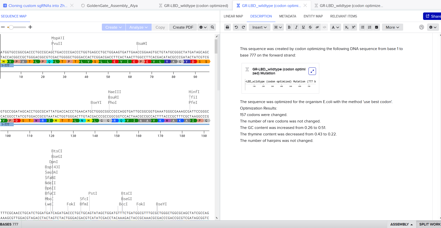
Final Project
DNA Twist Order
https://benchling.com/s/seq-6RNV8iFWmkamF8jpNHMm?m=slm-N50w63IrNM8qnjByu2LE