Week 11 HW: Bioproduction and Cloud Labs
Part A: Pixel Artwork Canvas | Collective Artwork
This was great fun and I kept open multiple tabs and in incognito to quickly fill up boxes (Thanks Georg for the trick!) and somehow managed having parts of my work in the final artwork. For reference I started making the ducks in the first quadrant and someone decided to take it ahead and keep them till the end :)
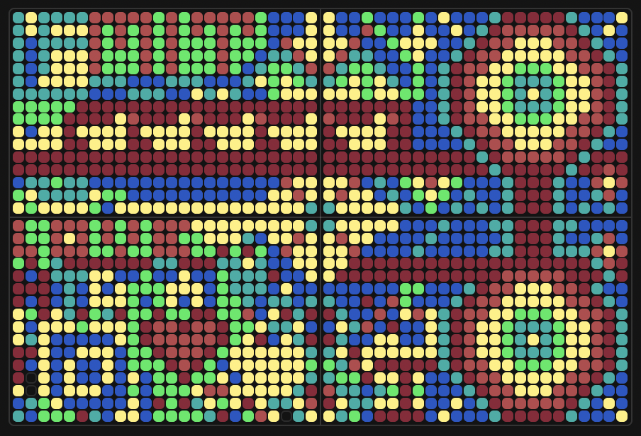
I ended up being #26 on the leaderboard too!

Something for the next year could be maybe a real-time display of who is hovering around and editing the board (something similar to when you can see where people are on google slides). But this also was super fun!
Part B: Cell-Free Protein Synthesis | Cell-Free Reagents
Referencing the cell-free protein synthesis reaction composition (the middle box outlined in yellow on the image above, also listed below), provide a 1-2 sentence description of what each component’s role is in the cell-free reaction.
BL21 (DE3) Star Lysate (includes T7 RNA Polymerase) provides the complete transcription and translation machinery (ribosomes, tRNAs, elongation factors, chaperones), with the DE3 strain supplying T7 RNA Polymerase to transcribe plasmid DNA under a T7 promoter into translatable mRNA.
Salts/Buffer
Potassium Glutamate (312.6 mM): The dominant monovalent salt that stabilizes ribosomes and mimics the ionic environment of the cytoplasm, supporting efficient translation. HEPES-KOH pH 7.5 (45.00 mM): Maintains the reaction at a stable, physiologically relevant pH throughout the 20-hour incubation, preventing enzymatic inactivation from pH drift. Magnesium Glutamate (7.0 mM): Supplies Mg²⁺, an essential cofactor for ribosome integrity, nucleotide stabilization, and polymerase activity. Potassium Phosphate 1.6:1 dibasic:monobasic (5.6 mM) & monobasic:dibasic (5.6 mM): This paired phosphate buffer system provides additional pH buffering and inorganic phosphate to support nucleotide regeneration and energy metabolism over the extended reaction time.
Energy / Nucleotide System
Ribose (77.4 mM): The central energy and nucleotide precursor in this NMP-Ribose system; cellular enzymes in the lysate use ribose to regenerate nucleotides and sustain energy metabolism across the full 20-hour reaction, enabling sustainable protein production. Glucose (6.9 mM): Supplements ribose as a carbon/energy source, feeding glycolytic and metabolic pathways in the lysate to help regenerate ATP and maintain energy charge. AMP (600.00 µM) & UMP (400.00 µM) & CMP (400.00 µM): These nucleoside monophosphates are the building blocks phosphorylated by lysate kinases into their triphosphate forms (ATP, UTP, CTP) for use in transcription; notably AMP is supplied at the highest concentration reflecting ATP’s dominant role in energy currency and transcription. GMP (0.00 µM): Notably absent/zero in this formulation, suggesting that guanosine nucleotides are instead derived entirely from the guanine base supplied separately via salvage pathways. Guanine (200 µM): A free nucleobase that is salvaged by lysate enzymes and converted to GMP and ultimately GTP, supplying the guanosine nucleotides needed for transcription initiation and ribosome function without adding GMP directly.
Translation Mix (Amino Acids)
17 Amino Acid Mix (4.10 mM): Provides the majority of the 20 standard amino acids as substrates for ribosomal peptide bond formation during translation. Tyrosine pH 12 (4.10 mM): Tyrosine’s poor solubility at neutral pH requires it to be dissolved at pH 12 and added separately to ensure it is present at sufficient concentration without crashing out of solution. Cysteine (4.00 mM): Added separately due to its chemical instability and oxidation sensitivity; its concentration is also independently tunable for proteins requiring precise redox conditions or disulfide bonds.
Nicotinamide (3.10 mM): A NAD⁺ precursor that replenishes the lysate’s nicotinamide cofactor pool, sustaining the redox reactions and energy regeneration enzymes that keep the reaction productive over the full 20-hour incubation.
Nuclease-Free Water: Brings the master mix to its final working volume without introducing RNases or DNases that would degrade the mRNA template or DNA plasmid, which would be particularly damaging over a long 20-hour reaction.
- Describe the main differences between the 1-hour optimized PEP-NTP master mix and the 20-hour NMP-Ribose-Glucose master mix. The most fundamental difference between the two formulations lies in their energy and nucleotide supply strategy. The 1-hour PEP/NTP system provides nucleotides in their fully phosphorylated triphosphate forms (ATP, GTP, CTP, UTP) along with phosphoenolpyruvate (PEP-Mono) and Maltodextrin 17 as immediate high-energy phosphate donors for rapid, front-loaded transcription and translation, whereas the 20-hour NMP-Ribose system supplies nucleotides as monophosphates (AMP, CMP, UMP) and relies on ribose and glucose as metabolic precursors that are gradually processed by lysate enzymes to regenerate energy over a sustained period. The 1-hour formulation also includes several additives like Spermidine, DMSO, cAMP, NAD, and Folinic Acid that work together to boost immediate transcriptional and translational efficiency, where for example spermidine stabilizes nucleic acids, cAMP activates metabolic pathways, and folinic acid supports one-carbon metabolism, while the 20-hour formulation simplifies this and relies solely on Nicotinamide to maintain redox cofactor pools throughout the longer reaction. There are also notable differences in salt concentrations between the two, with the 1-hour mix using slightly higher Potassium Glutamate (330.47 mM vs. 312.6 mM) and more HEPES (80 mM vs. 45 mM), which likely reflects optimization for a short burst of high activity rather than the more stable ionic environment needed to keep enzymes functional across a full 20-hour incubation.
Part C: Planning the Global Experiment | Cell-Free Master Mix Design
Given the 6 fluorescent proteins we used for our collaborative painting, identify and explain at least one biophysical or functional property of each protein that affects expression or readout in cell-free systems.
Here are the key biophysical or functional properties for each of the six fluorescent proteins used in the collaborative painting:
1. sfGFP (Superfolder GFP) sfGFP was specifically engineered with folding and solubility-enhancing mutations that allow it to fold robustly even when fused to poorly folded polypeptides, and it shows increased thermal stability and superior resistance to chemical denaturants compared to conventional GFPs. In a cell-free context, this makes sfGFP a highly reliable reporter since its robust folding characteristics mean it can mature efficiently even in the relatively unstructured, open environment of a lysate-based reaction.
2. mRFP1 mRFP1 is reported to be a somewhat slowly-maturing monomer, though it still matures more than 10 times faster than its tetrameric predecessor DsRed, with lower extinction coefficient, quantum yield, and photostability as tradeoffs. In a cell-free system, its relatively slow maturation and reduced brightness compared to newer RFPs means that significant incubation time is needed before a strong red fluorescence signal can be detected, which is particularly relevant when interpreting endpoint reads of a short reaction.
3. mKO2 (monomeric Kusabira-Orange 2) mKO2 is a mutant of mKO1 that was specifically engineered to feature rapid maturation while maintaining the brilliance and pH stability of the parent Kusabira-Orange protein. However, it does exhibit moderate acid sensitivity, which is worth considering in cell-free systems where pH can drift during extended incubation, potentially quenching the orange fluorescence signal over time.
4. mTurquoise2 mTurquoise2’s maturation kinetics are complex and cannot be captured by a single exponential, and in vivo characterization placed it among the slowest-maturing cyan fluorescent proteins tested, requiring a two-step maturation model. Despite this slow maturation being a potential limitation for short cell-free reactions, mTurquoise2 compensates with an exceptionally high quantum yield of 0.93, making it one of the brightest cyan fluorescent proteins available and capable of providing strong signal even at low expression levels.
5. mScarlet-I The single amino acid substitution T74I in mScarlet-I results in a marked acceleration of maturation compared to the parent mScarlet, though at the cost of a moderate decrease in quantum yield (0.54) and fluorescence lifetime (3.1 ns), both of which still remain higher than those of all previously engineered bright mRFPs. This faster maturation makes mScarlet-I particularly well suited for cell-free reactions, as the red fluorescence signal accumulates more quickly and can be reliably detected within the timeframe of the incubation.
6. Electra2 Electra2 is a blue fluorescent protein derived from mRuby3 (itself derived from the sea anemone Entacmaea quadricolor), and intracellular brightness measurements showed it to be over 2-fold brighter than mTagBFP2. However, like other eqFP611-derived proteins, aggregate formation is a known property of Electra2 across multiple organisms and expression contexts, which in a cell-free system could reduce the effective soluble fluorescent protein concentration and lead to an underestimation of actual expression yield if aggregates are not accounted for during fluorescence readout.
- Create a hypothesis for how adjusting one or more reagents in the cell-free mastermix could improve a specific biophysical or functional property you identified above, in order to maximize fluorescence over a 36-hour incubation.
mScarlet-I — Exhibits oxygen-dependent chromophore maturation, meaning that as molecular oxygen is progressively depleted over a long incubation, late-translated mScarlet-I molecules may fail to fully mature and remain non-fluorescent despite being successfully synthesized.
As mScarlet-I chromophore maturation requires molecular oxygen and is sensitive to reducing conditions, one solution could be increasing nicotinamide to sustain the NAD⁺/NADH redox balance over the full 36-hour window, while simultaneously reducing cysteine concentration to prevent excess reducing equivalents from competing with the oxidation step required for chromophore cyclization.
In order to validate and optimize this hypothesis, the following experimental set could be performed:
Sample 1 — Control
- Nicotinamide: 3.10 mM
- Cysteine: 4.00 mM
Sample 2 — Increased nicotinamide only
- Nicotinamide: 6.00 mM
- Cysteine: 4.00 mM
Sample 3 — Reduced cysteine only
- Nicotinamide: 3.10 mM
- Cysteine: 2.00 mM
Sample 4 — Combined nicotinamide increase and cysteine reduction
- Nicotinamide: 6.00 mM
- Cysteine: 2.00 mM
- The second phase of this lab will be to define the precise reagent concentrations for your cell-free experiment.
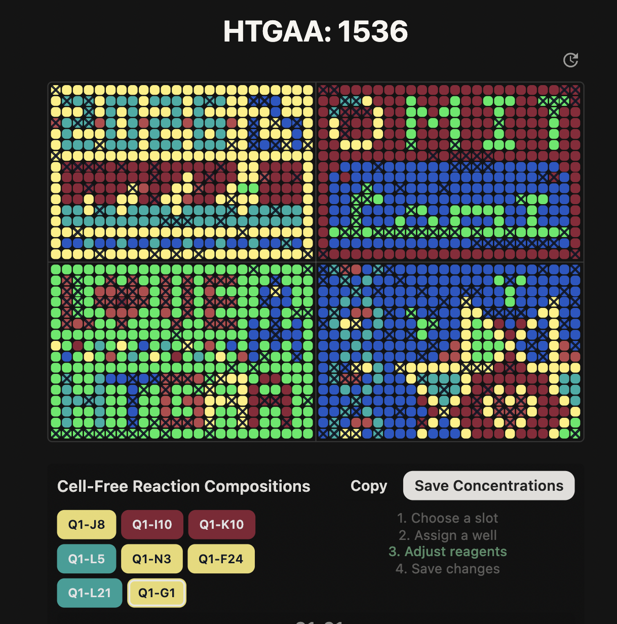
For this experiment, 8 wells were designed across two fluorescent protein hypotheses, both building on the 20-hour NMP-Ribose master mix preset as a baseline. The first set of 4 wells targets mScarlet-I, which requires molecular oxygen for chromophore maturation and is sensitive to reducing conditions over extended incubations. The hypothesis is that increasing nicotinamide concentration will sustain the NAD⁺/NADH redox balance across the full 36-hour reaction, supporting the oxidative environment needed for late-translated mScarlet-I molecules to fully mature. Wells J8 (control, 3.125 mM), I10 (4.500 mM), K10 (6.000 mM), and L5 (8.000 mM) form a nicotinamide gradient to identify the optimal concentration for maximizing red fluorescence endpoint readout. The second set of 4 wells targets mKO2, which exhibits pH sensitivity that can cause fluorescence loss as the reaction environment acidifies over time. The hypothesis is that strengthening the buffer system will maintain pH 7.5 throughout the 36-hour incubation, preserving mKO2 fluorescence intensity. Wells N3 (control), F24 (HEPES 60 mM), L21 (HEPES 75 mM), and G1 (HEPES 60 mM + potassium phosphate dibasic/monobasic both at 7.500 mM) test increasing buffer capacity both through HEPES alone and in combination with elevated phosphate support.