Week 9 HW: Cell-Free Systems
Homework Part A: General and Lecturer-Specific Questions
General homework questions
- Cell-free protein synthesis offers much greater flexibility and experimental control than traditional in vivo systems because it removes the constraints of cell viability, membrane transport, and metabolic regulation. In CFPS, components can be precisely tuned (DNA template concentration, ions, chaperones, cofactors), and toxic or unstable proteins can be produced without affecting living cells. Two cases where CFPS is especially advantageous:
- Toxic proteins (e.g., membrane-disrupting peptides or nucleases) that would kill host cells in vivo.
- Rapid prototyping of genetic constructs, where many variants need to be tested quickly without cloning or cell line generation.
- A CFPS system typically contains:
- Cell extract (E. coli, wheat germ, or rabbit reticulocyte lysate): Provides ribosomes, tRNAs, aminoacyl-tRNA synthetases, and translation machinery.
- DNA or mRNA template: Encodes the target protein; serves as the blueprint for transcription/translation.
- Energy system (ATP regeneration components): Supplies ATP/GTP required for transcription and translation.
- Amino acids: Building blocks for protein synthesis.
- Salts and cofactors (Mg²⁺, K⁺, etc.): Stabilize ribosomes and enzymes.
- Nucleotides (NTPs): Required for transcription of mRNA from DNA.
- Optional additives (chaperones, membrane mimics, redox agents): Improve folding and functionality of expressed proteins.
- Importance of energy regeneration Protein synthesis is extremely energy-intensive; each peptide bond consumes multiple ATP/GTP equivalents. Without regeneration, ATP is rapidly depleted, stopping translation.
To maintain continuous ATP supply, one method is:
- Phosphocreatine + creatine kinase system Phosphocreatine acts as a phosphate reservoir, and creatine kinase regenerates ATP from ADP continuously. Alternative systems include:
- PEP (phosphoenolpyruvate) + pyruvate kinase
- maltodextrin-based slow energy release systems
- Prokaryotic vs eukaryotic CFPS systems Prokaryotic (e.g., E. coli extract):
- Fast, high yield, cost-effective
- Best for simple cytosolic proteins
Example protein: GFP (Green Fluorescent Protein) Reason: GFP folds efficiently in bacterial systems without complex post-translational modifications.
Eukaryotic (e.g., rabbit reticulocyte or wheat germ extract):
- Supports disulfide bonds, glycosylation (limited), and complex folding
- Slower but more physiologically relevant Example protein: human erythropoietin (EPO) Reason: EPO requires proper disulfide bond formation and glycosylation for stability and activity.
- Membrane proteins are challenging because they:
- aggregate in aqueous solution
- require lipid environments for correct folding
Design strategy:
- Add nanodiscs or liposomes to mimic membranes
- Include detergents (e.g., DDM) to stabilize hydrophobic regions
- Use slow expression rates to prevent aggregation
- Co-express chaperones (e.g., Sec translocon components or DnaK system)
Challenges:
- Misfolding and aggregation
- Low yield
- Toxicity to extract components
- (1) Problem: Template instability or poor transcription
- Cause: DNA degradation or weak promoter
- Fix: Use linear DNA protection (GamS protein), switch to stronger promoter (T7), or use circular plasmid templates
(2) Problem: Energy depletion
- Cause: ATP runs out too quickly
- Fix: Improve regeneration system (e.g., phosphocreatine system or slow-release substrates)
(3) Problem: Protein misfolding or aggregation
- Cause: lack of folding assistance or membrane environment
- Fix: Add chaperones, lower temperature, or include nanodiscs/detergents
Homework question from Kate Adamala
Design of a Synthetic Minimal Cell: Environmental Inflammation Detector
- Function a. What would your synthetic cell do? Input and output
The synthetic minimal cell is designed as an inflammation-sensing therapeutic vesicle.
Input: Pro-inflammatory cytokines (TNF-α and IL-6) Output: Anti-inflammatory peptide (e.g., IL-10 mimetic peptide or a short inhibitory cytokine fragment)
The synthetic cell detects inflammatory signals in a tissue environment and responds by producing and releasing an anti-inflammatory therapeutic molecule to restore immune balance.
b. Could this be realized by cell-free Tx/Tl alone?
No, not efficiently.
Cell-free TX/TL systems could produce the anti-inflammatory peptide, but without encapsulation they would:
lack spatial control, diffuse uncontrollably, and be rapidly degraded in biological fluids.
Encapsulation is essential to:
localize response at inflammation sites, create a threshold-based sensing system, and protect the transcription–translation machinery. c. Could this be realized by a genetically modified natural cell?
Yes, macrophages or engineered HEK cells could theoretically perform this function.
However:
immune cells already have complex endogenous cytokine networks, tuning specificity and reducing off-target immune activation is difficult, and safety risks (overactivation or immune rejection) are high.
A synthetic minimal cell offers a modular, orthogonal, and safer alternative.
d. Desired outcome
In the presence of elevated TNF-α / IL-6, the synthetic cell:
activates internal gene circuits, produces anti-inflammatory peptides, releases them locally, and reduces inflammatory signaling in surrounding tissue.
The system behaves like a programmable immunomodulatory “drug factory”.
- Design of Synthetic Cell Components a. Membrane composition
The membrane is composed of:
DOPC (1,2-dioleoyl-sn-glycero-3-phosphocholine) – structural lipid Cholesterol – membrane stability and rigidity control DOPE (helper lipid) – enhances membrane fusion and protein insertion Optional PEGylated lipids – to reduce immune clearance
This creates a stable giant unilamellar vesicle (GUV)-like synthetic cell membrane.
b. Encapsulated components
Inside the synthetic cell:
Cell-free TX/TL system (E. coli-based extract) ATP regeneration system (phosphocreatine + creatine kinase) DNA circuits: TNF-α sensing module (aptamer or receptor-based transcriptional switch) IL-6 sensing module AND/threshold logic promoter Gene encoding: anti-inflammatory peptide (IL-10 mimetic) secretion peptide (e.g., α-hemolysin pore or signal peptide system) c. TX/TL system choice
A bacterial cell-free system (E. coli extract) is sufficient because:
no glycosylation is required for peptide output fast expression kinetics are beneficial well-characterized regulatory parts (T7 promoter, riboswitches) can be used
A mammalian system is not necessary unless full cytokine glycoproteins are required.
d. Communication with environment
The synthetic cell communicates via:
membrane protein pores (e.g., α-hemolysin, aHL) for small molecule exchange surface-displayed cytokine-binding aptamers or receptors for sensing TNF-α / IL-6
Mechanism: Cytokines bind to membrane receptors/aptamers Signal triggers internal TX/TL gene expression Peptide is produced Peptide diffuses out through pores or membrane leakage control
- Experimental Details a. Lipids and genes Lipids: DOPC (dioleoylphosphatidylcholine) DOPE (dioleoylphosphatidylethanolamine) Cholesterol DSPE-PEG2000 (optional stabilization lipid) Genes / genetic components: T7 RNA polymerase system (core TX/TL driver) TNF-α aptamer-based sensor module IL-6 responsive promoter circuit α-hemolysin (HlyA) gene (membrane pore formation) IL-10 mimetic peptide coding sequence Optional regulatory logic: toehold switches or riboswitch-based AND gate b. Measurement of system function
Function is measured using: ELISA assays for released IL-10 mimetic peptide Fluorescent reporter replacement (GFP or mCherry) in prototype systems Microfluidic inflammation-on-chip platforms Dose–response curves measuring: TNF-α concentration vs output peptide level IL-6 concentration vs output activation threshold
Additional validation: time-resolved fluorescence kinetics comparison against non-encapsulated TX/TL controls Summary
This synthetic minimal cell functions as a programmable immunological regulator, capable of sensing inflammatory cytokines and responding with localized therapeutic peptide production. Compared to natural cells, it offers modularity, safety, and precise tunability, while maintaining biological realism through encapsulated TX/TL systems and lipid vesicle architecture.
Homework question from Peter Nguyen
Application Field: Architecture (Living Responsive Building Materials)
A freeze-dried cell-free embedded wall system that activates with humidity to sense mold risk and actively produces antifungal and air-purifying enzymes inside building materials.
How it works
The system consists of freeze-dried cell-free transcription/translation (TX/TL) packets embedded inside porous architectural materials such as gypsum panels, mycelium composites, or biopolymer-based wall coatings. When ambient humidity increases (e.g., due to leaks, condensation, or flooding), water diffuses into the material and rehydrates the system, activating gene expression. The cell-free system is programmed with genetic circuits that sense moisture-associated chemical signals (e.g., fungal metabolites or pH changes) and respond by producing antifungal enzymes (such as chitinases) or antimicrobial peptides. These molecules then diffuse locally within the material, preventing mold growth and gradually restoring a healthy indoor environment. In more advanced versions, the system could also express fluorescent reporter proteins to visually indicate hidden water damage inside walls.
Societal challenge / market need
Buildings worldwide suffer from hidden moisture damage and mold growth, which leads to structural degradation, expensive repairs, and serious respiratory health issues. Current solutions are passive (insulation barriers) or reactive (manual inspection and remediation), meaning problems are often detected too late. This system provides continuous, autonomous environmental monitoring and mitigation inside building materials themselves, reducing maintenance costs and improving indoor air quality and public health.
Addressing limitations of cell-free systems
Cell-free systems are typically limited by being single-use, moisture-activated, and prone to degradation over time, but these limitations can be turned into design features in architecture. First, freeze-drying ensures long shelf stability, and embedding in hydrophobic–hydrophilic microdomains allows controlled activation only when moisture thresholds are exceeded. Second, the system is designed as a distributed modular network of many micro-reactors, so partial activation still provides functional coverage even if some units are exhausted. Third, encapsulation in protective polymer or lipid-based microcapsules can slow resource depletion and protect enzymes from environmental stress. Finally, redundancy and replaceable material panels (like “living tiles”) allow exhausted sections to be swapped during building maintenance cycles, making the system practical for real-world use.
Homework question from Ally Huang
- Background
Long-duration space missions expose astronauts to microgravity, radiation, and confined habitats that disrupt immune function and increase infection risk. In closed spacecraft environments, microbial growth on surfaces and within life-support systems is also difficult to monitor and control. Traditional lab diagnostics are too resource-heavy for space use. Therefore, there is a need for compact, freeze-dried, on-demand biosensing systems that can detect biological contamination or stress signals in real time. Cell-free systems such as BioBits® offer a lightweight, stable platform for performing molecular diagnostics directly in space environments.
- Molecular / genetic target
Bacterial 16S rRNA gene from E. coli and a GFP reporter gene under a synthetic pathogen-responsive regulatory circuit.
- Relevance to space biology problem
The 16S rRNA gene serves as a universal marker for bacterial presence and can be used to detect microbial contamination in spacecraft environments. By coupling detection of this sequence to a BioBits® cell-free gene expression system that drives GFP production, microbial contamination can be translated into a visible fluorescent signal. This is particularly important in space habitats, where microbial monitoring must be fast, portable, and low-resource. Early detection of bacterial growth helps prevent biofilm formation in life-support systems and reduces infection risk for astronauts in closed environments such as the ISS or future deep-space missions.
- Hypothesis / research goal
We hypothesize that a freeze-dried BioBits® cell-free system engineered with a genetic circuit responsive to bacterial 16S rRNA sequences can function as a reliable microbial detection platform under space-relevant conditions. Upon rehydration with environmental samples, the system will produce GFP in proportion to the concentration of bacterial genetic material, allowing quantitative or semi-quantitative detection. The goal is to create a rapid, low-mass, and equipment-minimal biosensor that enables real-time monitoring of microbial contamination in spacecraft. This approach leverages the stability and modularity of cell-free systems to overcome limitations of traditional cell-based diagnostics in space.
- Experimental plan
Freeze-dried BioBits® reactions will be rehydrated with samples containing known concentrations of bacterial DNA targeting the 16S rRNA gene. Experimental conditions will include high, medium, and low DNA concentrations, along with negative controls lacking bacterial DNA and controls containing non-target human DNA to test specificity. Where needed, miniPCR® will be used to amplify target sequences prior to detection. GFP expression will be measured using the P51 Molecular Fluorescence Viewer, and fluorescence intensity will be used to quantify detection sensitivity and response dynamics. Data will be analyzed to determine detection threshold, dynamic range, and specificity of the system.
Homework Part B: Individual Final Project
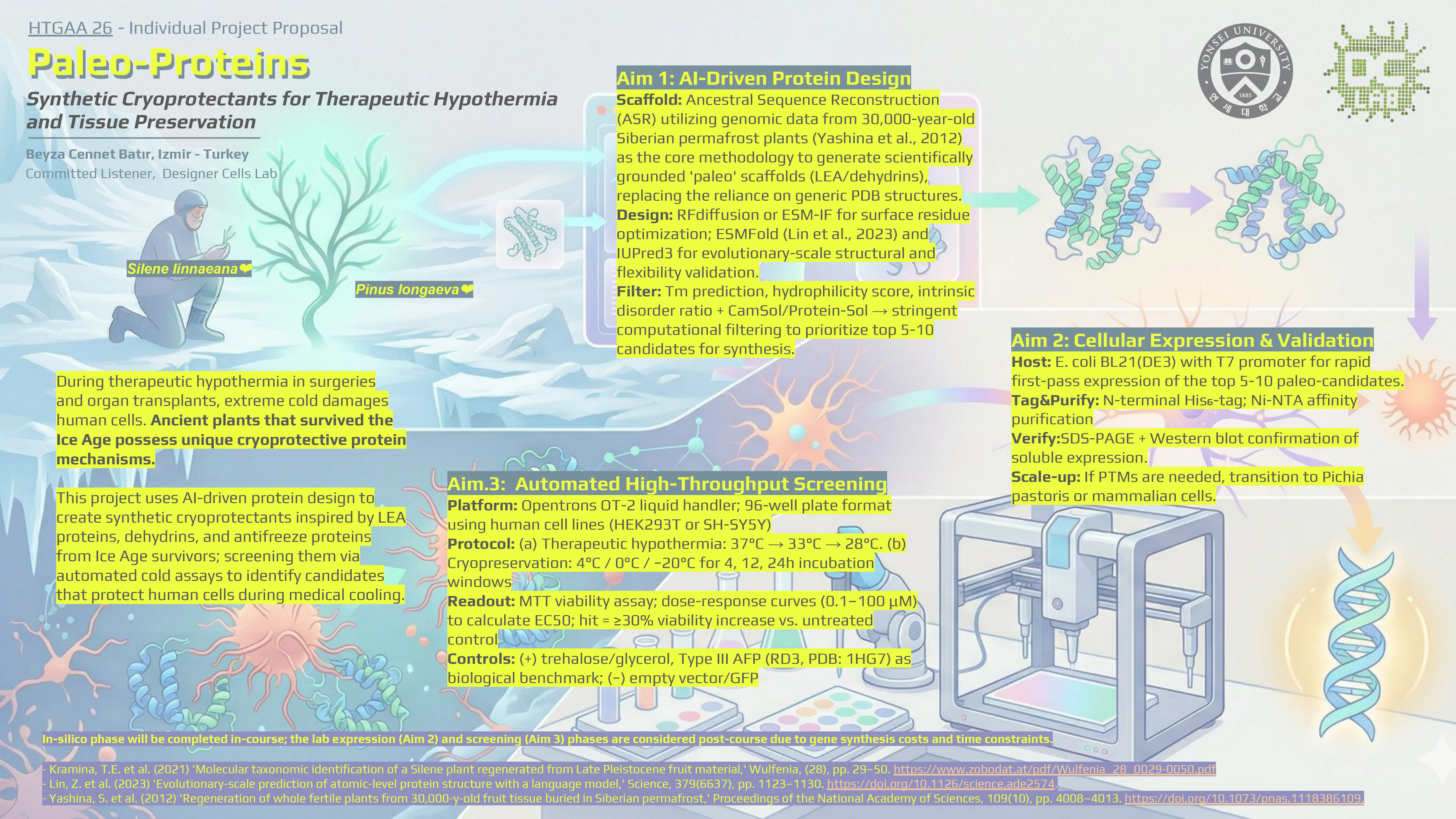
- Beyza Batır - One Final Project Idea cover image
- Please use this directing link to see my submitted form.
- Individual Final Project
- Order Details of the order from Twist Bioscience: https://drive.google.com/drive/folders/1RdyDg39u1akXjmPxIKRLrWrsKfAhuHpx