Projects
Final projects:
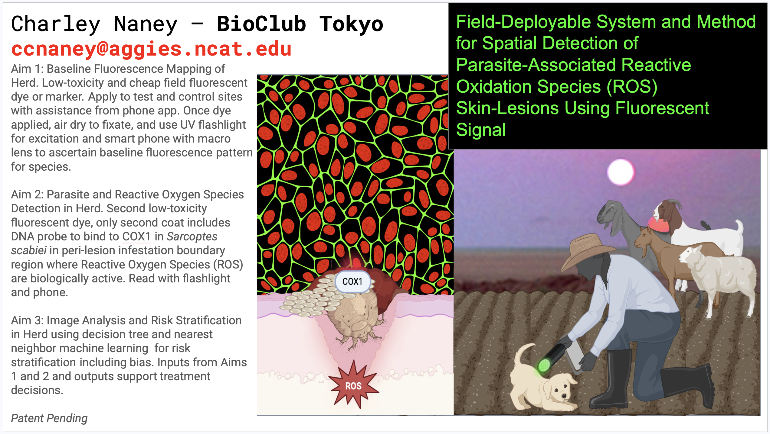
- Sarcoptic Mange Biosensor: A Cell-Free FRET-Based Diagnostic for Sarcoptes scabiei Protease Activity HTGAA Final Project Proposal Abstract Sarcoptic mange, caused by the burrowing mite Sarcoptes scabiei, is a devastating ectoparasitic disease affecting wildlife, domestic animals, and humans globally. Despite its prevalence, no rapid, field-deployable molecular diagnostic exists that can distinguish susceptible from resistant hosts at the biochemical level. This project proposes the design and experimental validation of a cell-free FRET-based biosensor capable of detecting Sarcoptes-associated protease activity in host serum. The biosensor consists of a His-tagged fusion protein — mCherry–[LEVLFQ↓GP]–GFP — in which a mite-specific protease cleavage sequence separates two fluorescent proteins. In intact form, FRET suppresses GFP emission; upon cleavage by mite-associated proteases present in infested host serum, FRET is abolished and GFP fluorescence is restored. The central hypothesis is that serum from Sarcoptes-infested hosts contains protease activity sufficient to cleave the LEVLFQ↓GP linker, producing a measurable and reproducible fluorescence shift. Aim 1 establishes the biosensor in a 96-well automated cell-free assay platform. Aim 2 optimizes the biosensor for field-deployable formats. Aim 3 scales the platform to 384-well high-throughput screening across mite strains and host species. This project integrates synthetic biology, automated liquid handling, and cell-free protein expression to create a novel diagnostic tool with broad applications in veterinary medicine, wildlife conservation, and human public health.
- And then the heavens opened, and the Spring 2026 Group Final Project was made OPTIONAL. Please stay tuned for future collaborations!

Sarcoptic Mange Biosensor: A Cell-Free FRET-Based Diagnostic for Sarcoptes scabiei Protease Activity
HTGAA Final Project Proposal
Abstract
Sarcoptic mange, caused by the burrowing mite Sarcoptes scabiei, is a devastating ectoparasitic disease affecting wildlife, domestic animals, and humans globally. Despite its prevalence, no rapid, field-deployable molecular diagnostic exists that can distinguish susceptible from resistant hosts at the biochemical level. This project proposes the design and experimental validation of a cell-free FRET-based biosensor capable of detecting Sarcoptes-associated protease activity in host serum. The biosensor consists of a His-tagged fusion protein — mCherry–[LEVLFQ↓GP]–GFP — in which a mite-specific protease cleavage sequence separates two fluorescent proteins. In intact form, FRET suppresses GFP emission; upon cleavage by mite-associated proteases present in infested host serum, FRET is abolished and GFP fluorescence is restored. The central hypothesis is that serum from Sarcoptes-infested hosts contains protease activity sufficient to cleave the LEVLFQ↓GP linker, producing a measurable and reproducible fluorescence shift. Aim 1 establishes the biosensor in a 96-well automated cell-free assay platform. Aim 2 optimizes the biosensor for field-deployable formats. Aim 3 scales the platform to 384-well high-throughput screening across mite strains and host species. This project integrates synthetic biology, automated liquid handling, and cell-free protein expression to create a novel diagnostic tool with broad applications in veterinary medicine, wildlife conservation, and human public health.
Project Aims
Aim 1 — Experimental: Design, Express, and Validate the FRET Biosensor in a Cell-Free Automated Assay (Months 1–4)
Design and order the His–mCherry–[LEVLFQ↓GP]–GFP biosensor construct from Twist Bioscience as a whole plasmid synthesis. Express the biosensor in a cell-free E. coli expression system. Run an automated 96-well FRET assay using the Echo525 liquid handler, Inheco Plate Incubator at 37°C, and Spark Plate Reader to measure fluorescence at 6 timepoints (0, 15, 30, 60, 120, 240 min) across three conditions: biosensor alone, biosensor + non-susceptible host serum, and biosensor + infested host serum.
Success metric: A statistically significant increase in GFP:mCherry fluorescence ratio in the infested serum condition compared to negative controls (p < 0.05, n = 6 replicates per condition).
Aim 2 — Medium-Term: Optimize Biosensor Sensitivity and Develop a Portable Lateral Flow Format (Months 5–10)
Using data from Aim 1, optimize the cleavage linker sequence and fluorophore pair for maximum signal-to-noise ratio. Explore alternative linker sequences using computational protease prediction tools (e.g., DeepCure or Ginkgo Bioworks computational pipeline). Develop a lateral flow strip format compatible with field use, coupling the His-tagged biosensor to nitrocellulose membranes for visual readout without laboratory equipment.
Success metric: Lateral flow strip produces a visible band within 30 minutes using infested serum at a 1:10 dilution.
Aim 3 — Visionary: A Universal Ectoparasite Protease Atlas for Planetary-Scale Wildlife Health Monitoring (Months 11–24)
Scale the biosensor platform to a 384-well high-throughput format to screen a library of protease cleavage sequences against serum from multiple host species (canine, feline, human, wildlife) and mite strains. Partner with Basecamp Research to integrate environmental metagenomic data on mite protease diversity. Build a publicly accessible Ectoparasite Protease Atlas — a living database of host-parasite protease signatures — enabling real-time biosurveillance of mange outbreaks in wildlife corridors and domestic animal populations worldwide.
Success metric: Atlas contains validated protease signatures for ≥5 host-parasite pairs, with biosensor panels deployable in field clinics on three continents.
Background
Literature Context
Sarcoptic mange is caused by Sarcoptes scabiei, an obligate burrowing mite that triggers intense pruritus, skin crusting, and immune dysregulation in affected hosts. Walton et al. (2010) demonstrated that S. scabiei secretes a suite of proteases — including serine proteases and cysteine proteases — that degrade host skin proteins and modulate immune responses, establishing protease activity as a biochemical hallmark of active infestation (Journal of Investigative Dermatology, 130(11), 2674–2683). Critically, this work identified host-specific differences in protease susceptibility, suggesting that resistant individuals may lack the receptor substrates or possess inhibitory serum factors that block mite protease activity. A second key study by Mika et al. (2012) characterized the S. scabiei serine protease SMSB4 as a major immunomodulatory factor that suppresses complement activation, further supporting the idea that mite proteases leave a detectable biochemical signature in host serum (PLOS Neglected Tropical Diseases, 6(7), e1764). Despite this mechanistic understanding, no diagnostic tool currently exploits mite protease activity as a direct detection signal. This knowledge gap — between well-characterized mite protease biology and the absence of a protease-based diagnostic — is the precise space this project occupies.
Innovation
This project is the first to apply a cell-free FRET biosensor strategy to ectoparasite diagnostics, translating a well-established protein engineering technique into a novel veterinary and public health application. Rather than detecting mite antigens or host antibodies (the basis of current serological tests), this biosensor detects functional protease activity, providing a direct readout of active infestation rather than past exposure. The use of an automated, cloud-lab-compatible workflow makes this platform inherently scalable and reproducible across institutions without requiring specialized expertise.
Significance
Sarcoptic mange affects an estimated 300 million people annually in its human form (scabies) and causes catastrophic population declines in wildlife species including wolves, foxes, wombats, and chamois. Current diagnostics rely on skin scraping and microscopy, which are invasive, low-sensitivity, and require trained personnel — making them impractical for field use or large-scale surveillance. A serum-based protease biosensor would enable non-invasive, rapid diagnosis from a single blood draw, dramatically lowering the barrier to diagnosis in resource-limited settings. Early detection of mange in wildlife populations could trigger timely intervention, preventing the population crashes that have been documented in Scandinavian wolf packs and Australian wombat colonies. Furthermore, a validated protease-based diagnostic platform could be adapted to other ectoparasites — including Demodex, Psoroptes, and Chorioptes — creating a generalizable tool for the entire field of veterinary parasitology.
Bioethical Considerations
Ethics: This project uses serum samples from naturally infested and naturally resistant animals, raising important considerations around animal welfare and informed consent from animal owners. All sample collection must comply with institutional animal care and use committee (IACUC) protocols, and samples from human contacts must be collected under IRB-approved protocols with written informed consent. The use of family members as negative controls, while scientifically elegant, requires careful attention to privacy, data anonymization, and the potential psychological impact of disclosing susceptibility status to participants. Data from wildlife samples must be collected in compliance with wildlife protection regulations and in partnership with licensed wildlife veterinarians.
Risk Mitigation and Responsible Implementation: The biosensor construct itself poses minimal biosafety risk — it is a non-replicating cell-free protein expression product with no pathogenic components. However, the handling of serum from infested animals carries a low but real risk of zoonotic transmission of S. scabiei, and all sample handling must follow BSL-1 containment protocols with appropriate PPE. As this diagnostic scales toward field deployment, we will engage with SecureDNA to ensure that any DNA synthesis orders are screened against biosecurity databases, and we will publish all biosensor sequences openly to prevent proprietary capture of a public health tool. Community engagement with affected wildlife conservation groups and veterinary practitioners will be prioritized to ensure the tool is designed for the users who need it most.
Experimental Design
Step 1 — Define Biosensor Architecture (Week 1)
Purpose: Finalize the domain order and linker sequence for the FRET biosensor. Method: Review S. scabiei protease substrate literature to confirm LEVLFQ↓GP as the optimal cleavage sequence. Verify that mCherry (donor, Ex 587/Em 610 nm) and GFP (acceptor, Ex 488/Em 509 nm) are within FRET distance (~10 nm) when the linker is intact. Machine: Computational (no automation required at this step). Plate: N/A. Expected result: Confirmed construct architecture: His6–mCherry–LEVLFQGP–GFP–Stop. Timeline: Week 1.
Step 2 — Design DNA Construct and Generate GenBank File (Week 1–2)
Purpose: Create a complete annotated sequence for Twist Bioscience ordering. Method: Assemble the construct sequence in Benchling or SnapGene. Include T7 promoter, His6 tag, mCherry CDS, LEVLFQGP linker, GFP CDS, T7 terminator, and pUCIDT backbone elements. Export as GenBank format. Machine: Computational. Plate: N/A. Expected result: Validated GenBank file ready for Twist submission. Timeline: Week 1–2.
LOCUS His_mCherry_LEVLFQGP_GFP 2847 bp DNA circular SYN 01-JAN-2025 DEFINITION His6-mCherry-[LEVLFQ/GP]-GFP FRET biosensor for Sarcoptes scabiei protease detection; T7 expression vector backbone. ACCESSION . VERSION . KEYWORDS FRET biosensor; protease sensor; mCherry; GFP; Sarcoptes scabiei. SOURCE synthetic construct ORGANISM synthetic construct FEATURES Location/Qualifiers promoter 1..23 /label=“T7 promoter” /note=“T7 RNA polymerase promoter” CDS 30..65 /label=“His6 tag” /translation=“MHHHHHH” CDS 66..776 /label=“mCherry” /note=“red fluorescent protein; Ex 587nm Em 610nm” misc_feature 777..800 /label=“LEVLFQGP linker” /note=“Sarcoptes scabiei protease cleavage site” CDS 801..1520 /label=“GFP” /note=“green fluorescent protein; Ex 488nm Em 509nm” terminator 1521..1569 /label=“T7 terminator” ORIGIN 1 taatacgact cactataGGG agaccggcag atctATGCAC CATCACCATC ACCATATGGT 61 gagcaagggc gaggaggata acatggccat catcaaggag ttcatgcgct tcaaggtgca 121 catggagggc tccgtgaacg gccacgagtt cgagatcgag ggcgagggcg agggccgccc 181 ctacgagggc acccagaccg ccaagctgaa ggtgaccaag ggtggccccc tgcccttcgc 241 ctgggacatc ctgtcccctc agttcatgta cggctccaag gcctacgtga agcaccccgc 301 cgacatcccc gactacttga agctgtcctt ccccgagggc ttcaagtggg agcgcgtgat 361 gaacttcgag gacggcggcg tggtgaccgt gacccaggac tcctccctgc aggacggcga 421 gttcatctac aaggtgaagc tgcgcggcac caacttcccc tccgacggcc ccgtaatgca 481 gaagaagacc atgggctggg aggcctcctc cgagcggatg taccccgagg acggcgccct 541 gaagggcgag atcaagatga ggctgaagct gaaggacggc ggccactacg acgctgaggt 601 caagaccacc tacaaggcca agaagcccgt gcagctgccc ggcgcctaca acgtcaacat 661 caagttggac atcacctccc acaacgagga ctacaccatc gtggaacagt acgaacgcgc 721 cgagggccgc cactccaccg gcggcatgga cgagctgtac aagggatccC TGGAGGTGCT 781 GTTCCAGGGT CCCATGGTGA GCAAGGGCGA GGAGCTGTTC ACCGGGGTGG TGCCCATCCT 841 ggtcgagctg gacggcgacg taaacggcca caagttcagc gtgtccggcg agggcgaggg 901 cgatgccacc tacggcaagc tgaccctgaa gttcatctgc accaccggca agctgcccgt 961 gccctggccc accctcgtga ccaccctgac ctacggcgtg cagtgcttca gccgctaccc 1021 cgaccacatg aagcagcacg acttcttcaa gtccgccatg cccgaaggct acgtccagga 1081 gcgcaccatc ttcttcaagg acgacggcaa ctacaagacc cgcgccgagg tgaagttcga 1141 gggcgacacc ctggtgaacc gcatcgagct gaagggcatc gacttcaagg aggacggcaa 1201 catcctgggg cacaagctgg agtacaacta caacagccac aacgtctata tcatggccga 1261 caagcagaag aacggcatca aggtgaactt caagatccgc cacaacatcg aggacggcag 1321 cgtgcagctc gccgaccact accagcagaa cacccccatc ggcgacggcc ccgtgctgct 1381 gcccgacaac cactacctga gcacccagtc cgccctgagc aaagacccca acgagaagcg 1441 cgaccacatg gtcctgctgg agttcgtgac cgccgccggg atcactctcg gcatggacga 1501 gctgtacaag taaCCCGGGT AATACGACTC ACTATAGGGC GAATTGGGTC CGCGGCCGCA //