Individual Final Project


Sarcoptic Mange Biosensor: A Cell-Free FRET-Based Diagnostic for Sarcoptes scabiei Protease Activity
HTGAA Final Project Proposal
Abstract
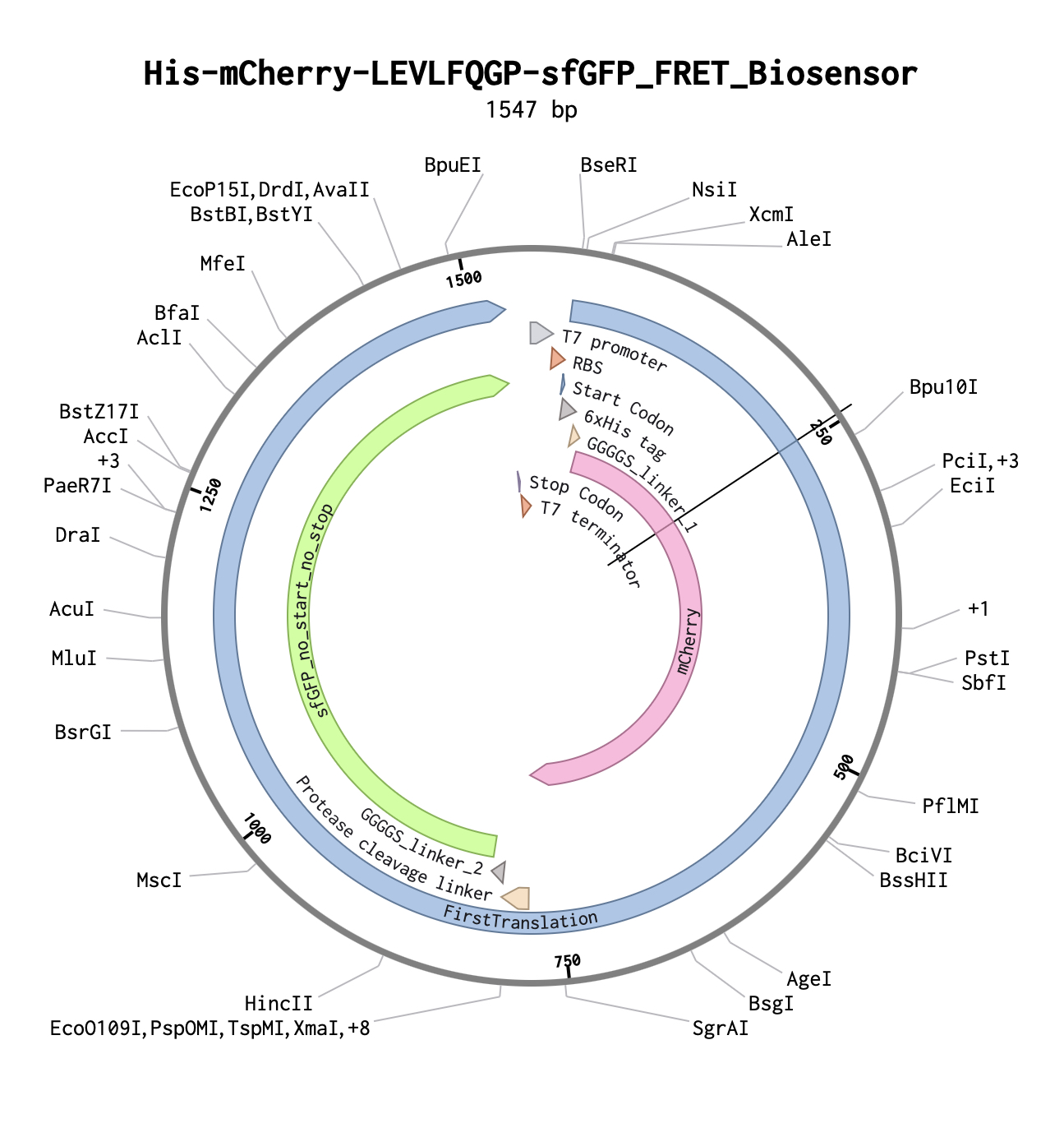
Sarcoptic mange, caused by the burrowing mite Sarcoptes scabiei, is a devastating ectoparasitic disease affecting wildlife, domestic animals, and humans globally. Despite its prevalence, no rapid, field-deployable molecular diagnostic exists that can distinguish susceptible from resistant hosts at the biochemical level. This project proposes the design and experimental validation of a cell-free FRET-based biosensor capable of detecting Sarcoptes-associated protease activity in host serum. The biosensor consists of a His-tagged fusion protein — mCherry–[LEVLFQ↓GP]–GFP — in which a mite-specific protease cleavage sequence separates two fluorescent proteins. In intact form, FRET suppresses GFP emission; upon cleavage by mite-associated proteases present in infested host serum, FRET is abolished and GFP fluorescence is restored. The central hypothesis is that serum from Sarcoptes-infested hosts contains protease activity sufficient to cleave the LEVLFQ↓GP linker, producing a measurable and reproducible fluorescence shift. Aim 1 establishes the biosensor in a 96-well automated cell-free assay platform. Aim 2 optimizes the biosensor for field-deployable formats. Aim 3 scales the platform to 384-well high-throughput screening across mite strains and host species. This project integrates synthetic biology, automated liquid handling, and cell-free protein expression to create a novel diagnostic tool with broad applications in veterinary medicine, wildlife conservation, and human public health.
Project Aims
Aim 1 — Experimental: Design, Express, and Validate the FRET Biosensor in a Cell-Free Automated Assay (Months 1–4)
Design and order the His–mCherry–[LEVLFQ↓GP]–GFP biosensor construct from Twist Bioscience as a whole plasmid synthesis. Express the biosensor in a cell-free E. coli expression system. Run an automated 96-well FRET assay using the Echo525 liquid handler, Inheco Plate Incubator at 37°C, and Spark Plate Reader to measure fluorescence at 6 timepoints (0, 15, 30, 60, 120, 240 min) across three conditions: biosensor alone, biosensor + non-susceptible host serum, and biosensor + infested host serum.
Success metric: A statistically significant increase in GFP:mCherry fluorescence ratio in the infested serum condition compared to negative controls (p < 0.05, n = 6 replicates per condition).
Aim 2 — Medium-Term: Optimize Biosensor Sensitivity and Develop a Portable Lateral Flow Format (Months 5–10)
Using data from Aim 1, optimize the cleavage linker sequence and fluorophore pair for maximum signal-to-noise ratio. Explore alternative linker sequences using computational protease prediction tools (e.g., DeepCure or Ginkgo Bioworks computational pipeline). Develop a lateral flow strip format compatible with field use, coupling the His-tagged biosensor to nitrocellulose membranes for visual readout without laboratory equipment.
Success metric: Lateral flow strip produces a visible band within 30 minutes using infested serum at a 1:10 dilution.
Aim 3 — Visionary: A Universal Ectoparasite Protease Atlas for Planetary-Scale Wildlife Health Monitoring (Months 11–24)
Scale the biosensor platform to a 384-well high-throughput format to screen a library of protease cleavage sequences against serum from multiple host species (canine, feline, human, wildlife) and mite strains. Partner with Basecamp Research to integrate environmental metagenomic data on mite protease diversity. Build a publicly accessible Ectoparasite Protease Atlas — a living database of host-parasite protease signatures — enabling real-time biosurveillance of mange outbreaks in wildlife corridors and domestic animal populations worldwide.
Success metric: Atlas contains validated protease signatures for ≥5 host-parasite pairs, with biosensor panels deployable in field clinics on three continents.
Background
Literature Context
Sarcoptic mange is caused by Sarcoptes scabiei, an obligate burrowing mite that triggers intense pruritus, skin crusting, and immune dysregulation in affected hosts. Walton et al. (2010) demonstrated that S. scabiei secretes a suite of proteases — including serine proteases and cysteine proteases — that degrade host skin proteins and modulate immune responses, establishing protease activity as a biochemical hallmark of active infestation (Journal of Investigative Dermatology, 130(11), 2674–2683). Critically, this work identified host-specific differences in protease susceptibility, suggesting that resistant individuals may lack the receptor substrates or possess inhibitory serum factors that block mite protease activity. A second key study by Mika et al. (2012) characterized the S. scabiei serine protease SMSB4 as a major immunomodulatory factor that suppresses complement activation, further supporting the idea that mite proteases leave a detectable biochemical signature in host serum (PLOS Neglected Tropical Diseases, 6(7), e1764). Despite this mechanistic understanding, no diagnostic tool currently exploits mite protease activity as a direct detection signal. This knowledge gap — between well-characterized mite protease biology and the absence of a protease-based diagnostic — is the precise space this project occupies.
Innovation
This project is the first to apply a cell-free FRET biosensor strategy to ectoparasite diagnostics, translating a well-established protein engineering technique into a novel veterinary and public health application. Rather than detecting mite antigens or host antibodies (the basis of current serological tests), this biosensor detects functional protease activity, providing a direct readout of active infestation rather than past exposure. The use of an automated, cloud-lab-compatible workflow makes this platform inherently scalable and reproducible across institutions without requiring specialized expertise.
Significance
Sarcoptic mange affects an estimated 300 million people annually in its human form (scabies) and causes catastrophic population declines in wildlife species including wolves, foxes, wombats, and chamois. Current diagnostics rely on skin scraping and microscopy, which are invasive, low-sensitivity, and require trained personnel — making them impractical for field use or large-scale surveillance. A serum-based protease biosensor would enable non-invasive, rapid diagnosis from a single blood draw, dramatically lowering the barrier to diagnosis in resource-limited settings. Early detection of mange in wildlife populations could trigger timely intervention, preventing the population crashes that have been documented in Scandinavian wolf packs and Australian wombat colonies. Furthermore, a validated protease-based diagnostic platform could be adapted to other ectoparasites — including Demodex, Psoroptes, and Chorioptes — creating a generalizable tool for the entire field of veterinary parasitology.
Bioethical Considerations
Ethics: This project uses serum samples from naturally infested and naturally resistant animals, raising important considerations around animal welfare and informed consent from animal owners. All sample collection must comply with institutional animal care and use committee (IACUC) protocols, and samples from human contacts must be collected under IRB-approved protocols with written informed consent. The use of family members as negative controls, while scientifically elegant, requires careful attention to privacy, data anonymization, and the potential psychological impact of disclosing susceptibility status to participants. Data from wildlife samples must be collected in compliance with wildlife protection regulations and in partnership with licensed wildlife veterinarians.
Risk Mitigation and Responsible Implementation: The biosensor construct itself poses minimal biosafety risk — it is a non-replicating cell-free protein expression product with no pathogenic components. However, the handling of serum from infested animals carries a low but real risk of zoonotic transmission of S. scabiei, and all sample handling must follow BSL-1 containment protocols with appropriate PPE. As this diagnostic scales toward field deployment, we will engage with SecureDNA to ensure that any DNA synthesis orders are screened against biosecurity databases, and we will publish all biosensor sequences openly to prevent proprietary capture of a public health tool. Community engagement with affected wildlife conservation groups and veterinary practitioners will be prioritized to ensure the tool is designed for the users who need it most.
Experimental Design
Step 1 — Define Biosensor Architecture (Week 1)
Purpose: Finalize the domain order and linker sequence for the FRET biosensor. Method: Review S. scabiei protease substrate literature to confirm LEVLFQ↓GP as the optimal cleavage sequence. Verify that mCherry (donor, Ex 587/Em 610 nm) and GFP (acceptor, Ex 488/Em 509 nm) are within FRET distance (~10 nm) when the linker is intact. Machine: Computational (no automation required at this step). Plate: N/A. Expected result: Confirmed construct architecture: His6–mCherry–LEVLFQGP–GFP–Stop. Timeline: Week 1.
Step 2 — Design DNA Construct and Generate GenBank File (Week 1–2)
Purpose: Create a complete annotated sequence for Twist Bioscience ordering. Method: Assemble the construct sequence in Benchling or SnapGene. Include T7 promoter, His6 tag, mCherry CDS, LEVLFQGP linker, GFP CDS, T7 terminator, and pUCIDT backbone elements. Export as GenBank format. Machine: Computational. Plate: N/A. Expected result: Validated GenBank file ready for Twist submission. Timeline: Week 1–2.
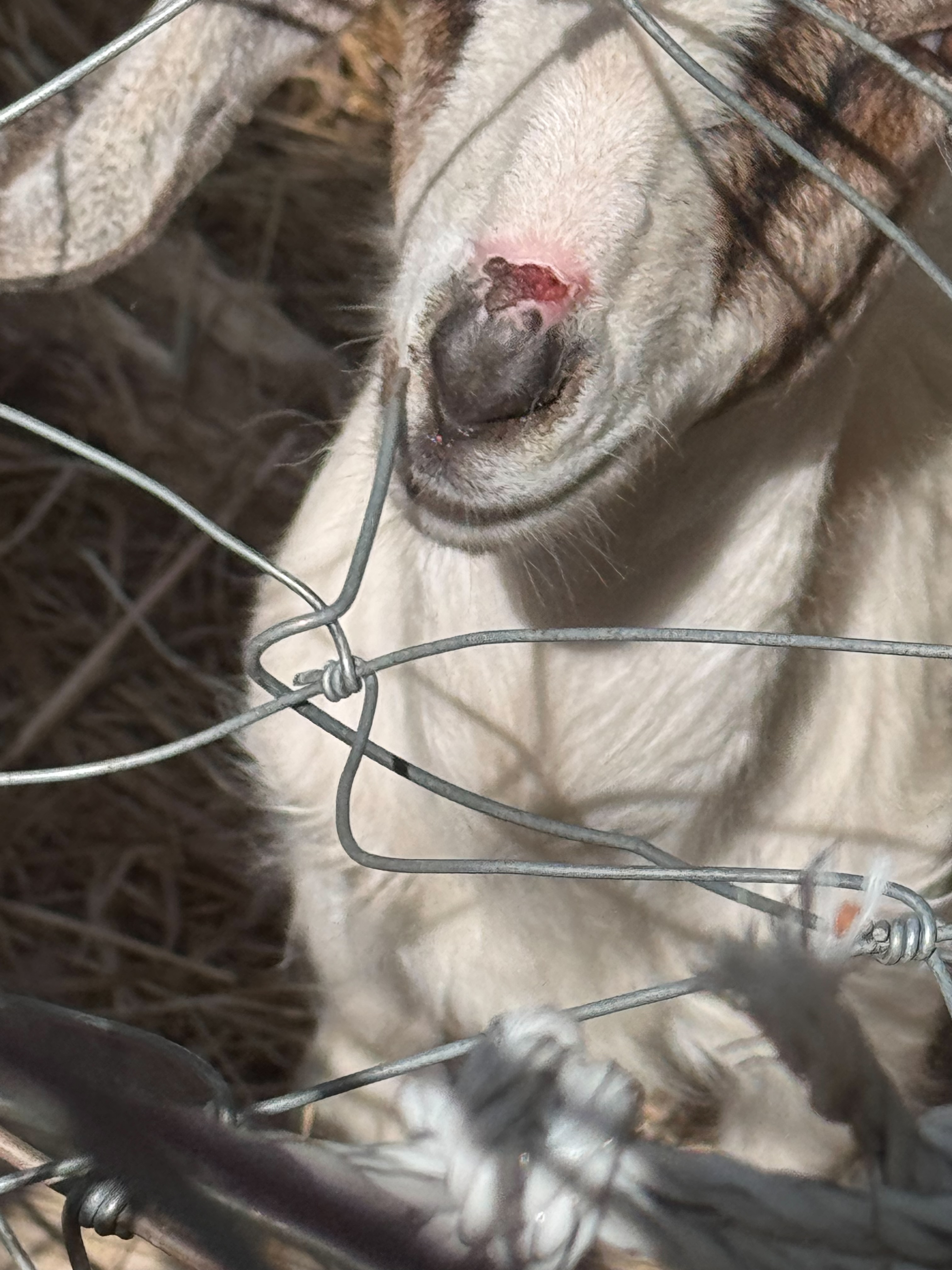
Sarcoptes scabiei Suspected Here!
HTGAA 2026 Final Project: Sarcoptes exploit host geometry – developing assay to measure redox landscape in stratum corneum of goat
By Charley Naney
SECTION 1: ABSTRACT
- Provide an abstract/summary for your project. (minimum 150 words) • Should be a self-contained description of the project • Should contain brief outline of:
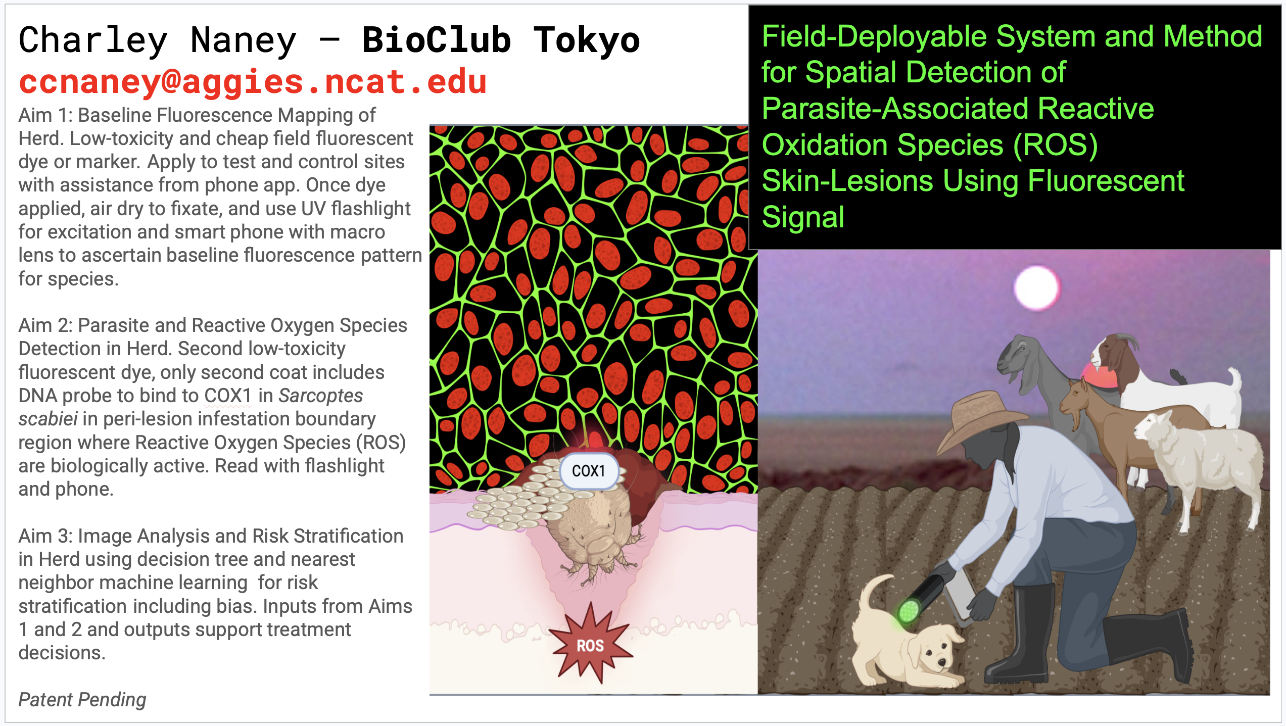
- Significance: The ectoparasitic mite Sarcoptes scabiei exploits the stratum corneum of mammalian skin, including hosts within the Bovidae family (subfamily Caprinae), notably the genera Capra (goats) and Ovis (sheep). While S. scabiei infects a broad range of mammals, this project focuses on caprine hosts (Capra), which provide a tractable and agriculturally relevant system. Goats and sheep share a common ancestor within Caprinae but diverged approximately 4–5 million years ago, adapting to distinct ecological geometries. Goats evolved in heterogeneous, vertical, and discontinuous mountain environments, whereas sheep adapted to more homogeneous, open, and flock-oriented landscapes. These divergent ecological pressures have shaped not only behavior and morphology but potentially the spatial structure of skin physiology, including barrier properties, microenvironmental heterogeneity, and host–parasite interactions. Sarcoptes scabiei is a globally distributed, highly contagious parasite of major veterinary and public health concern. Cross-host transmission is frequently observed in practice: infestations in sheep may spread to goats, dogs, and humans in shared environments, reflecting the long cohabitation of these species under both natural and artificial selection over the past 8,000–10,000 years. This deep ecological and evolutionary entanglement suggests that Sarcoptes scabiei operates within host-derived microenvironments that are conserved enough to permit transmission, yet variable enough to shape infection dynamics. This project introduces a spatially resolved diagnostic concept: rather than sampling lesions indiscriminately, it targets the peri-lesional boundary, hypothesized to represent a localized maximum in reactive oxygen species (ROS) generated by host immune activity. By developing a small-scale assay—potentially leveraging fluorescent ROS probes—to detect this spatial oxidative signature, I aim to create a rapid, geometry-informed approach to identifying mite-associated lesions. While species-level discrimination of Sarcoptes lineages may not be feasible within the project timeline, this approach can be strengthened through triangulation, integrating spatial ROS signals with observational and survey-based metadata (host species, lesion morphology, transmission context). Together, these elements may yield a more robust and field-deployable diagnostic framework. Beyond immediate application, this work addresses a broader evolutionary and biophysical question: how parasites exploit host tissue geometry to create stable microenvironments, and how those environments can be detected through their spatial redox signatures. In this sense, Sarcoptes scabiei infection becomes not only a veterinary problem, but a model system for understanding how geometry, evolution, and immune dynamics converge in living tissue.
- Broad objectives: I will develop spaitially resolved diagnostic assay for detecting Sarcoptes scabiei infection, or evidence of burrows in epidermal layers of skin, using fluorescent ROS probes capable of detecting oxidative stress signature in caprine skin. This approach will include farmer education and a survey for targeting host and environmental metadata, and skin targeting of peri-lesional microenvironments. Testing data can later be combined with transcriptomic sequencing, as other researchers are starting to do to develop putative genes that can be analyzed to develop more exact assay-based biomarkers for diagnosis, prognosis, and more targeted therapy with effective surveillance, as only a recovering Epidemiologist, goat and sheep farmer, returned to Evolutionary Microbiology can dream up.
- Hypotheses: Sarcoptes scabiei infection or infestation, need to decide on best state variable terminology here, is a pathogenic process involving reorganization of the geometry of the stratum corneum. The mites burrow to feed, make shelter, and lay eggs which results in a diffusion-constrained microenvironment ideal for assaying host immune responses surrounding, including what I hypothesize to be a localized region with maximum concentration of ROS distributed within a peri-lesional boundary.
- Specific aims
- Null Hypothesis: In probable Sarcoptes scabiel infested goats, ROS levels will be uniformly distributed across lesion core, peri-lesional boundary tissue, with no spaitial enrichment at lesion boundary.
Alternative Hypothesis: In probable Sarcoptes scabiel infested goats, a localized maximum of ROS at the peri-lesional boundary in caprine skin will be present.
- Methods to be employed • Use lay language (i.e., understandable by the general public) as much as possible SECTION 2: PROJECT AIMS
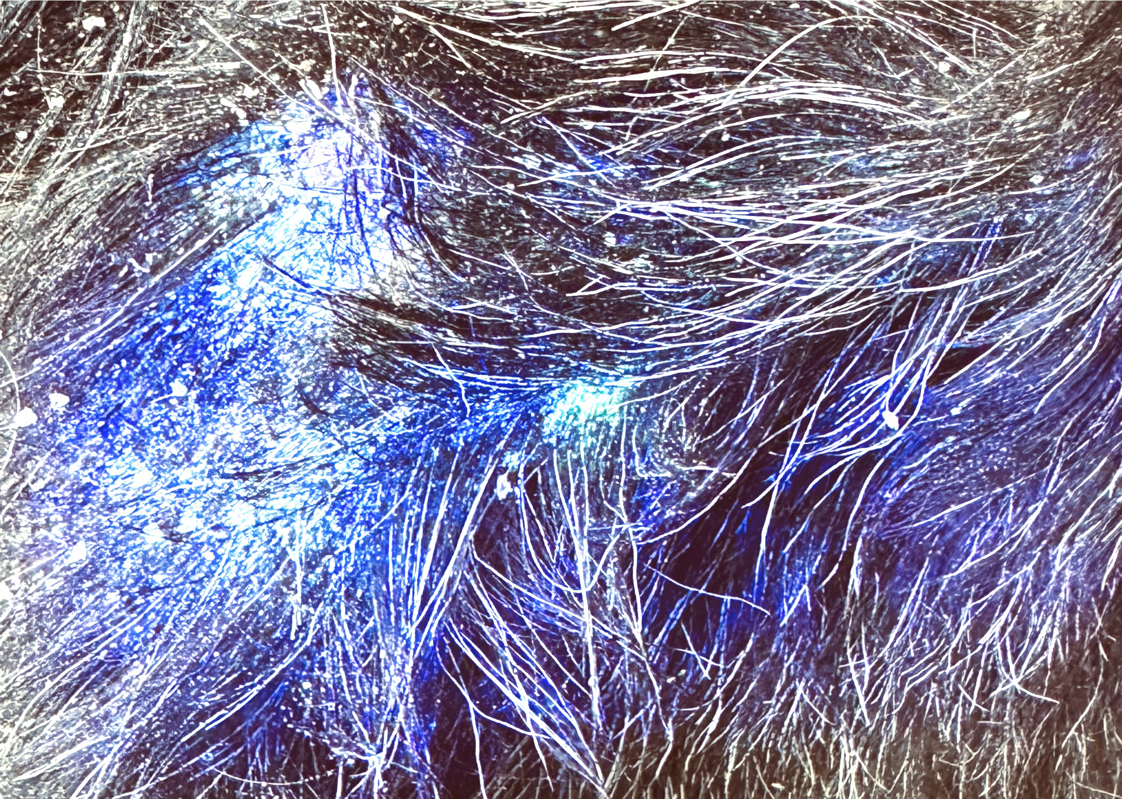
- Outline three aims of your final project (min. 3 sentences, at least one for each aim) Aim 1: Baseline Fluorescence Mapping of Herd The subject (i.e., goat, sheep, other mammal frequently in-contact with herd) is visually inspected using system for suspected lesions indicative of possible infestation. A low toxicity fluorescent dye or marker may be applied to skin surface to survey skin surface to establish baseline imaging conditions. A portable light source (i.e., UV or visible excitation light) will be coupled with imaging device (i.e., smartphone camera with accessory macro lens) will be used capture images of skin regions where first coat of fluorescent die label is applied. This region can include suspected lesion or control region. These images are then analyzed to establish baseline fluorescence patterns and instructions to improve collection of baseline images. Aim 2: Reactive Oxygen Species Detection in Herd Low toxicity in-field application of fluorescent probe capable of interacting with reactive oxygen species is applied to the skin surface, including the lesion and surrounding peri-lesional region. The sample will not be fixed with formaldehyde, a known carcinogen, but instead will be air dried with optional portable fan to accelerate non-chemical fixation process. Excitation will then be reached with appropriate light source, at which point scan and capture of ROS signals will be completed. Spatial variation in fluorescence intensity will then be analyzed to identify biologically active regions, including potential peri-lesional infestation boundaries. Aim 3: Image Analysis and Risk Stratification in Herd Captured images from Aim 1 and Aim 2 are analyzed using computational methods, including intracellular artificial neural network circuit for analogue machine learning modeler to identify patterns of fluorescence intensity associated with peri-lesion activity. The system will classify individual animals in herd based on risk levels to identify individuals requiring further testing as well as guiding further testing and treatment. In some embodiments, the system may be deployed to integrate data across multiple animals to support herd-level decision-making.
State or link any methods/experimental protocols/OpenTrons protocols/DNA or protein designs/protein design tools or models/Twist orders you will use
Quality Control and Validation This is a scientific em-BOLD-iment project , so before system is packaged for any farmer with a smart phone and a goat, an appropriate confirmatory analysis using cutting edge laboratory methods will be performed. Analysis will be conducted using automation and high-throughput systems. The validation protocol will be developed with guidance from MIT HTGAA 2026. Validation steps may be used to correlate field-detected fluorescence signals with molecular or biological markers of infestation.
- Briefly expand upon the significance of your final project. (min. 5 sentences) • Examples of topics to discuss
- There are several ways my project solves a pressing problem in the world. First and foremost, it provides any farmer with a smart phone and my kit, which I hope they can buy at Walmart or Tractor Supply, or they can pick one up for free where such conveniences are not available. The kit will include low-toxicity fluorescence, snap on macro lens, uv flashlight, QR code with registration for an App. They can then snap photo of their cat, and prompt will say not goat. They find goat, prompt correct animal, take photo here but part hair first, ect. This tool will empower them to test their intuition about an animal in the herd that they think may be infested with Sarcoptes scabiei without spending hundreds or thousands of dollars to expose every animal in the herd to toxic treatments for prevention or to consult a vet before they know if they have a problem. For example, “sulfur lime,” which is one of the treatments I am referring to, coats the entire animal in reactive oxygen species. In contrast, S. scabiei is highly localized and as an obligate parasite with a very long evolutionary tail will move from one susceptible animal to the next at a leisurely pace. Granted when enough animals are infested it can be a hard chain of transmission to break, but early detection and localized remediation is effective. Early detection is critical in this regard. Another challenge is the random way it enters herd, often because neighboring farmers are in the same boat. Then there are all the intermediate hosts between herds. The only viable alternative to my solution is marrying a vet or having a kid go to vet school, but my kit is a lot cheaper.
Epilogue Sarcoptes scabiei Cured Here!
No one likes to read the ending before it’s fully written. However in this case if I have a stroke or something in the next two weeks finishing my homework and project documentation it’s important to note that even before the methods were completely executed the approach worked. I have eliminated more than 95% of the Sarcoptes scabiei in my own herd using targeted real-time biosurveillance and the right waves of light. Ultimately, this project will demonstrate this statement biologically and statistically but I already have enough clinical evidence to report with confidence that the intervention strategy worked. I had goats with horns falling off and infected wounds in their nose that were recirculating air flow visibly and audibly. Each animal was an infestation puzzle. The herd was an infestation puzzle. Zoonotic transmission across species was an infestation puzzle. Infestation across domestic and wild animals was an infestation puzzle. All of those puzzles were solved with this project and it is not completed yet, which means efficacy will only improve. None of this existed before I developed this project for HTGAA 2026 and the world will be a better place when it is completed.
Before
After