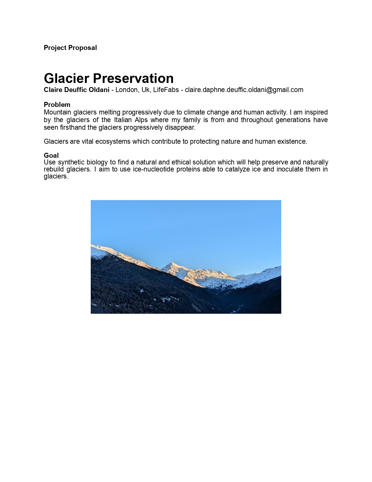
I am a French and Italian textile designer specialized in weaving. I have a maximalist and edgy style with a strong interest in unusual materials and an experimentative approach to weaving.
I am now completing an MA in Biodesign at Central Saint Martins where I aim to develop new innovative materials combining design and science. I am very curious about biology and genetics.
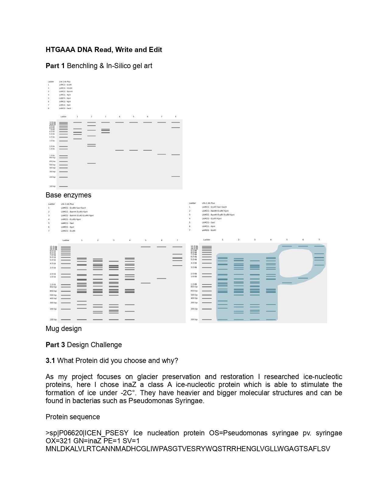
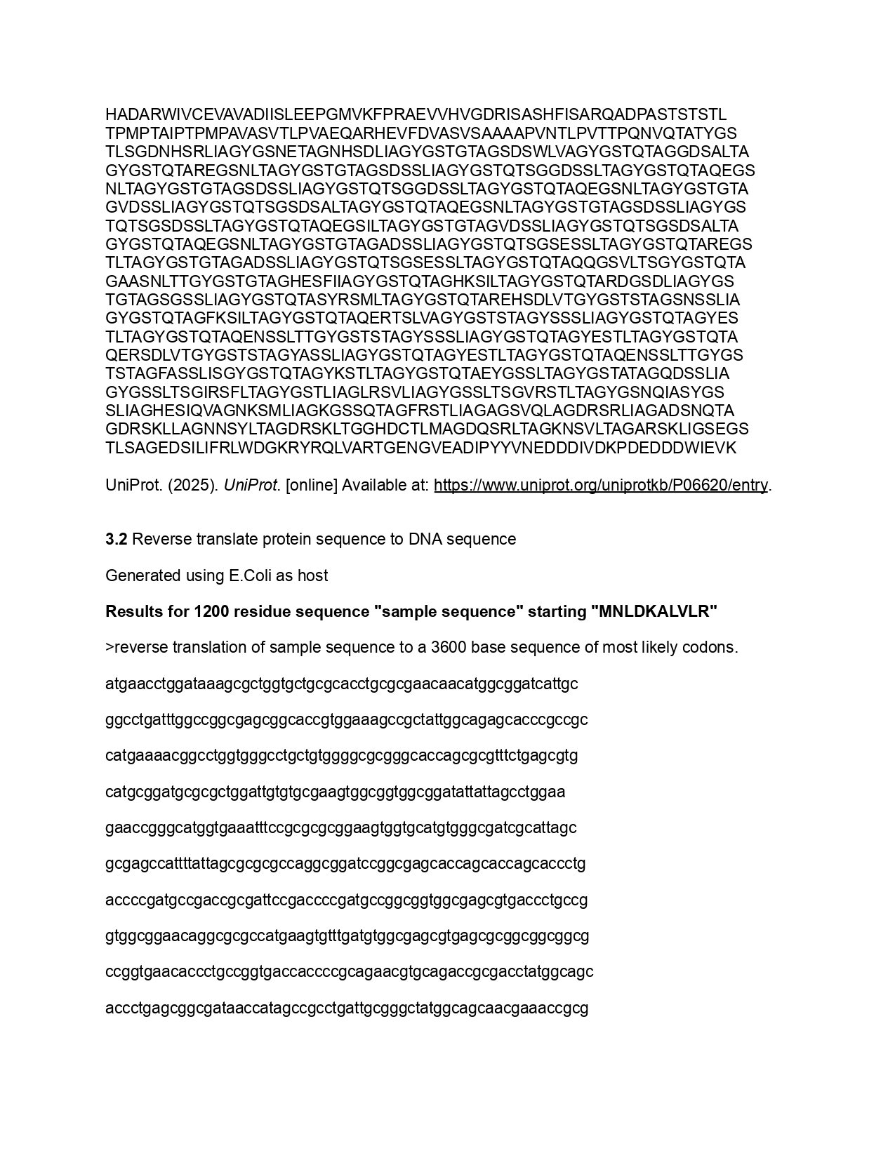
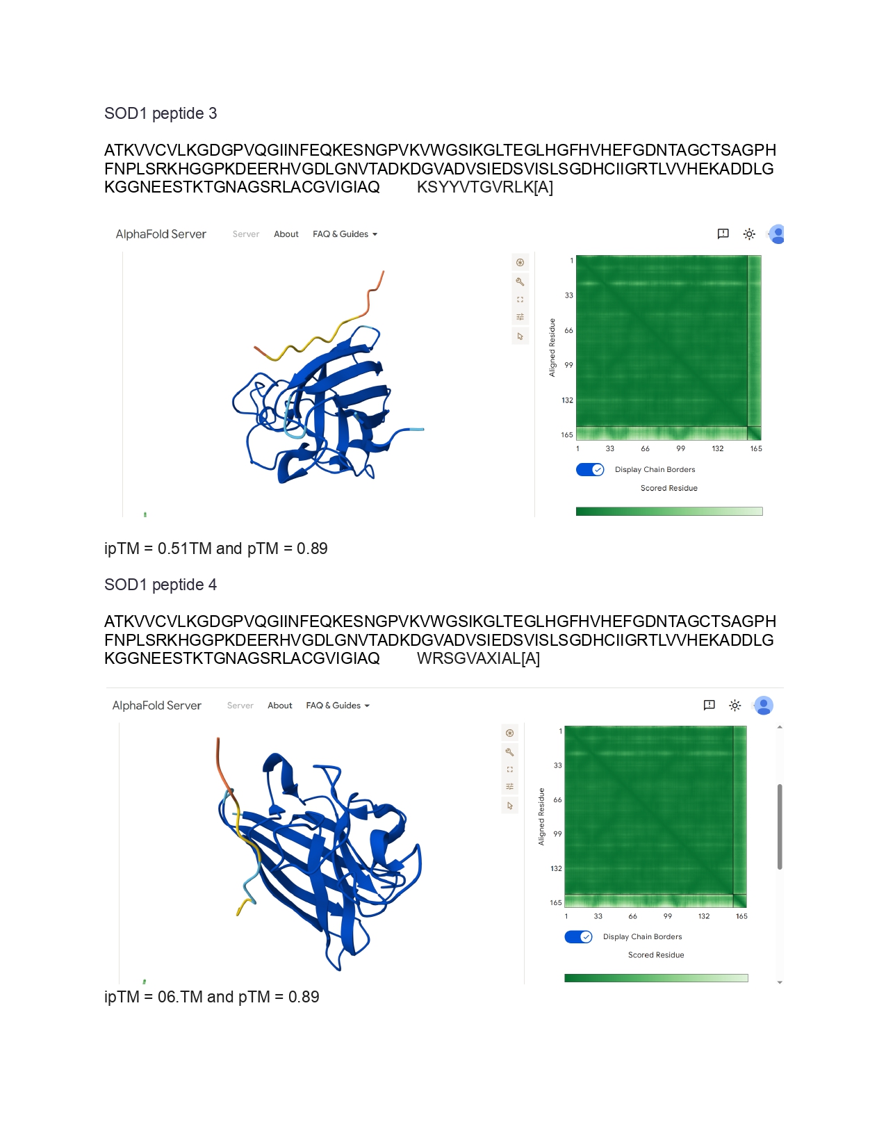
HTGAA Week 6 Genetic circuit part 1 DNA Assembly
1.What are some components in the Phusion High-Fidelity PCR Master Mix and what is their purpose?
The Phusion High-Fidelity PCR Master Mix is a concentrated DNA polymerase solution containing a high fidelity reaction buffer, MgCI (Magnesium chloride) and dNTPs ( deoxynucleoside triphosphates). It is used in PCR as polymerase solution can help fill the gaps in the sequence cloned during Gibson or HiFi assembly. The reaction buffer serves as a stabilizer and MgCI and dNTPs serve as building blocks for PCR. Reference list BioChain Institute Inc. (2024). Biochain Institute Inc. [online] Biochain Institute Inc. Available at: https://www.biochain.com/blog/using-dntp-in-polymerase-chain-reaction-pcr/. New England Biolabs (2026). [online] Neb.com. Available at: https://www.neb.com/en-gb/products/m0531-phusion-high-fidelity-pcr-master-mix-with-hf-buffer [Accessed 23 Mar. 2026].
HTGAA Week 7 Genetic circuits part 2 Part 1: Intracellular Artificial Neural Networks
1.What advantages do IANNs have over traditional genetic circuits, whose input/output behaviors are Boolean functions? The advantage of IANNs over Boolean genetic circuits is their analog sensing and nonlinear processing capabilities, while traditional genetic circuits function as an On and Off system the IANNs can work in a continuous way due to its broader and stronger range of nonlinear inputs. Additionally, the IANNs have better pattern recognition and generalization technology, they can detect more complex patterns in data and can organise them better than the Boolean can.The IANNs are also less likely to fail and have a better error tolerance than the Boolean which is very susceptible to fail if there is a single issue with a gate. Moreover, the IANNs have a better scalable capacity due to the protein splicing mechanism allowing to create a multiple input-output circuit. The IANN technology can also better adapt and evolve to its environment compared to the Boolean which has a fixed function in its environment. Finally, IANNs have a strong memory quality that can be passed through to subsequent generations within the cell due stoichiometric cleavage and splicing which is irreversible.
HTGAA Week 9 Cell Free Systems Part A General and Lecture specific questions
General Homework Questions
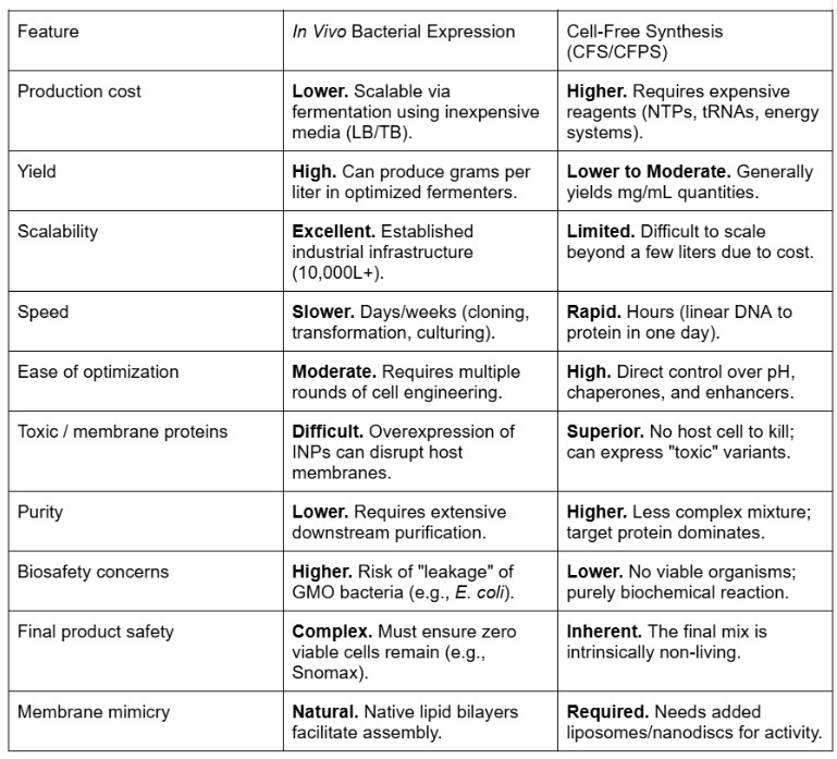
1.Explain the main advantages of cell-free protein synthesis over traditional in vivo methods, specifically in terms of flexibility and control over experimental variables. Name at least two cases where cell-free expression is more beneficial than cell production.
Time : CFPS can be executed in very little time, a couple of hours however in vivo methods will take a few days to a few weeks. Time is a key part of efficient research as less time is wasted waiting to synthesize proteins to experiment on. System : CFPS is an open access system as it has no membrane and gives direct access to the molecule which one wants to work on, it offers more flexibility. It also allows for better control over the synthesis as one can specifically choose which components to input in a protein and better predict protein folding, this simplifies monitoring as well. Versus an in vivo method which has a membrane and therefore a closed system, this means the host and its other components will also react to any modification making it harder to control and direct, the cell’s functioning can often get in the way of the synthesis or create unpredicted issues. Tolerance: CFPS can tolerate high rates of toxic and difficult proteins as it is non living, however, in vivo technologies are more sensitive to toxins as it is likely to harm the host. CFPS also works using non-natural components offering a wider spectrum of possibilities. Complexity : CFPS is a simple PCR based procedure while in vivo requires more complex cloning and transformation steps. Reference List Silverman, A. D., et al. (2020). Quarto: A User’s Guide to Cell-Free Protein Synthesis. Nature Reviews Genetics. Gregorio, N. E., et al. (2019). Cell-free microbial synthesis of proteins. Frontiers in Bioengineering and Biotechnology.
HTGAA Week 10 Advanced imaging & measurement technology Final Project
1.Please identify at least one (ideally many) aspect(s) of your project that you will measure. It could be the mass or sequence of a protein, the presence, absence, or quantity of a biomarker, etc.
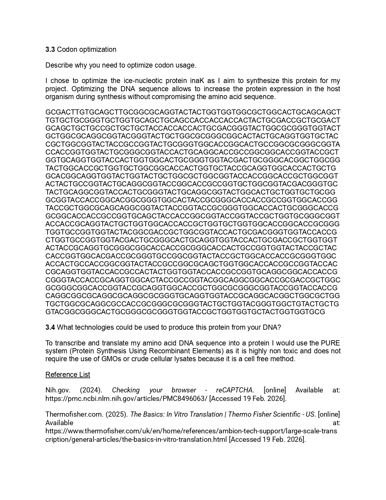
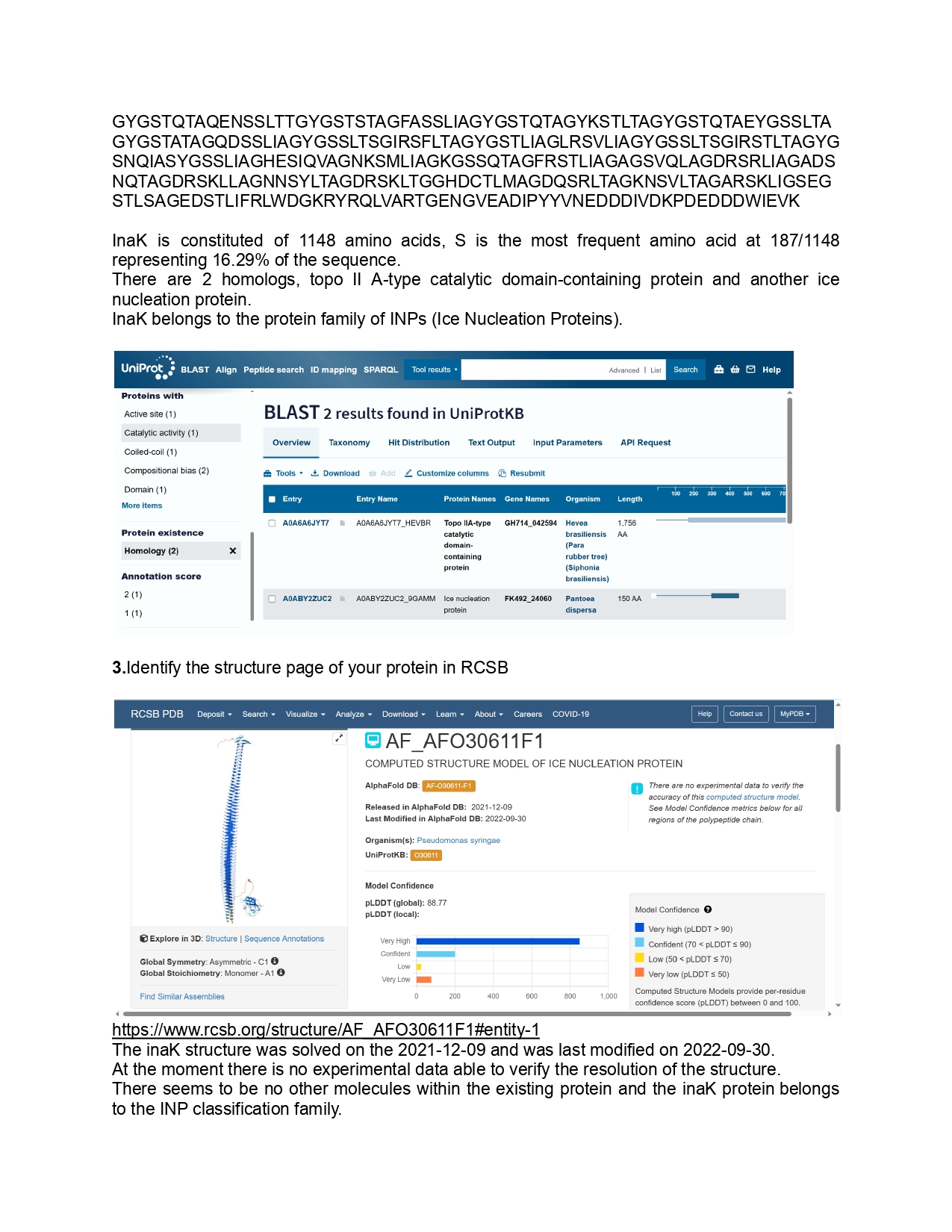
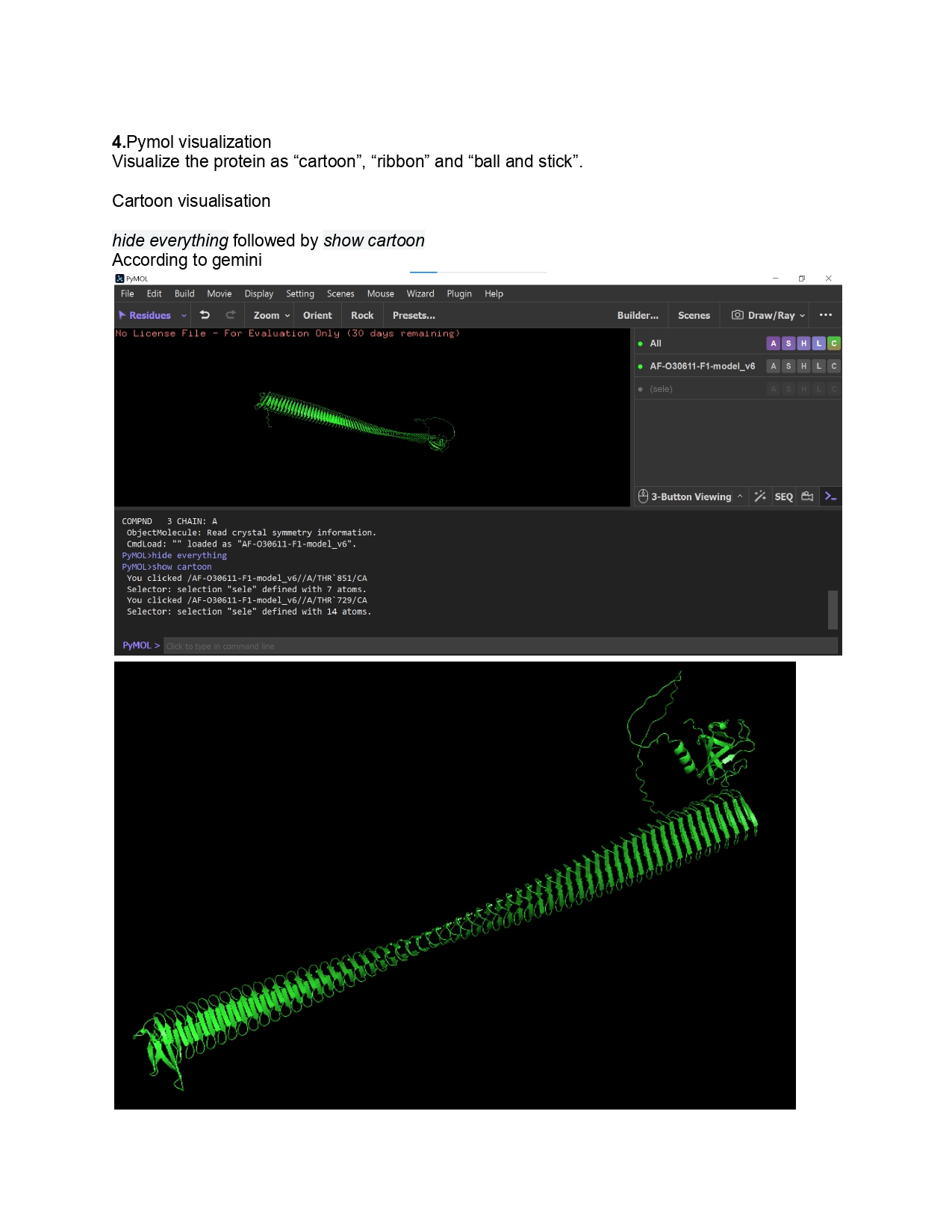
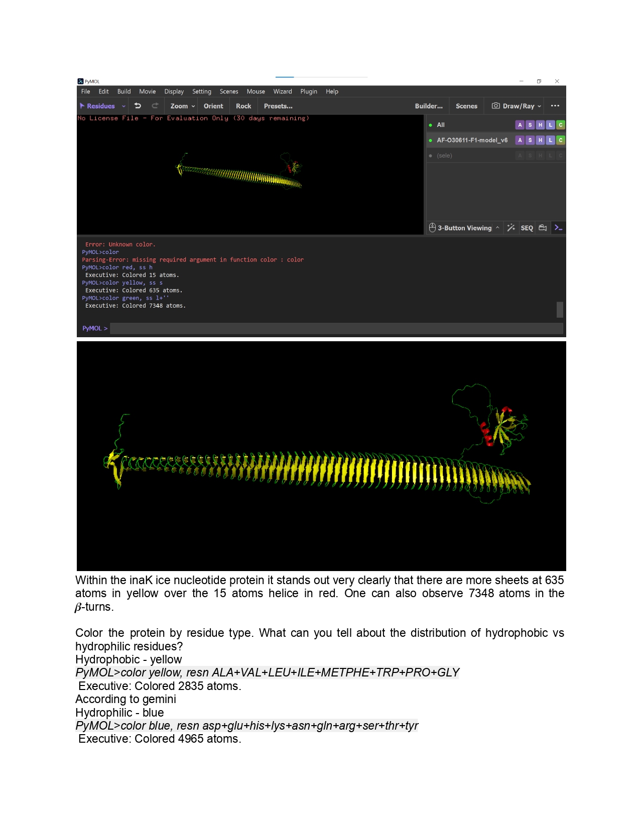
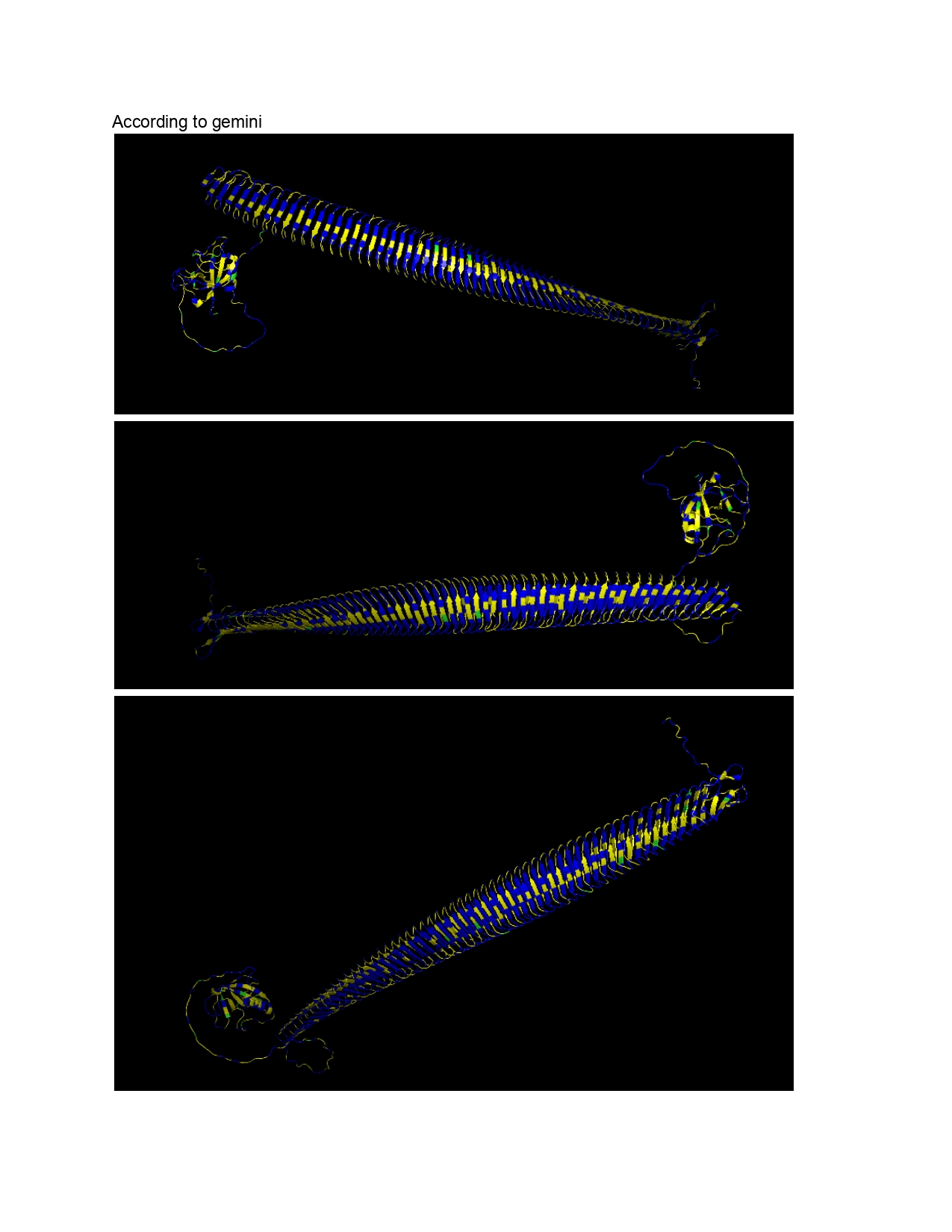
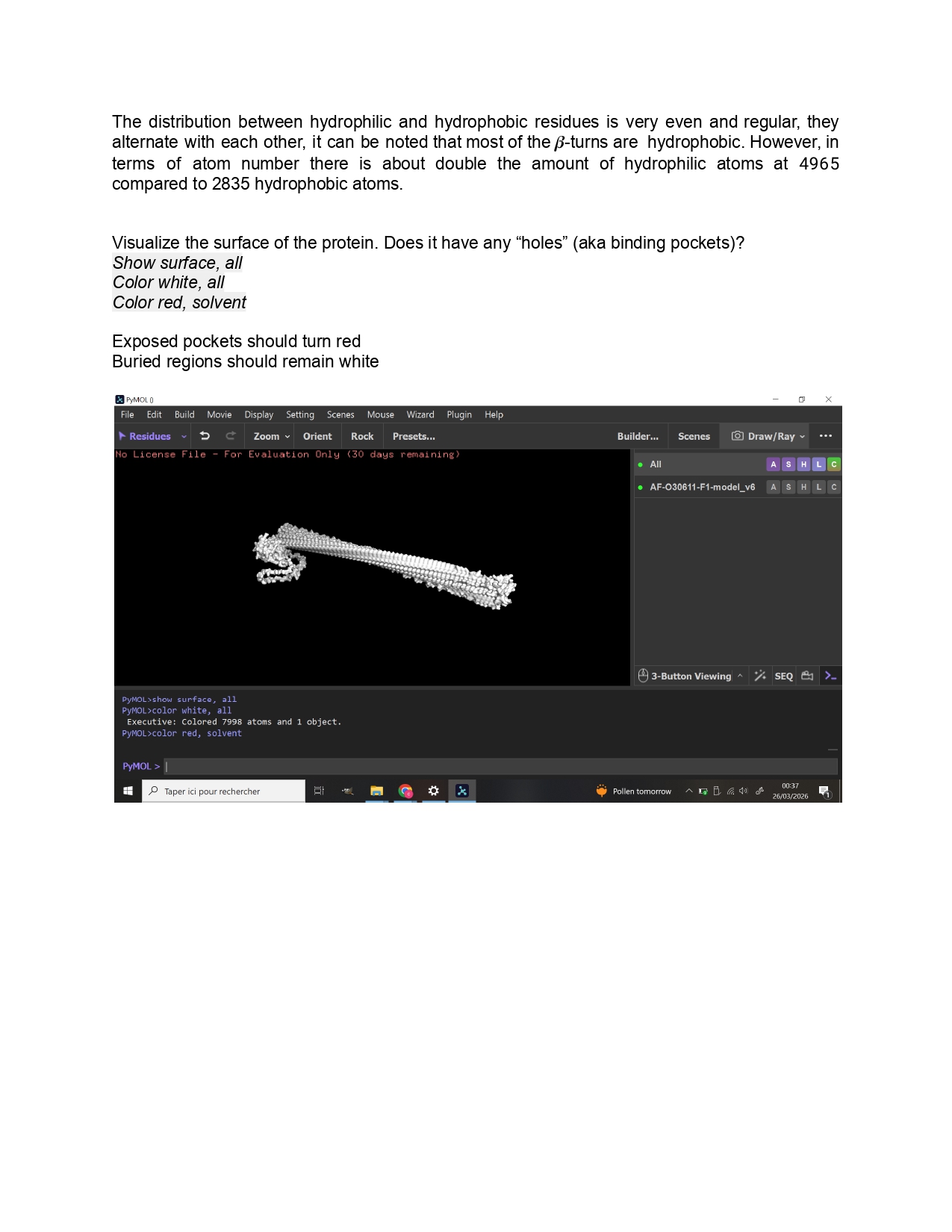
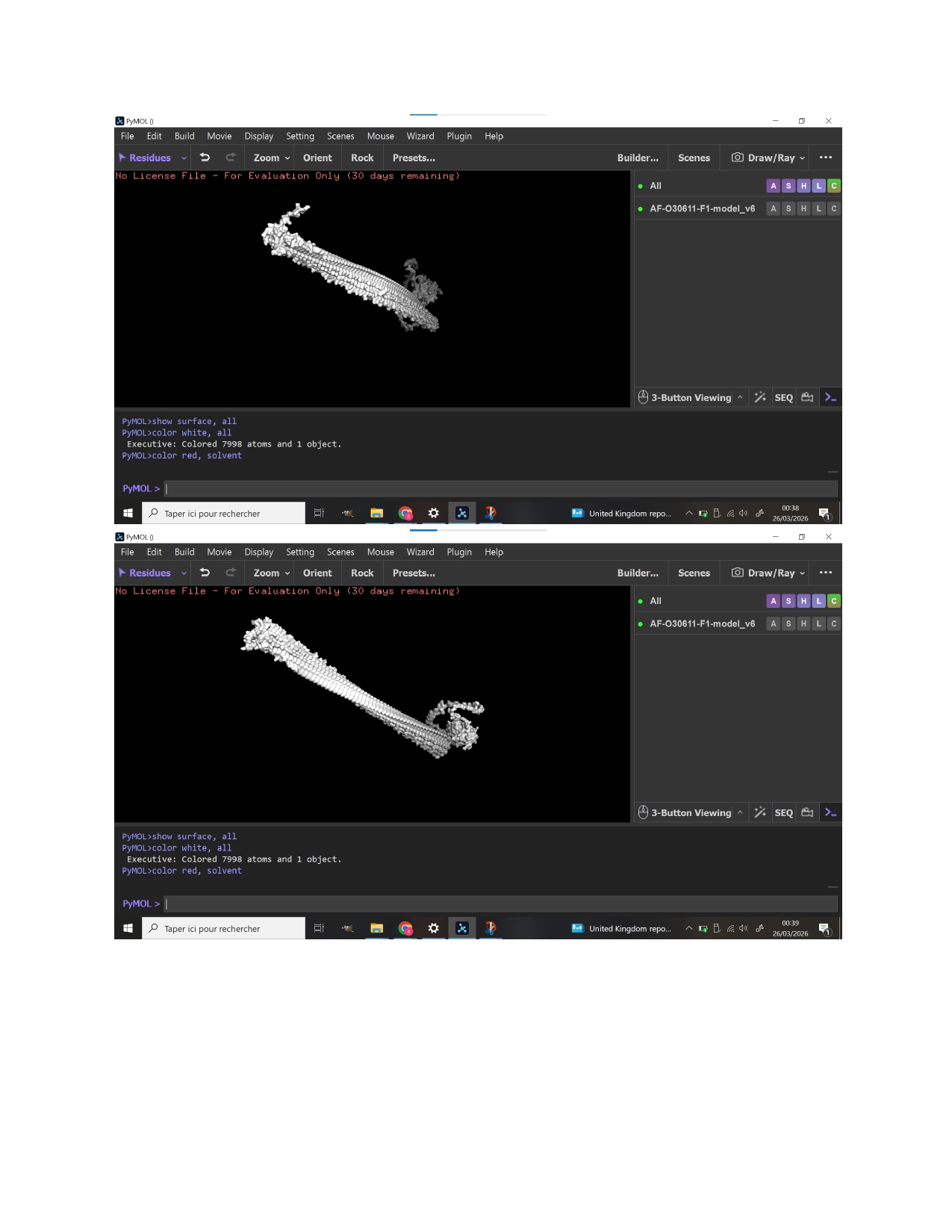
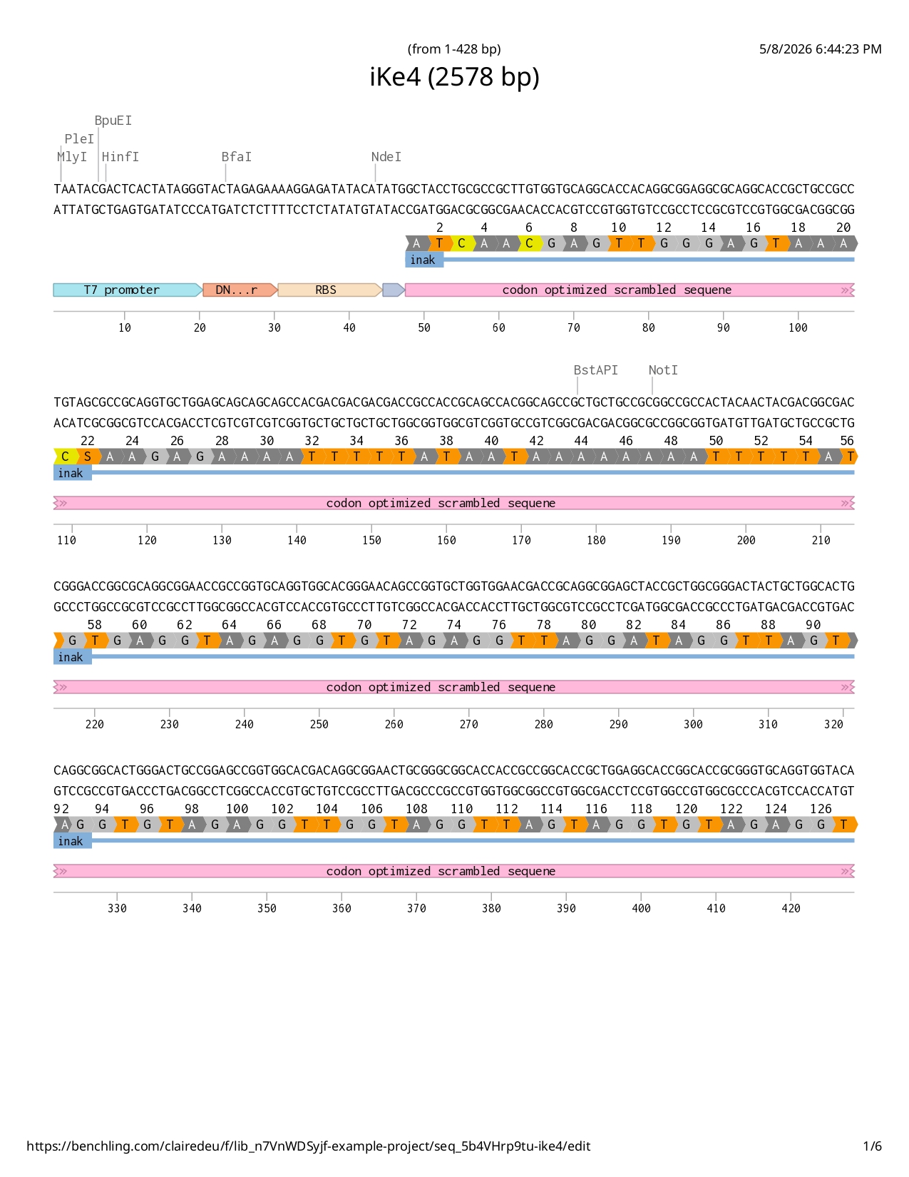
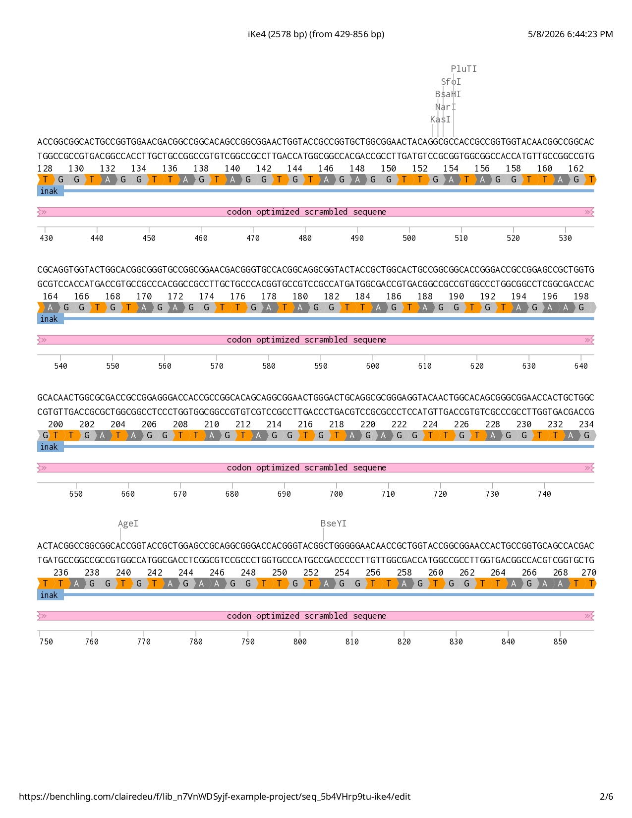
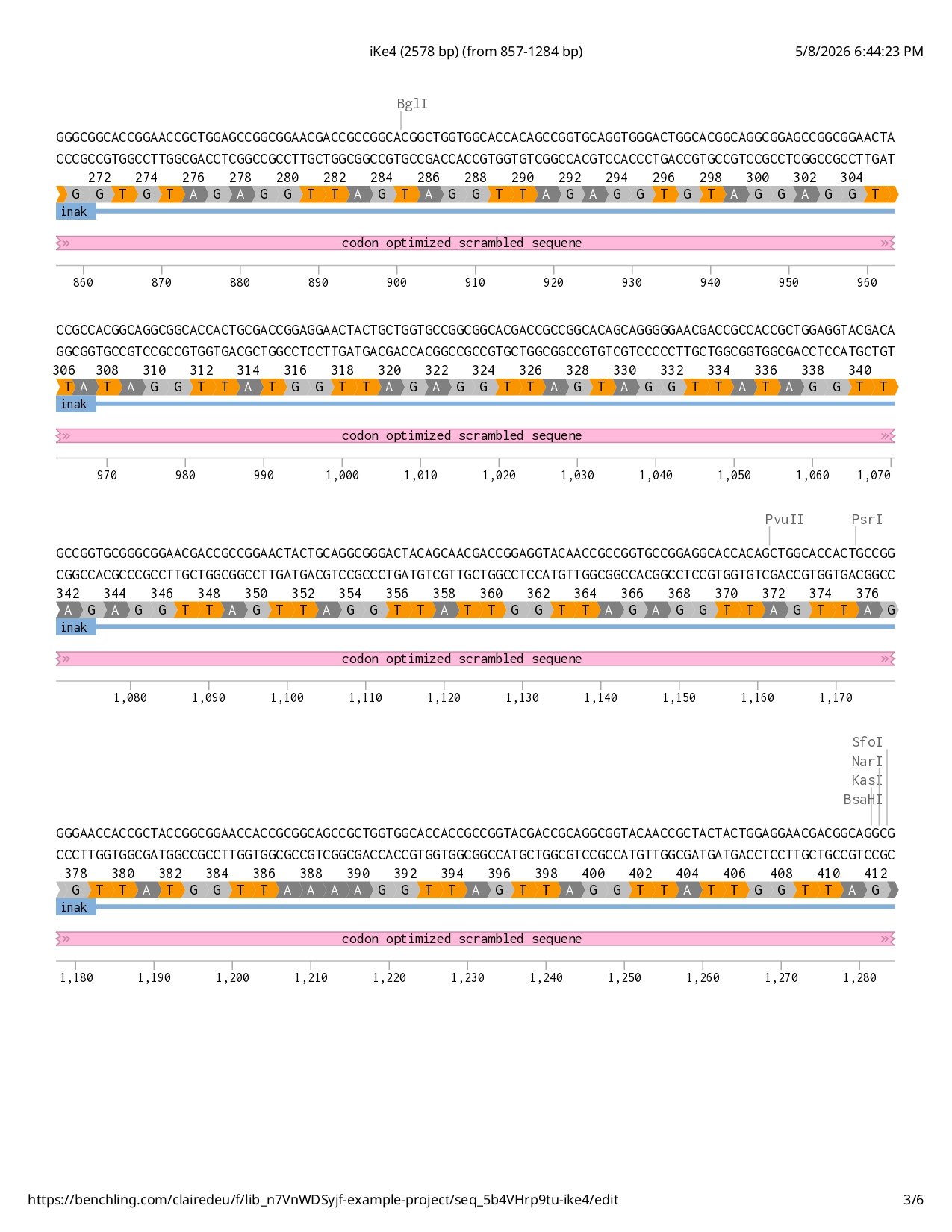
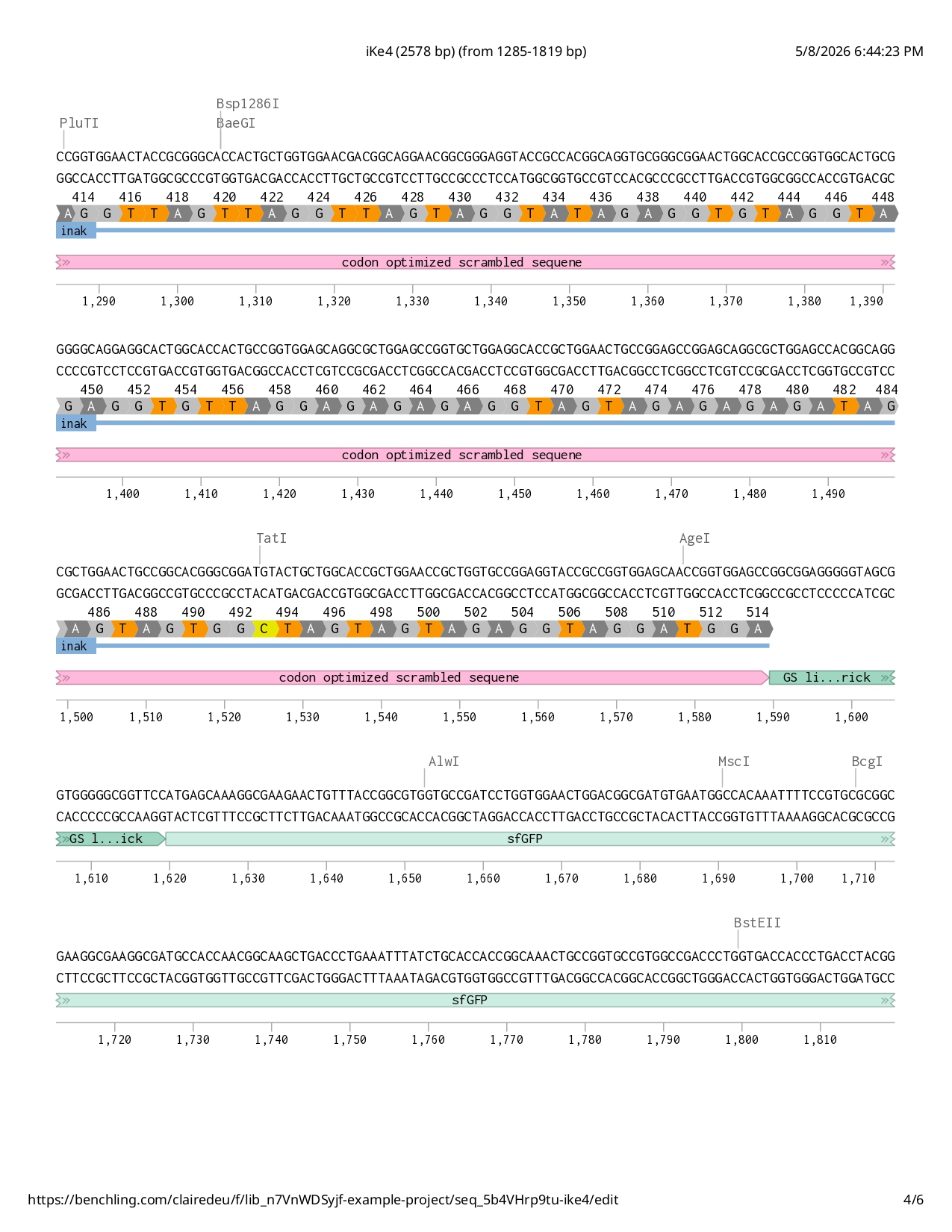
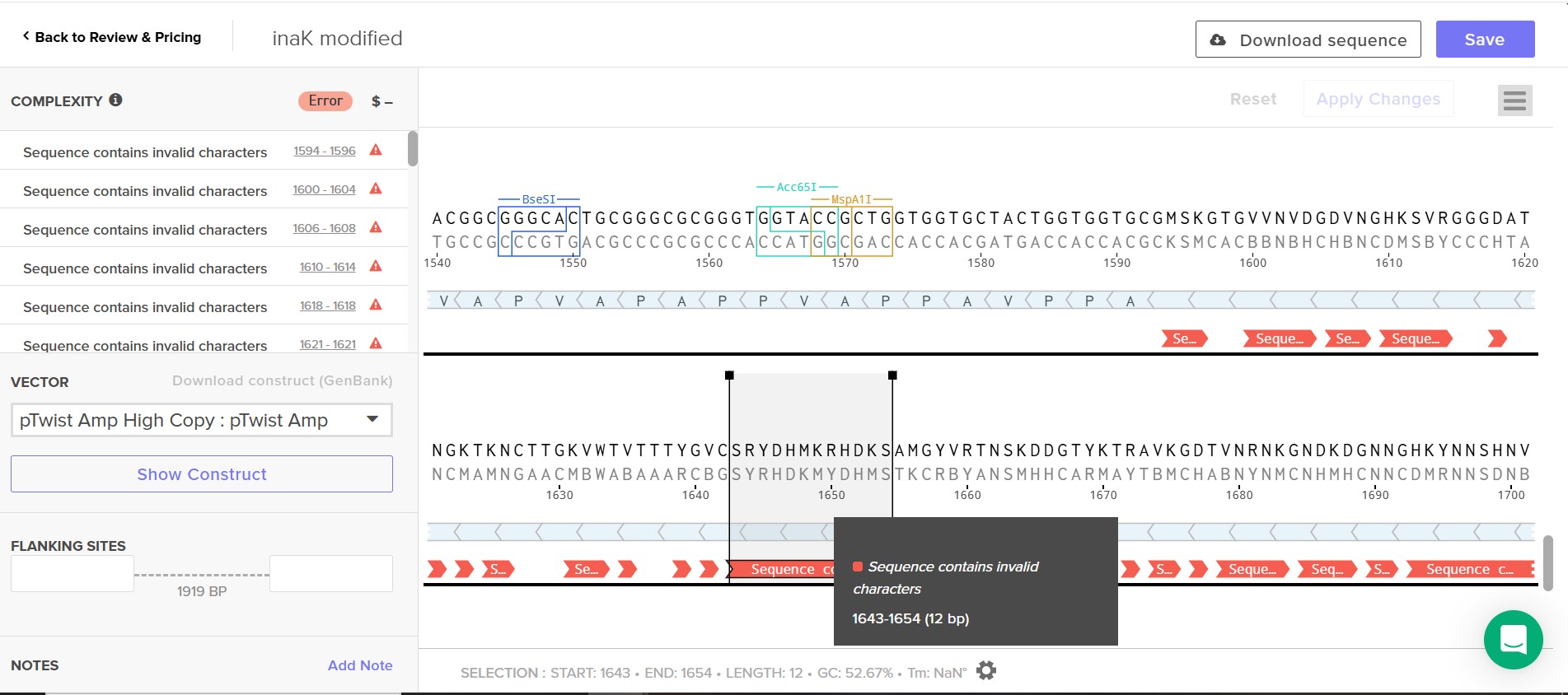
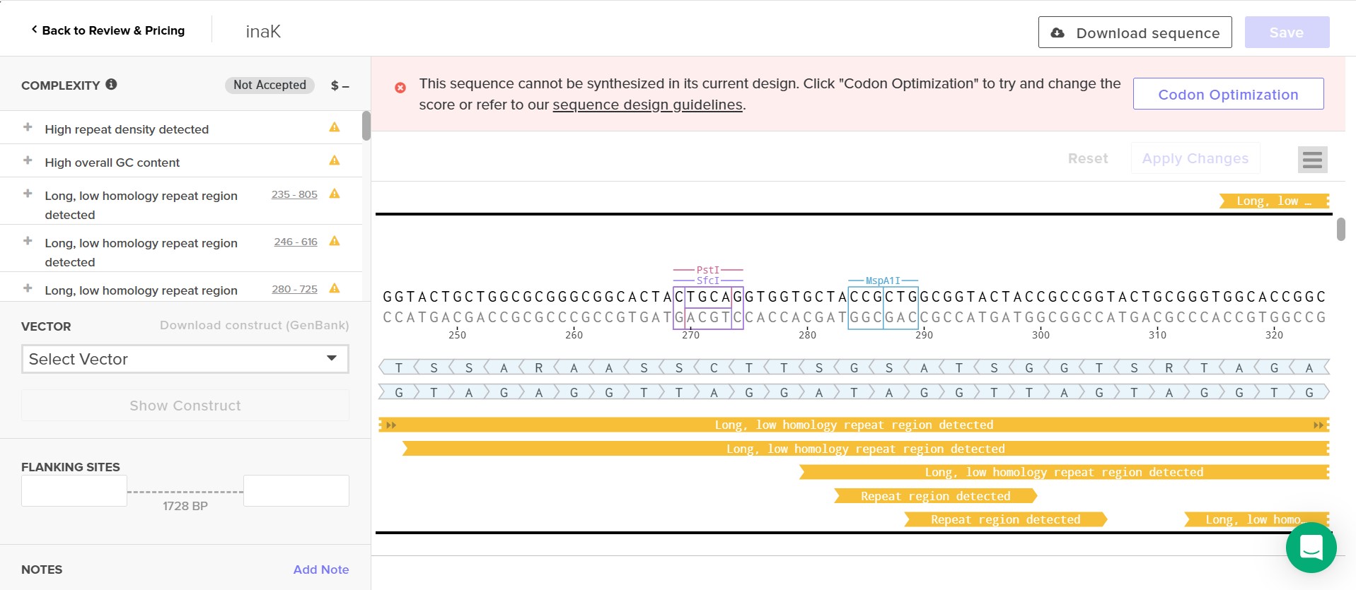
As for this project I aim to use inaK for ice production I would like to measure the ice nucleation ratio and efficiency of the inaK protein. Additionally, I would like to measure the temperatures inaK can resist to, on its own and as a supplement to an ice sample. If my initial experiments are successful I would like to measure the inaK ratio innoculated into ice to find the most optimal inaK quantity needed.
Week 11 Bioproduction & Cloud Lab Part A - The 1.536 pixel art work canvas, collective artwork
1.Contribute at least one pixel to the global artwork
I added early on a pixel towards the top left corner. I do not have much to say about this section of the work except maybe understanding the full purpose of this exercise.
Subsections of Homework
Week 1 HW: Principles and Practices
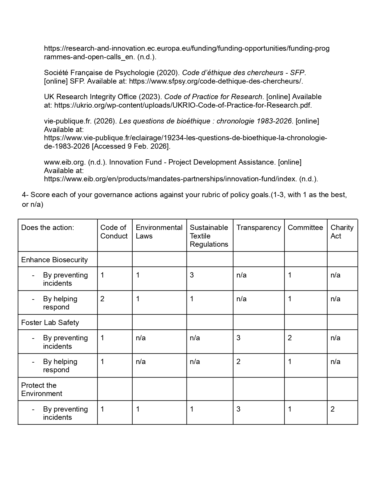
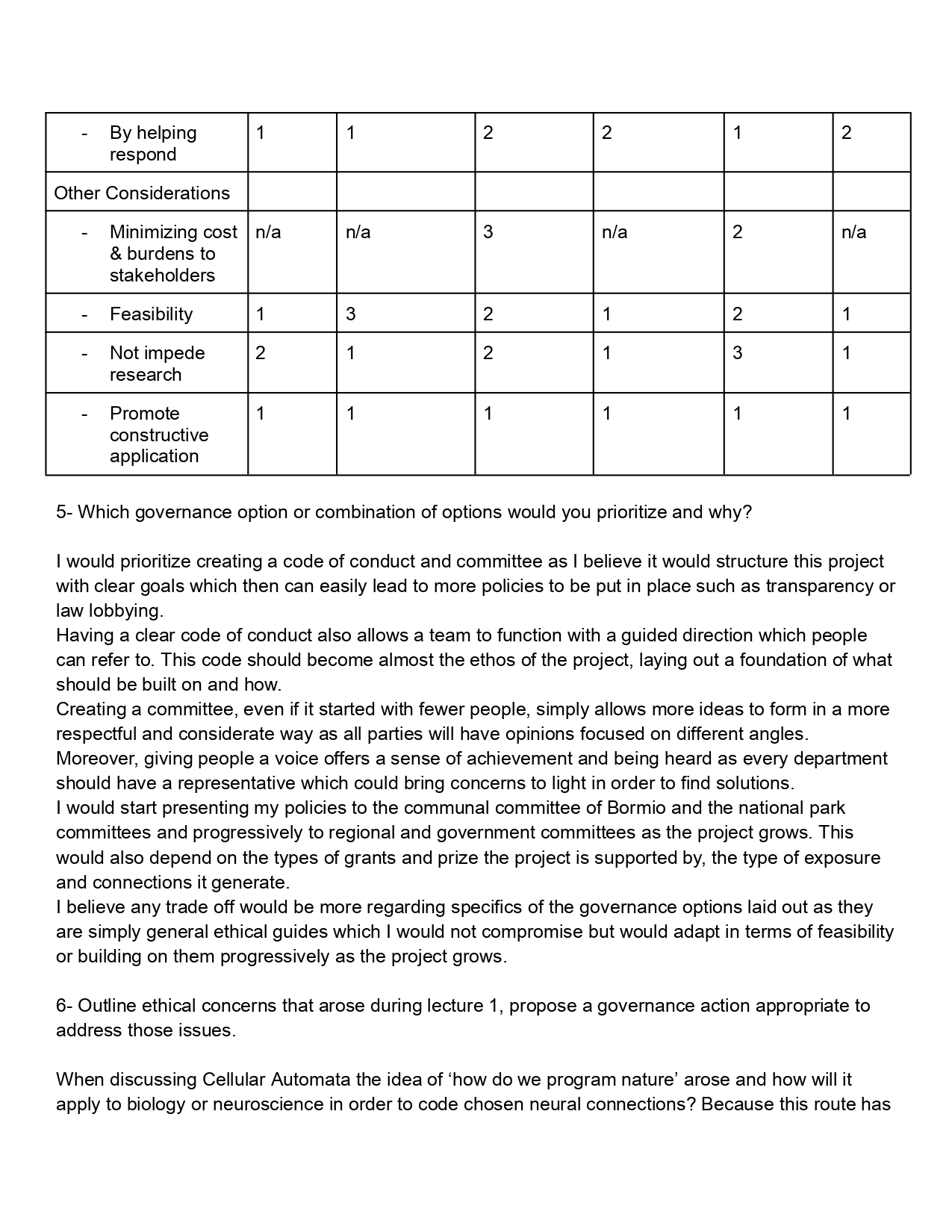
Does the option:
Option 1
Option 2
Option 3
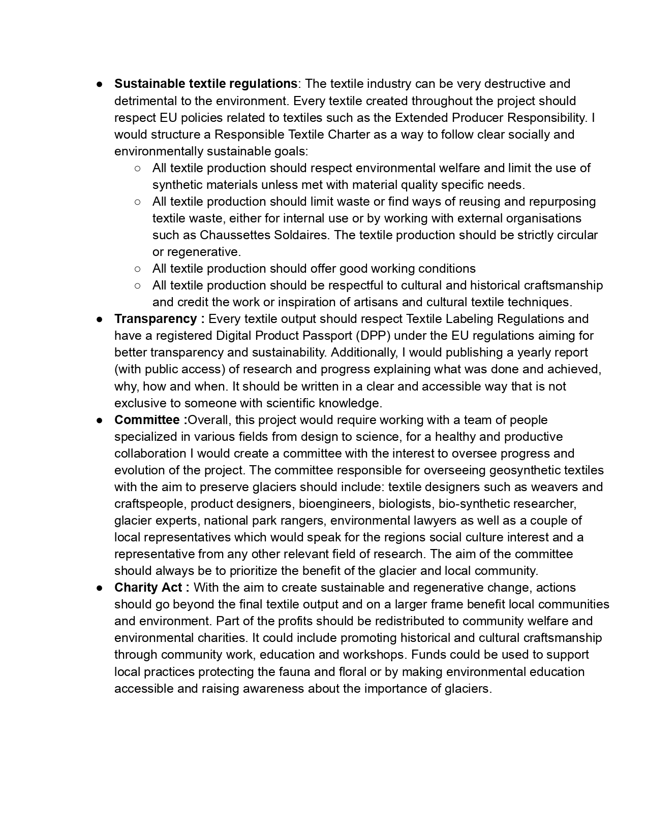
Enhance Biosecurity
• By preventing incidents
• By helping respond
Foster Lab Safety
• By preventing incident
• By helping respond
Protect the environment
• By preventing incidents
• By helping respond
Other considerations
• Minimizing costs and burdens to stakeholders
• Feasibility?
• Not impede research
• Promote constructive applications
Week 2 HW:DNA Read, Write & Edit
Week 3 HW:Lab Automation
Week 4 HW: Protein Design Part 1
Week 5 HW: Protein Design Part 2
Week 6 HW: Genetic Circuits Part 1
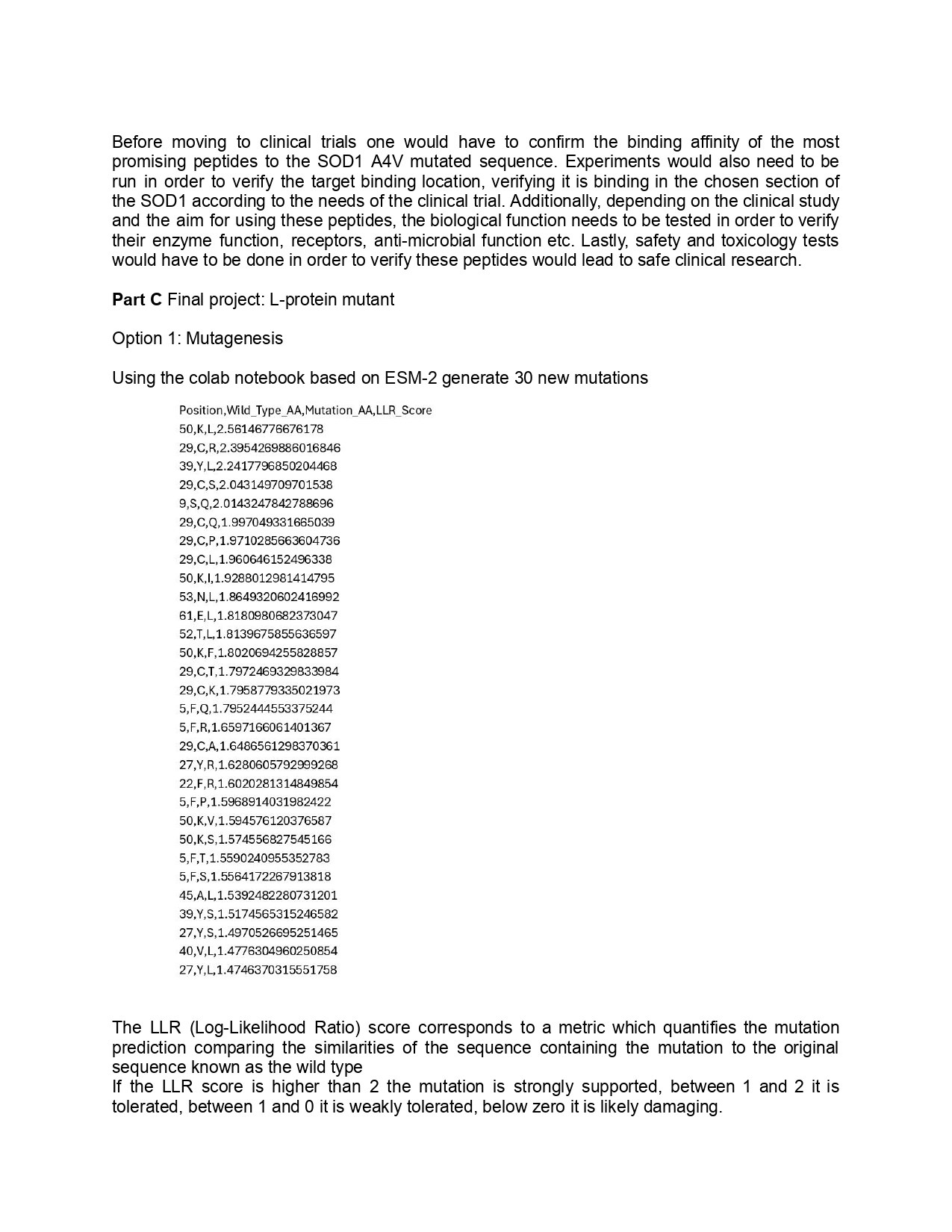
HTGAA Week 6 Genetic circuit part 1
DNA Assembly
1.What are some components in the Phusion High-Fidelity PCR Master Mix and what is their purpose?
The Phusion High-Fidelity PCR Master Mix is a concentrated DNA polymerase solution containing a high fidelity reaction buffer, MgCI (Magnesium chloride) and dNTPs ( deoxynucleoside triphosphates). It is used in PCR as polymerase solution can help fill the gaps in the sequence cloned during Gibson or HiFi assembly. The reaction buffer serves as a stabilizer and MgCI and dNTPs serve as building blocks for PCR.
Reference list
BioChain Institute Inc. (2024). Biochain Institute Inc. [online] Biochain Institute Inc. Available at: https://www.biochain.com/blog/using-dntp-in-polymerase-chain-reaction-pcr/.
New England Biolabs (2026). [online] Neb.com. Available at: https://www.neb.com/en-gb/products/m0531-phusion-high-fidelity-pcr-master-mix-with-hf-buffer [Accessed 23 Mar. 2026].
2.What are some factors that determine primer annealing temperature during PCR?
The primer annealing temperature during PCR will be determined by the melting temperature ( Tm) of primers and according to the primer design guidelines in the protocol the binding region 18-22 bp at a 52-58°C Tm allows for primers pairs to have 5°C between each other.
3.There are two methods from this class that create linear fragments of DNA: PCR, and restriction enzyme digests. Compare and contrast these two methods, both in terms of protocol as well as when one may be preferable to use over the other.
The aim of PCR is to amplify a specific DNA sequence in order to duplicate it a large amount of times and restriction enzyme digests uses enzymes to cut DNA of a chosen sequence sites into smaller sequence fragments. The PCR allows to copy the DNA while the restriction enzyme digest allows to cut DNA (insert and vector, so the plasmid) to create compatible fragment sides enabling better ligation, they are often used as a combination in cloning.
PCR uses heat-stable DNA polymerase and primers in order to stimulate a DNA replication and the restriction enzyme digests uses endonuclease enzymes to break phosphodiester bonds.
Restriction enzyme digests tend to be the preferred method as it is an easier protocol, it is a single step incubation protocol and less machinery. PCR requires precise primers, binders, restriction digests and temperatures, there are a lot more steps to the protocol and a slight lack of precision in any part of the protocol or the media and the PCR might be incorrect. However, the PCR does produce very large amounts of DNA sequencing.
4.How can you ensure that the DNA sequences that you have digested and PCR-ed will be appropriate for Gibson cloning?
To make sure a DNA sequence is suitable for the Gibson cloning one can verify that the DNA sequence has homologous primers and high-fidelity amplification in order to check that there are overlapping homologous sequences which will allow for a seamless fragment assembly.
Primer overlaps should have 20-40 homologous bp, preferably with a higher ratio of GC amino acids which favor stable annealing, between fragments an overlap of 15-30 bp is sufficient but the more there are fragments the longer the overlap sections should be.
5.How does the plasmid DNA enter the E. coli cells during transformation?
There are two main ways for E. coli to enter the cells during transformation:
On one hand, a heat shock, a chemical transformation where heating the cell and DNA mixture abruptly forms a thermal current allowing to shape temporary pores in the bacterial cell letting the plasmid pass through.
On the other hand, electroporation, creating those same pores in the bacterial cell for the plasmid to pass through but this time using high electrical voltage.
6.Describe another assembly method in detail (such as Golden Gate Assembly)
Explain the other method in 5 - 7 sentences plus diagrams (either handmade or online).
Another DNA assembly method is the CPEC, Circular Polymerase Extension Cloning, an in vivo and in vitro technology that uses PCR to amplify and extend and overlap sequence fragments. Due to its overlapping quality it allows for inserts or assembly without using restriction enzymes. This technology relies on homologs, the fragments are first amplified in PCR with homologous ends between them, the homologous region give the possibility to anneal and extend each section by the DNA polymerase through a second PCR round, then the DNA can be recombined creating larger DNA sequences which can then be introduced to the plasmid.
Reference list
Bitesize Bio. (2019). CPEC– a Quick and Inexpensive Cloning Strategy. [online] Available at: https://bitesizebio.com/44113/cpec-a-quick-and-inexpensive-cloning-strategy/.
Chao, R., Yuan, Y. and Zhao, H. (2014). Recent advances in DNA assembly technologies. FEMS Yeast Research, p.n/a-n/a. doi:https://doi.org/10.1111/1567-1364.12171.
Asimov Kernel N/A as we were not given access
Week 7 HW: Genetic Circuits Part 2
HTGAA Week 7 Genetic circuits part 2
Part 1: Intracellular Artificial Neural Networks
1.What advantages do IANNs have over traditional genetic circuits, whose input/output behaviors are Boolean functions?
The advantage of IANNs over Boolean genetic circuits is their analog sensing and nonlinear processing capabilities, while traditional genetic circuits function as an On and Off system the IANNs can work in a continuous way due to its broader and stronger range of nonlinear inputs. Additionally, the IANNs have better pattern recognition and generalization technology, they can detect more complex patterns in data and can organise them better than the Boolean can.The IANNs are also less likely to fail and have a better error tolerance than the Boolean which is very susceptible to fail if there is a single issue with a gate. Moreover, the IANNs have a better scalable capacity due to the protein splicing mechanism allowing to create a multiple input-output circuit. The IANN technology can also better adapt and evolve to its environment compared to the Boolean which has a fixed function in its environment. Finally, IANNs have a strong memory quality that can be passed through to subsequent generations within the cell due stoichiometric cleavage and splicing which is irreversible.
Reference list
Ana Halužan Vasle and Miha Moškon (2024). Synthetic biological neural networks: From current implementations to future perspectives. Biosystems, 237, pp.105164–105164. doi:https://doi.org/10.1016/j.biosystems.2024.105164.
Anastassov, S., Filo, M. and Khammash, M. (2024a). Inteins: A Swiss army knife for synthetic biology. Biotechnology Advances, 73, p.108349. doi:https://doi.org/10.1016/j.biotechadv.2024.108349.
Anastassov, S., Filo, M. and Khammash, M. (2024b). Inteins: A Swiss army knife for synthetic biology. Biotechnology Advances, 73, p.108349. doi:https://doi.org/10.1016/j.biotechadv.2024.108349.
Claus Kadelka, Taras-Michael Butrie, Hilton, E., Kinseth, J. and Haris Serdarevic (2024). A meta-analysis of Boolean network models reveals design principles of gene regulatory networks. Science Advances, 10(2). doi:https://doi.org/10.1126/sciadv.adj0822.
Gao, Y., Wang, L. and Wang, B. (2023). Customizing cellular signal processing by synthetic multi-level regulatory circuits. Nature communications, [online] 14(1). doi:https://doi.org/10.1038/s41467-023-44256-1.
Ilia, K. and Del Vecchio, D. (2022). Squaring a Circle: To What Extent Are Traditional Circuit Analogies Impeding Synthetic Biology? GEN Biotechnology, 1(2), pp.150–155. doi:https://doi.org/10.1089/genbio.2021.0014.
Karkalos, N.E. and Markopoulos, A.P. (2017). Modeling of hard machining. [online] Available at: https://www.sciencedirect.com/topics/mathematics/artificial-neural-network.
Wang, H., Wang, L., Zhong, B. and Dai, Z. (2022). Protein Splicing of Inteins: A Powerful Tool in Synthetic Biology. Frontiers in Bioengineering and Biotechnology, 10. doi:https://doi.org/10.3389/fbioe.2022.810180.
2.Describe a useful application for an IANN; include a detailed description of input/output behavior, as well as any limitations an IANN might face to achieve your goal.
The IANN genetic circuit can be use in cancer theranostics as they have the ability to process multiple biomarkers simultaneously and within a tumour can clearly identify and distinguish a cancerous cell from a healthy one. Additionally, the IANN is able to recognise the cancer patterns and can trigger the synthesis of an anti-cancer drug.
The IANN circuit used for cancer theranostics requires many inputs in order to increase precision:
MicroRNAs which allow to classify cancerous cells
TSAs which are Tumor Specific Antigens
TAAs which are Tumor Associated Antigens, they work specifically on the cancerous tissue
TME which is a Tumor Microenvironment
Enzymes
NIR which is an external stimuli such as light, magnetic field or ultrasounds which server a switch to trigger the therapy is a specific area of the body
The outputs for an IANN circuit used for cancer theranostics presents itself as a localized response:
Apoptosis triggering which leads to the programmed cell death induced by the drug delivery
Drug delivery of anticancer agents in a controlled and chosen way and area
Bioimaging through fluorescence, NIR signals or MRI
PTT or PDT heat generated chemical reaction which kills tumor cells
Creation of immune stimulatory proteins and antibodies
The IANN faces many limits however in cancer theranostics use, firstly, it is a very costly technology and is difficult to scale as it requires using and reproducing many inorganic nanoparticles in a consistent way.
One of the main issues the IANN presents is the biosafety issue as it uses inorganic nanoparticles which lack biodegradability which cause systemic toxicity and long-term retention in the liver, spleen and kidney which can lead to further toxic side effects. Furthermore, because some nanoparticles do not degrade it can cause premature drug release or loss of diagnostic functionality. The IANN also does not provide very clear imagery. Moreover, the IANN faces difficulties penetrating deep cancerous tissue, this physiological barrier issue is made worse by the binding of nanoparticles to the cancerous cells on the outer edge of a tumour which restrict even more the penetration into inner areas of the tumor tissue. Lastly, IANN can lead to skin discoloration because of its high concentration of metallic nanoparticles.
Reference List
Aminolroayaei, F., Shahbazi‐Gahrouei, D., Shahbazi‐Gahrouei, S. and Rasouli, N. (2021). Recent nanotheranostics applications for cancer therapy and diagnosis: A review. IET Nanobiotechnology, 15(3), pp.247–256. doi:https://doi.org/10.1049/nbt2.12021.
Ausländer, S. and Fussenegger, M. (2016). Engineering Gene Circuits for Mammalian Cell–Based Applications. Cold Spring Harbor Perspectives in Biology, 8(7), p.a023895. doi:https://doi.org/10.1101/cshperspect.a023895.
Brijendra Kumar Kashyap, Singh, V., Manoj Kumar Solanki, Kumar, A., Janne Ruokolainen and Kavindra Kumar Kesari (2023). Smart Nanomaterials in Cancer Theranostics: Challenges and Opportunities. ACS omega, 8(16), pp.14290–14320. doi:https://doi.org/10.1021/acsomega.2c07840.
Chen, H. and Zhao, Y. (2018). Applications of Light-Responsive Systems for Cancer Theranostics. ACS Applied Materials & Interfaces, 10(25), pp.21021–21034. doi:https://doi.org/10.1021/acsami.8b01114.
Chen, J., Fu, S., Zhang, C., Liu, H. and Su, X. (2022). DNA Logic Circuits for Cancer Theranostics. Small, 18(20). doi:https://doi.org/10.1002/smll.202108008.
Jiang, L., Fu, Z., Ye, B., Feng, X., Chen, Z., Chen, Q., Long, Y., Wang, S. and Deng, G. (2025). Metal nanoparticles in cancer theranostics: from synthesis to tumor microenvironment-responsive applications. Drug Delivery, 32(1). doi:https://doi.org/10.1080/10717544.2025.2565480.
Kang, S., Gil, Y.-G., Min, D.-H. and Jang, H. (2020). Nonrecurring Circuit Nanozymatic Enhancement of Hypoxic Pancreatic Cancer Phototherapy Using Speckled Ru–Te Hollow Nanorods. ACS Nano, 14(4), pp.4383–4394. doi:https://doi.org/10.1021/acsnano.9b09974.
Li, Y., Zhang, X., Wang, J., Wang, K., Li, B., Qiao, X., He, W., Cai, J., Liu, D. and Yang, L.-L. (2025). Leveraging adenosine triphosphate for cancer theranostics. Theranostics, [online] 15(10), pp.4708–4733. doi:https://doi.org/10.7150/thno.106291.
Moon Sung Kang, Kwon, M., Jang, H.-S., Jeong, S., Han, D.-W. and Ki Su Kim (2022). Biosafety of inorganic nanomaterials for theranostic applications. Emergent materials, 5(6), pp.1995–2029. doi:https://doi.org/10.1007/s42247-022-00426-3.
Sergeeva, O.V., Luo, L. and Guiseppi-Elie, A. (2025). Cancer theragnostics: closing the loop for advanced personalized cancer treatment through the platform integration of therapeutics and diagnostics. Frontiers in Bioengineering and Biotechnology, 12. doi:https://doi.org/10.3389/fbioe.2024.1499474.
Tânia F.G.G. Cova, Freitas, D. and Odebrecht, S. (2019). Computational Approaches in Theranostics: Mining and Predicting Cancer Data. 11(3), pp.119–119. doi:https://doi.org/10.3390/pharmaceutics11030119.
Weranga Rajapaksha, Riya Khetan, Ian, Blencowe, A., Garg, S., Albrecht, H. and Gillam, T.A. (2024). Future theranostic strategies: emerging ovarian cancer biomarkers to bridge the gap between diagnosis and treatment. Frontiers in Drug Delivery, 4. doi:https://doi.org/10.3389/fddev.2024.1339936.
Xie, Z., Wroblewska, L., Prochazka, L., Weiss, R. and Benenson, Y. (2011). Multi-input RNAi-based logic circuit for identification of specific cancer cells. Science (New York, N.Y.), [online] 333(6047), pp.1307–11. doi:https://doi.org/10.1126/science.1205527.
Zuo, Y., Li, P., Wang, W., Xu, C., Xu, S., Herman, Sun, J., Jin, G., Wang, W., Ryan, Jacky and Tang, B.Z. (2024). Tumor Site‐Specific In Vivo Theranostics Enabled by Microenvironment‐Dependent Chemical Transformation and Self‐Amplifying Effect. Advanced Science, 12(4), pp.e2409506–e2409506. doi:https://doi.org/10.1002/advs.202409506.
3.Draw a diagram for an intercellular multilayer perceptron where layer 1 outputs an endoribonuclease that regulates a fluorescent protein output in layer 2.
Part 2 Fungal Materials
1.What are some examples of existing fungal materials and what are they used for? What are their advantages and disadvantages over traditional counterparts?
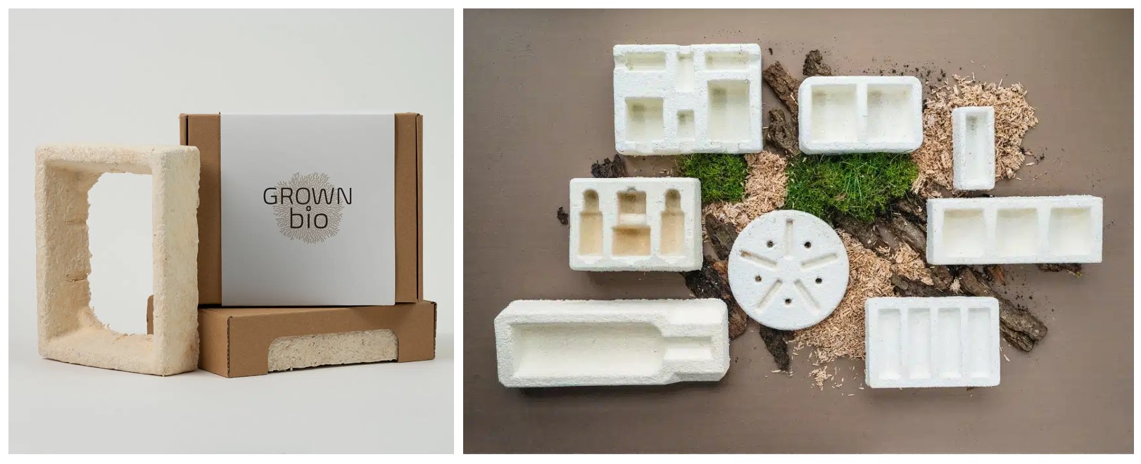
One of the most common examples of fungal materials today is mycelium packaging, mycelium is the main fungal strand used in biodesign. It is able to grow within a mold and once cooked it dies but the shape given is preserved.
Mycelium is non harmful to the environment and biodegradable which has inspired many companies to launch mycelium packaging with the aim to replace and reduce plastic use in packaging. Mycelium grows rapidly but still requires the time to grow compared to plastic packaging which can be produced instantly. Mycelium is a living organism and holds a higher risk in production. Mycelium materials however require no chemical input which is better for the environment, the producer and the consumer, eliminating the risk of chemicals and microplastics. Furthermore, mycelium material packaging is currently more costly than producing plastic but the cost gap is reducing as mycelium materials are being scaled up according to a mycelium packaging market report of 2025.
Companies such as Grown Bio already offer viable alternatives to packaging of all sorts. They offer a range of packaging of all shapes and qualities (some with reinforced protective design) which one could buy directly or they offer the possibility to grow one’s own packaging.
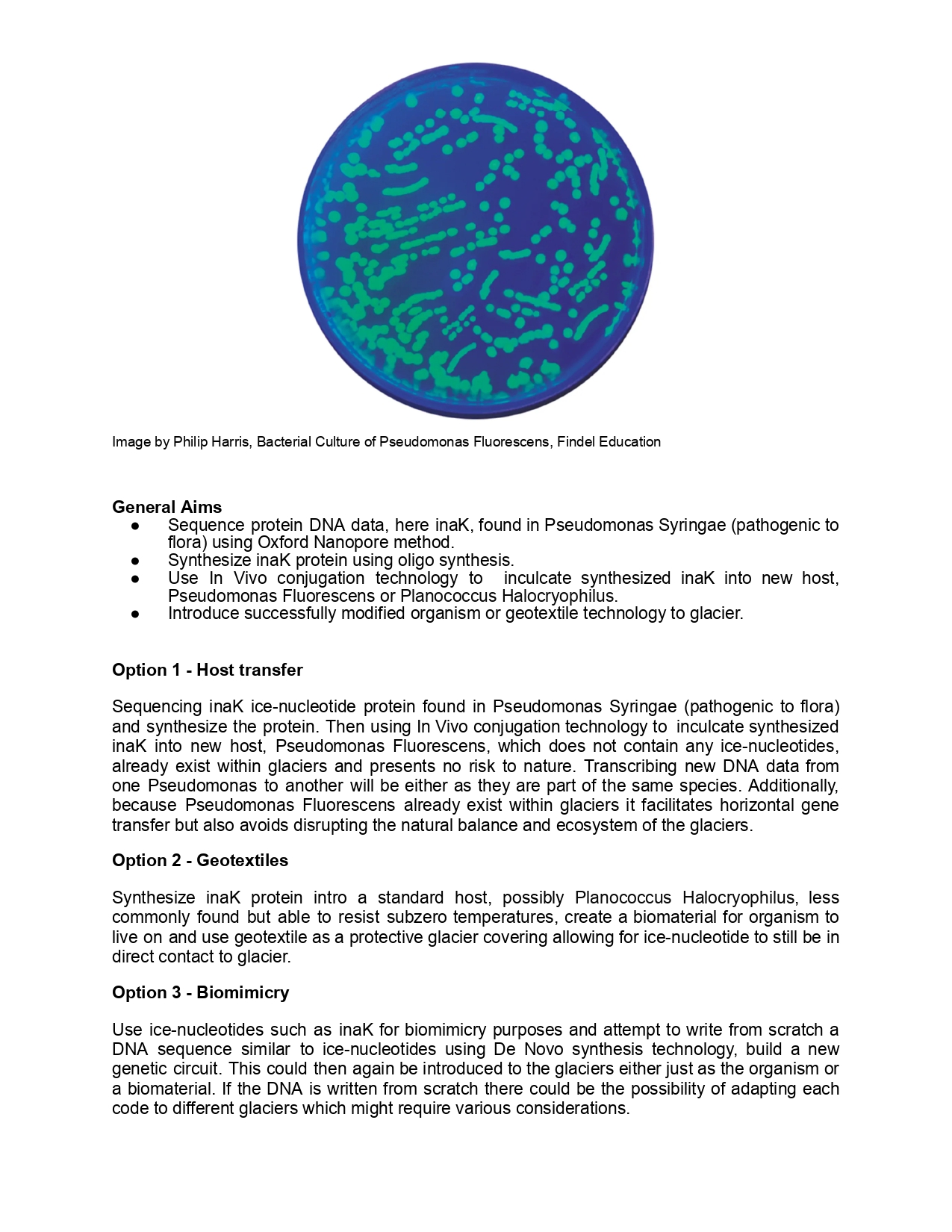
2.What might you want to genetically engineer fungi to do and why? What are the advantages of doing synthetic biology in fungi as opposed to bacteria?
In the continuation of mycelium packaging I would love to explore whether one could genetically modify the mycelium to have active properties as well. Maybe mycelium packaging could also have cold properties allowing to store products which require to be kept in the cold. Many mycelium species, such as the most commonly used oyster mushroom (Pleurotus ostreatus), can withstand freezing temperatures and simply go into a dormant state. Typically in packaging mycelium would be killed with heat to stop its growth and preserve its shape, however, could the mycelium be kept alive, genetically modified with ice nucleation proteins, put in a dormant state because of the freezing temperature which would still stun its growth and therefore preserve its shape and then be used a cold packaging system, which in its end of life could still be biodegradable or contribute actively to nature.
It appears there would be two ways to genetically modify mycelium to produce cold. Mycelium is already used successfully as a host in synthetic biology. Additionally, it seems that one could introduce ice nucleotides to the genetic code of mycelium DNA and the mycelium would accept it. An experiment of adding ice nucleotide proteins to water which was then fed to mycelium has already been done, with the aim to study freezing in mycelium (Schwidetzky et al., 2023). Secondly, certain mycelium strands (including the commonly used oyster mushroom) already appear to contain ice nucleotide allowing them to resist freezing temperatures, one could explore genetically modifying the mycelium to express this protein in a more active way allowing it to produce a freezing quality.
Fungi can produce complex molecules and proteins better than bacteria, they are also able to produce much more enzymes than bacteria making purification processes easier. Fungi are also eukaryotic organisms which allows them to perform complex post-translational modifications like protein folding offer a bigger potential for synthetic biology. Additionally, fungi naturally produce more secondary metabolites than bacteria such as terpenoids, polyketides and alkaloids which are commonly used in pharmaceutical research and development. Moreover, fungi genomes naturally contain biosynthetic gene clusters (BGCs) which are used in synthetic biology to engineer new-to-nature chemicals.
Fungi can be easier to work with as they are able to live off a wider range of feedstock and grow rapidly as well as being robust cultures able to adapt to harsh environments and withstand a range of PH levels or a range of temperatures.
Reference list
Awasthi, S., Alam, M.I. and Pal, D.B. (2025). Importance of Utilizing Fungus Rather Than Bacteria for Biomass Valorization. Fungal Biology, pp.107–140. doi:https://doi.org/10.1007/978-3-031-82599-6_5.
CATALEX BIO. (2025). Fungal vs Bacterial Enzymes: Industrial Applications & Selection Guide | Catalex Bio Enzyme Manufacturer & Supplier. [online] Available at: https://catalexbio.com/fungal-vs-bacterial-enzymes-comparison-guide/.
Cordero, B., Ellie Rose Mattoon, Ramos, Z. and Casadevall, A. (2023). The hypothermic nature of fungi. PNAS, 120(19). doi:https://doi.org/10.1073/pnas.2221996120.
Eufemio, R.J., Rojas, M., Shaw, K., de Almeida Ribeiro, I., Guo, H.-B., Renzer, G., Belay, K., Liu, H., Suseendran, P., Wang, X., Fröhlich-Nowoisky, J., Pöschl, U., Bonn, M., Berry, R.J., Molinero, V., Vinatzer, B.A. and Meister, K. (2026). A previously unrecognized class of fungal ice-nucleating proteins with bacterial ancestry. Science Advances, 12(11). doi:https://doi.org/10.1126/sciadv.aed9652.
Garg, S. (2025a). The importance of fungal biotechnology for sustainable applications. Trends in Biotechnology, [online] 0(0). doi:https://doi.org/10.1016/j.tibtech.2025.06.010.
Garg, S. (2025b). The importance of fungal biotechnology for sustainable applications. Trends in Biotechnology, [online] 0(0). doi:https://doi.org/10.1016/j.tibtech.2025.06.010.
Hinneburg, H., Gu, S. and Naseri, G. (2025). Fungal Innovations—Advancing Sustainable Materials, Genetics, and Applications for Industry. Journal of Fungi, 11(10), p.721. doi:https://doi.org/10.3390/jof11100721.
Jo, C., Zhang, J., Tam, J.M., Church, G.M., Khalil, A.S., Segrè, D. and Tang, T.-C. (2023a). Unlocking the magic in mycelium: Using synthetic biology to optimize filamentous fungi for biomanufacturing and sustainability. Materials Today Bio, 19, p.100560. doi:https://doi.org/10.1016/j.mtbio.2023.100560.
Jo, C., Zhang, J., Tam, J.M., Church, G.M., Khalil, A.S., Segrè, D. and Tang, T.-C. (2023b). Unlocking the magic in mycelium: Using synthetic biology to optimize filamentous fungi for biomanufacturing and sustainability. Materials Today Bio, 19, p.100560. doi:https://doi.org/10.1016/j.mtbio.2023.100560.
Ma, D., Yang, G., Mu, L. and Li, C. (2011). Tolerance of ectomycorrhizal fungus mycelium to low temperature and freezing–thawing. Canadian Journal of Microbiology, 57(4), pp.328–332. doi:https://doi.org/10.1139/w11-001.
Moreno-Giménez, E., Mónica Gandía, Zara Sáez, Manzanares, P., Yenush, L., Orzáez, D., Marcos, J.F. and Garrigues, S. (2023). FungalBraid 2.0: expanding the synthetic biology toolbox for the biotechnological exploitation of filamentous fungi. Frontiers in Bioengineering and Biotechnology, 11. doi:https://doi.org/10.3389/fbioe.2023.1222812.
Nielsen, J. (2013). Production of biopharmaceutical proteins by yeast. Bioengineered, 4(4), pp.207–211. doi:https://doi.org/10.4161/bioe.22856.
Raymond, J.A. and Janech, M.G. (2009). Ice-binding proteins from enoki and shiitake mushrooms. Cryobiology, 58(2), pp.151–156. doi:https://doi.org/10.1016/j.cryobiol.2008.11.009.
Sanitá Lima, M. and Coutinho de Lucas, R. (2022). Co-cultivation, Co-culture, Mixed Culture, and Microbial Consortium of Fungi: An Understudied Strategy for Biomass Conversion. Frontiers in Microbiology, 12. doi:https://doi.org/10.3389/fmicb.2021.837685.
Schwidetzky, R., Ingrid, Bothen, N., Backes, A.T., DeVries, A.L., Bonn, M., Fröhlich-Nowoisky, J., Molinero, V. and Meister, K. (2023a). Functional aggregation of cell-free proteins enables fungal ice nucleation. Proceedings of the National Academy of Sciences of the United States of America, 120(46). doi:https://doi.org/10.1073/pnas.2303243120.
Schwidetzky, R., Ingrid, Bothen, N., Backes, A.T., DeVries, A.L., Bonn, M., Fröhlich-Nowoisky, J., Molinero, V. and Meister, K. (2023b). Functional aggregation of cell-free proteins enables fungal ice nucleation. Proceedings of the National Academy of Sciences of the United States of America, 120(46). doi:https://doi.org/10.1073/pnas.2303243120.
Shankar, M.P., Hamza, A., Khalad, A., Shanthi, G., Kuppireddy, S. and Kumar, D.S. (2024). Engineering mushroom mycelium for a greener built environment: Advancements in mycelium-based biocomposites and bioleather. Food Bioscience, [online] 62, p.105577. doi:https://doi.org/10.1016/j.fbio.2024.105577.
Week 9 HW: Cell Free Systems
HTGAA Week 9 Cell Free Systems
Part A General and Lecture specific questions
General Homework Questions
1.Explain the main advantages of cell-free protein synthesis over traditional in vivo methods, specifically in terms of flexibility and control over experimental variables. Name at least two cases where cell-free expression is more beneficial than cell production.
Time : CFPS can be executed in very little time, a couple of hours however in vivo methods will take a few days to a few weeks. Time is a key part of efficient research as less time is wasted waiting to synthesize proteins to experiment on.
System : CFPS is an open access system as it has no membrane and gives direct access to the molecule which one wants to work on, it offers more flexibility. It also allows for better control over the synthesis as one can specifically choose which components to input in a protein and better predict protein folding, this simplifies monitoring as well. Versus an in vivo method which has a membrane and therefore a closed system, this means the host and its other components will also react to any modification making it harder to control and direct, the cell’s functioning can often get in the way of the synthesis or create unpredicted issues.
Tolerance: CFPS can tolerate high rates of toxic and difficult proteins as it is non living, however, in vivo technologies are more sensitive to toxins as it is likely to harm the host. CFPS also works using non-natural components offering a wider spectrum of possibilities.
Complexity : CFPS is a simple PCR based procedure while in vivo requires more complex cloning and transformation steps.
Reference List
Silverman, A. D., et al. (2020). Quarto: A User’s Guide to Cell-Free Protein Synthesis. Nature Reviews Genetics.
Gregorio, N. E., et al. (2019). Cell-free microbial synthesis of proteins. Frontiers in Bioengineering and Biotechnology.
2.Describe the main components of a cell-free expression system and explain the role of each component.
There are four core components to a CFPS system which are the catalytic engine, the instruction template, the energy source and the biochemical blocks.
At first a “soup” is created by breaking a cell pen and removing debris, this gives us the heart of the system which provides us with the molecules required for transcription and translation like ribosomes or tRNAs.
The DNA instruction template is the blueprint for the CFPS and it is very flexible as it can function with a plasmid but it can also work using PCR linear PCR products which allows to reduce cloning time in the preparatory stages.
Compared to most synthesis technologies requiring a lot of energy to keep the cell alive and the ribosomes active, CFPS can be fueled by ATPs and GTPs which are high energy sources.
Biochemical building blocks and buffers stabilize the CFPS reaction by adding to the solution amino acids to build the chain, the RNA polymerase and often magnesium or potassium salts which can help stabilize the protein folding.
Reference List
Silverman, A. D., Karim, A. S., & Jewett, M. C. (2020). Quarto: A User’s Guide to Cell-Free Protein Synthesis. Nature Reviews Genetics.
Hodgman, C. E., & Jewett, M. C. (2012). Cell-free protein synthesis: The state of the art. Biotechnology and Bioengineering.
Caschera, F., & Noireaux, V. (2014). Synthesis of 2.3 mg/ml of GFP with an all-E. coli cell-free transcription-translation system. Biological Engineering.
Shimizu, Y., et al. (2001). Cell-free translation reconstituted with purified components. Nature Biotechnology.
Tinafar, A., et al. (2019). A Manual of Cell-Free Protein Synthesis Systems. Frontiers in Bioengineering and Biotechnology.
3.Why is energy provision regeneration critical in cell-free systems? Describe a method you could use to ensure continuous ATP supply in your cell-free experiment.
Protein synthesis is a very energy taxing process. An issue in CFPS is the ATP “leaks” caused by enzymes breaking down enzymes to ADP (without the ribosome using it) and inorganic phosphate, the regeneration of energy allows to preserve fuel for the synthesis rather than it being wasted by enzymes.
A method of not just adding an excess of ATP is adding a substrate fuel which could be Phosphoenolpyruvate (PEP) and Pyruvate Kinase (PK). The substrate PEP acts as a battery which contains the phosphate group. Once the ribosome processes the ATP into an ADP the PK is able to directly collect the phosphate from the PEP and link it back onto the ADP changing it back to an ATP creating a steady loop preserving ATP levels consistent till all the PEP is processed.
Reference List
Silverman, A. D., et al. (2020). Nature Reviews Genetics.
Caschera, F., & Noireaux, V. (2014). Biological Engineering.
4.Compare prokaryotic versus eukaryotic cell-free expression systems. Choose a protein to produce in each system and explain why.
Both prokaryotic and eukaryotic cell free systems aim to produce protein from a DNA template.
Prokaryotic systems have high speed and yield rates while eukaryotic systems are slower and produce less proteins.
In prokaryotic systems the transcription and translation happen in the same place, they are coupled, eukaryotic systems are decoupled and can require extra steps.
While the eukaryotic cell free expression is quite costly the prokaryotic based system is inexpensive and simpler to set up and run.
However, the eukaryotic system can offer advanced protein folding and more complex modifications as it is able to mimic human-like post-translational modification. The prokaryotic cannot offer natural modifications and creates basic protein folding.
Unless an experiment requires human-like testing I would choose the prokaryotic cell free expression system as it is simpler, less expensive and more prolific. Additionally, if an experiment requires more stability and better control, the fact that prokaryotic systems cannot offer modifications might also make it more predictable and easier to work with.
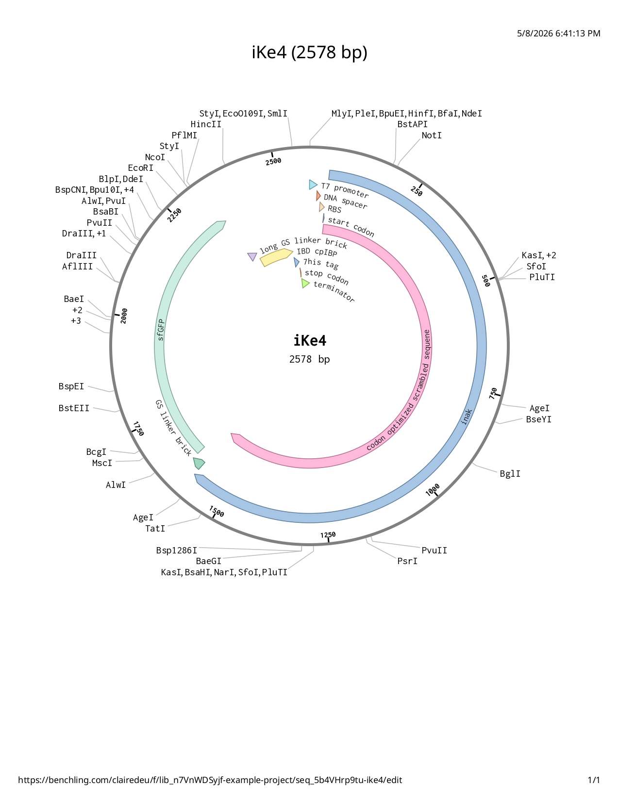
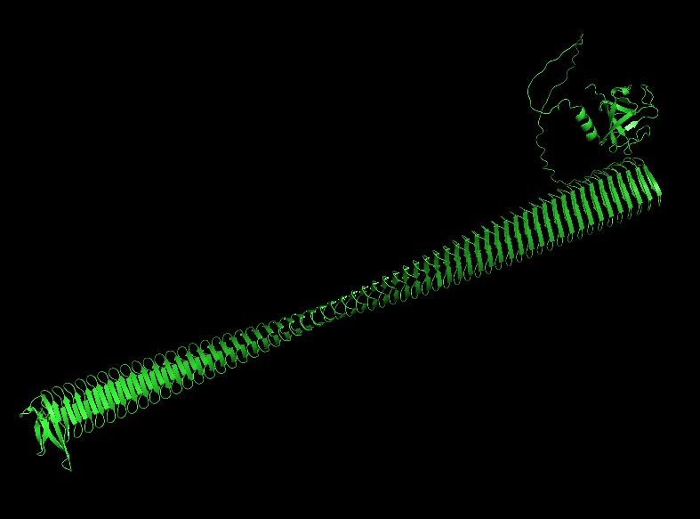
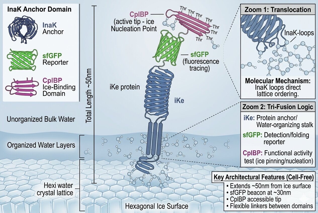
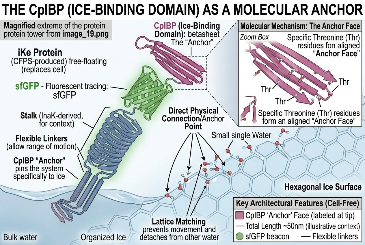
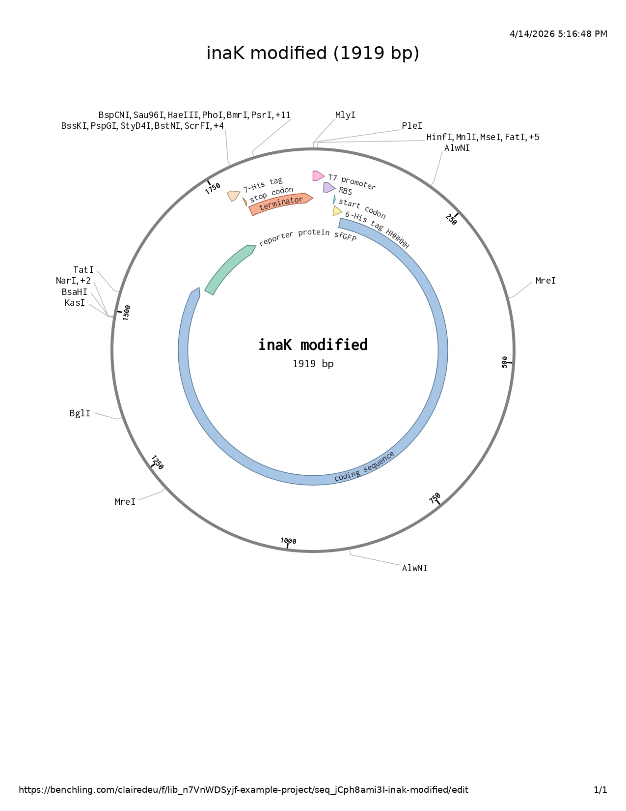
The INP inaK protein I have been working with would be suitable for prokaryotic cell-free expression as it is a highly repetitive protein which is commonly found in Pseudomonas Syringae, a prokaryote, but if I use an E. coli extract then the codon bias is the same as the native bacteria so the ribosome will read with ease the repetitive portions of the sequence. Moreover, inaK only requires a membrane to anchor to, it does need additional complex compounds. Furthermore, I would aim for a high concentration of inaK for my project in order to produce more ice, which then is more suitable to the high yield of prokaryotic systems.
For a eukaryotic cell-free expression system I would choose a complex human protein such as the tPA, tissue plasminogen activator (a clot-buster), which have more complex modifications and toxins, it would benefit from a more advanced and control protein folding method.
Globally, if a protein has a bacterial origin or has a simple structure and needs to be produced in a large quantity then the prokaryotic systems are more suitable. Eukaryotic systems are useful for complex and human or mammal proteins.
Reference List
Zemella, A., et al. (2015). Cell-Free Protein Synthesis: Pros and Cons of Prokaryotic and Eukaryotic Systems. ChemBioChem.
Silverman, A. D., et al. (2020). Quarto: A User’s Guide to Cell-Free Protein Synthesis. Nature Reviews Genetics.
Endo, Y., & Sawasaki, T. (2006). A cell-free protein synthesis system for high-throughput proteomics. Journal of Structural and Functional Genomics. (Focusing on Wheat Germ advantages).
5.How would you design a cell-free experiment to optimize the expression of a membrane protein? Discuss the challenges and how you would address them in your setup.
I will be using inaK as it is a membrane protein and use a prokaryotic system.
The first issue I will be faced with is that the inaK N-terminal is highly hydrophobic and might create aggregates if I don’t offer it a lipid surface to link to.
I would start with a circular plasmid sourced using Twist and Benchling.
For the expression system I might use an E. coli based S30 extract because the inaK is a bacterial protein and the codon bias and translation speed using the E. coli ribosomes will be naturally optimized for its repetitive sequence. In this case, the transcription and translation will be coupled to ensure the protein folds as it is produced.
I would include PEP and PK for energy regeneration.
Next, as I need to provide the inaK with a new membrane to latch on to I could select a liposome host membrane which might mimic their initial membrane closely, it is structured as small bubbles of phospholipids which should help the first hydrophobic issue.
To determine where exactly to produce the inaK I might have to run a few optimization matrix experiments whether tuning temperature, optimizing lipid to protein ratio to avoid aggregations or a diluted solution, and magnesium and potassium concentrations as membrane proteins are sensitive to charge and a wrong ratio could make the ribosome collapse.
In the end I would have to evaluate the success ratio of my system. The purpose of the inaK is to freeze water, I could place a drop of the cell free solution on a cold plate and observe which solution (supposing I would run a few) would freeze at the highest temperature.
Reference List
Hartmann, et al. (2022). Overcoming bottlenecks for in vitro synthesis of ice nucleating protein InaZ.
Silverman, A. D., et al. (2020). Quarto: A User’s Guide to Cell-Free Protein Synthesis. Nature Reviews Genetics.
Henrich, et al. (2015). Analyzing the specialized lipid environment of membrane proteins.
6.Imagine you observe a low yield of your target protein in a cell-free system. Describe three possible reasons for this and suggest a troubleshooting strategy for each.
A low yield result can be common in CFPS.
This can be caused by energy exhaustion, this system is very energy consuming and the PEP might have been consumed too rapidly or the ATP consuming enzymes too active in the sample leading to the ribosome to not have enough energy to produce enough protein. To resolve this a higher concentration of the energy substrate could be used.
The DNA template might also be damaged or degraded, causing a blueprint issue. If a linear PCR template is used rather than a plasmid it can happen that enzymes might interfere or damage the ends of the template causing no proteins to be created. Using a controlled plasmid should help avoid this issue, it is possible to make a linear PCR into a circular plasmid through ligations.
There might also be an ionic imbalance within the solution which can be due to an unstable ribosome or DNA sequence, this imbalance might work for certain proteins but maybe not the one studied. For instance, mRNA and DNA are very dependent on magnesium and potassium which requires finding the perfect ion balance for the protein in the solution. Adjusting the magnesium ratios can help, this can be done by running multiple tubes with different magnesium percentages.
Reference List
Silverman, A. D., et al. (2020). Quarto: A User’s Guide to Cell-Free Protein Synthesis. Nature Reviews Genetics.
Sun, Z. Z., et al. (2013). Linear DNA for rapid prototyping. ACS Synthetic Biology.
Caschera, F., & Noireaux, V. (2014). Synthesis of 2.3 mg/ml of GFP. Biological Engineering.
Questions from Kate Adamala
Design an example of a useful synthetic minimal cell as follows:
1.Pick a function and describe it.
a.What would your synthetic cell do? What is the input and what is the output?
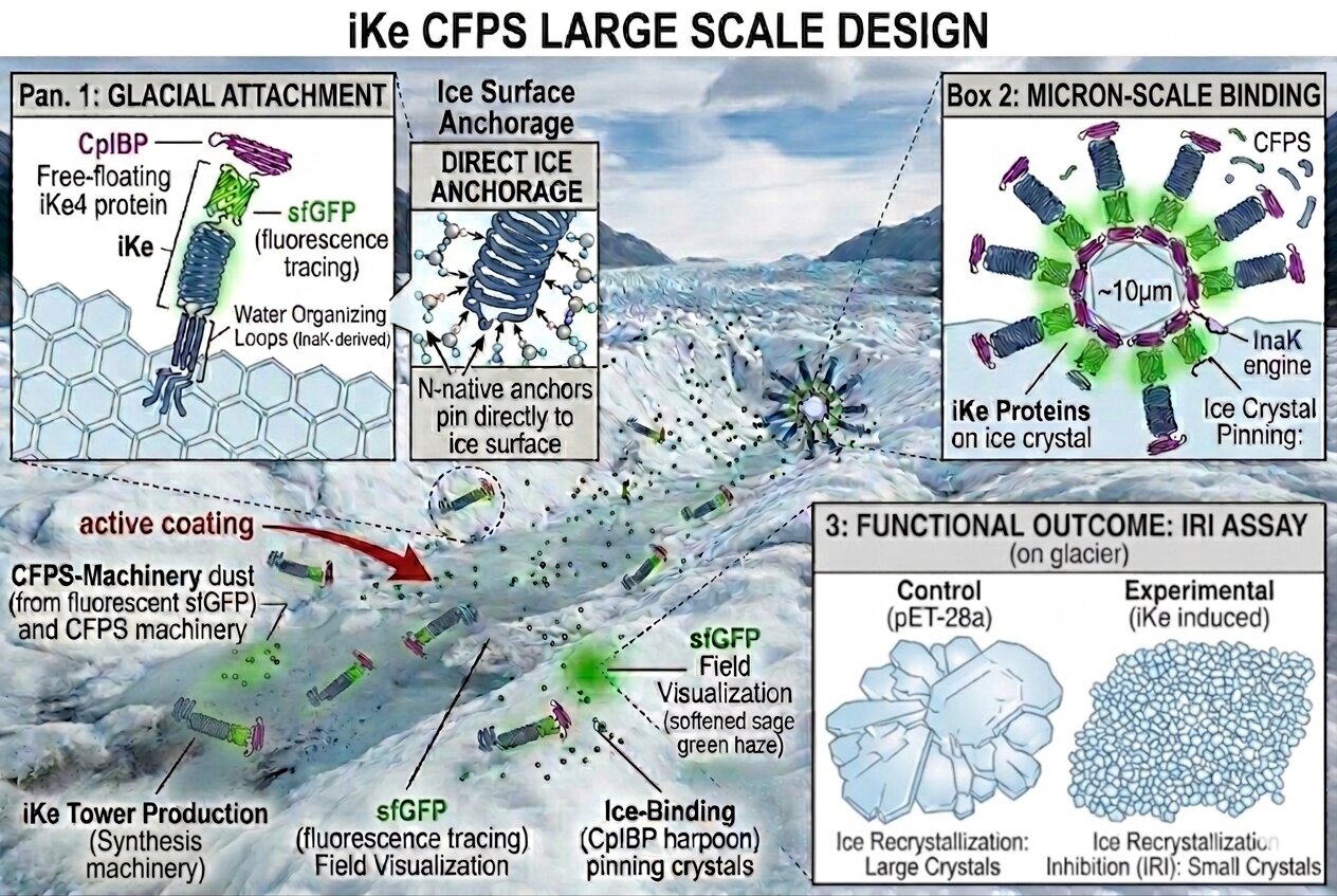
My synthetic cell design will aim to be used to synthesize inaK (using a prokaryotic cell design) and allow it to more efficiently catalyze ice. Using CFPS for inaK synthesis enables me to control and organise the proteins to sit on the membrane surface in high concentration. Ice nucleation is a surface dependent process and through CFPS I could target the ice nucleation in active sites or clusters which will be more effective than loose individual proteins.
The input is what I would give the cell to stay active. I would input a codon optimized plasmid containing the inaK sequence serving as genetic instructions. I would also input the standard set of 20 amino acids in order to build the inaK protein chains. I would input an ATP for energy (with PEP and PK to have energy regeneration). Finally, I would input magnesium and potassium salts to stabilize the internal soup of the cell.
If I am designing a prokaryotic cell design then the transcription and translation are coupled and happen simultaneously.
The output of this design creates a functional change, primarily the folded inaK proteins anchors to this new membrane and forms an ice active surface. The proteins are physically aligned in a new structure allowing for the crystal lattice of surrounding water molecules. Finally, there would be inorganic phosphate and heat outputs which will be a result of the ATP function.
Reference List
Noireaux, V., & Libchaber, A. (2004). A vesicle bioreactor as a step toward an artificial cell. PNAS.
Silverman, A. D., et al. (2020). Quarto: A User’s Guide to Cell-Free Protein Synthesis. Nature Reviews Genetics.
b.Could this function be realized by cell-free Tx/Tl alone, without encapsulation?
This design could be realized by cell-free Tx/Tl without encapsulation but it will change the function of the synthesis. If I remove the membrane then the inaK depending on it to efficiently catalyze ice would significantly change. The ice nucleation would decrease as I would lose the opportunity to design and arrange clusters but the protein can still technically be synthesized using the 20 amino acids and the DNA template and it would still catalyze ice to some extent. The main restriction will be that the hydrophobic N-terminal will have no membrane to latch onto and it will create disorder aggregates disturbing the ice formation.
As much as this synthesis can technically be realized without encapsulation it would not be beneficial or very useful to the experiment.
Reference List
Schmid, S., et al. (2016). Probing the ice-nucleation activity of the Pseudomonas syringae InaK protein. (Discussing the importance of membrane context).
Noireaux, V., & Libchaber, A. (2004). A vesicle bioreactor as a step toward an artificial cell. PNAS.
c.Could this function be realized by genetically modified natural cell?
Yes this function could be realized in a genetically modified natural cell, E. coli was used as a living cell before CFPS became a common technique. Here the inaK could be inserted as plasmid into a lab strain of E. coli to serve as a host. The advantage would be that it is energy self-sustainning as it is living,however, it has a higher complexity rate as there are many other interfering proteins and it has a high bio-safety risk as living GMOs can escape and multiply.
A cell free system would need a controlled energy input but has a simpler and safer function. The synthesis efficiency is also much higher and more robust in CFPS than in natural cell.
Reference List
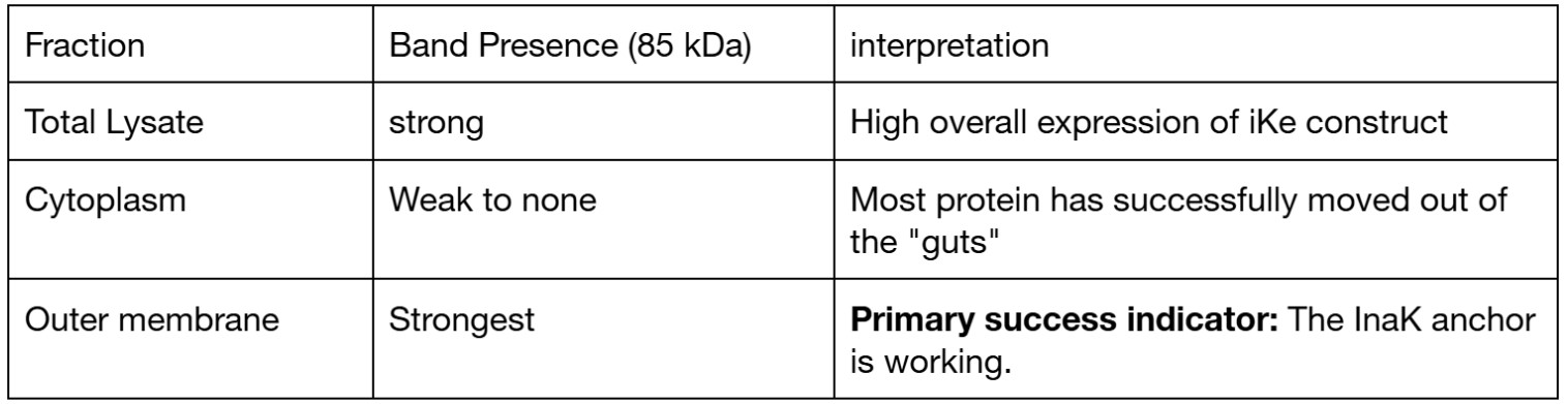
In a Natural Cell: The surface of a living E. coli is crowded with lipopolysaccharides (LPS), flagella, and other proteins. These can “get in the way” of the InaK anchors, potentially lowering the density of the ice-active patches.
In a Synthetic Cell: You can create a “naked” lipid bilayer. This allows the InaK proteins to pack together tightly without any biological “clutter,” often leading to a more efficient ice-nucleation point.
d.Describe the desired outcome of your synthetic cell operation.
Ideally the inaK protein would catalyze ice in a more efficient and organized manner which could be used in glaciers to help preserve and rebuild their natural ice formation and be more resistant to warmer temperatures.
2.Design all components that would need to be part of your synthetic cell.
a.What would be the membrane made of?
To create a suitable cell and membrane for the inaK protein and its ice nucleation function I would start with a primary lipid mix of 70% POPE (palmitoyl oleoyl sn glycero phosphoethanolamine), helping creating necessary curvature and lateral pressure in membrane thanks to its cone shape, and 30% POPG (palmitoyl oleoyl sn glycero phosphoglycerol), provides negatively charged lipids to help inaK proteins be inserted in the membrane. The common mixture for mimicking an E.coli or Pseudomonas (inaK is commonly found in Pseudomonas Syringae) membrane is composed of PE (phosphatidylethanolamine) and PG (phosphatidylglycerol). This method would prevent aggregation of inaK as the hydrophobic N-terminal cannot insert itself into too stiff of a membrane. Furthermore, the lateral pressure of the membrane will be imitated and this tension helps push the protein together to cluster the inaK and enable ice nucleation at a higher temperature.
Reference List
Schmid et al. (2016) found that the ice-nucleation activity of InaK is highly dependent on being embedded in a lipid bilayer rather than just being free-floating, specifically noting that PE/PG mixes provided the most “natural” environment for bacterial anchors.
b.What would you encapsulate inside? Enzymes, small molecules.
In order for the cell not to just be a lipid membrane bubble I will add a variety of synthetic cell internal organs which ideally would focus on improving the speed of the transcription and translation system as the inaK is a long and repetitive protein.
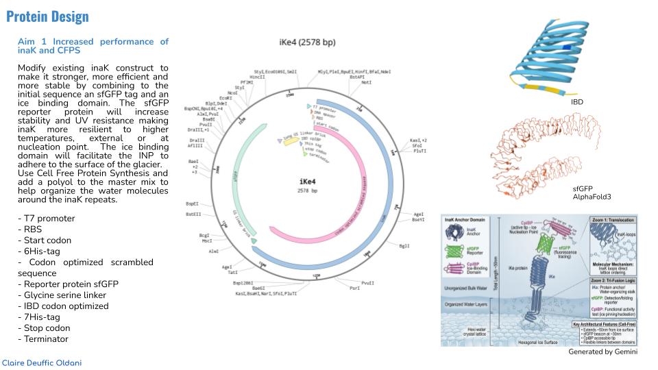
The cell will encapsulate the genetic blueprint, the DNA plasmid containing the inaK codon optimized gene sequence. Additionally, there should be a strong promoter and double terminator to process large amounts of mRNA better and to ensure the RNA polymerase end in a specific chosen section preventing useless loss of energy.
I could add a PURE system to the cell replacing a cell lysate, this is a purified protein synthesis mix including 10S ribosomes (builds the protein), T7 RNA polymerase (turns DNA to mRNA), 36 essential enzymes to charge tRNA and translate information ( aminoacyl, IF,EF,RF) and tRNAs (transfer RNAs moving amino acids to the ribosome).
Moreover, I will add the energy source for the PURE system to work. This will include the set of 20 amino acids to build the inaK protein chain, NTPs (ATP, GTP, CTP and UTP) as energy for building the protein sequence and the secondary energy substrate, here creatine phosphate combined with creatine kinase commonly used in PURE systems to keep the ATP charged.
To ensure the protein gets to the membrane wall correctly and efficiently I might add SRPs (signal recognition particles) which will connect to the inaK and guide to the membrane and chaperones (DnaK, DnaJ and Grpe) which help with protein folding and prevent the inaK anchor to create clumps before reaching the membrane.
Finally, to stabilize the cell I will add magnesium salts.
I used Gemini to help me organize my information.
Reference List
Noireaux, V., & Libchaber, A. (2004). A vesicle bioreactor as a step toward an artificial cell. PNAS. This paper provides the foundational protocol for encapsulating T7-based expression systems inside POPE/POPG vesicles.
Cui, Y., Chen, X., Wang, Z. and Lu, Y. (2022) ‘Cell-free PURE system: evolution and achievements’, Biodesign Research, 2022, art. no. 9847014. doi: 10.34133/2022/9847014.
c.Which organism your Tx/Tl system will come from? Is bacterial OK, or do you need a mammalian system for some reason? (hint: for example, if you want to use small molecule modulated promotors, like Tet-ON, you need mammalian)
For the inaK CFPS I have chosen a prokaryotic system using an E. coli based PURE method, because it is bacterial the codon language between the Pseudomonas prokaryotic bacteria origin of the inaK and the synthetic prokaryotic cell will match and the ribosome function will be more efficient. The Tx/TI system will then be coupled which works better for a membrane protein and here my hydrophobic anchor will find the membrane immediately avoiding complications. Additionally, in this case a mammalian system might struggle with the repetitiveness of my sequence.
Using a prokaryotic E. coli PURE design will give me a higher yield.
Reference List
Cui, Y., et al. (2022). ‘Cell-free PURE system: evolution and achievements’, Biodesign Research. This paper confirms that the E. coli-based PURE system is the most effective defined platform for expressing and inserting bacterial membrane proteins.
d.How will your synthetic cell communicate with the environment? (hint: are substrates permeable? or do you need to express the membrane channel?)
The cell will communicate mainly with its external environment through the protein action itself, the inaK has an ice nucleating function which will translate as a physical frozen output, the surrounding water molecules will also align and will themselves freeze.
Further membrane modifications could also allow for more communication with its environment whether through adding pores to the membrane to create an ATP reactive cell if it detects surrounding ATPs, if not it would stay dormant.
3.Experimental details
a.List all lipids and genes. (bonus: find the specific genes; for example, instead of just saying “small molecule membrane channel” pick the actual gene.)
inaK: protein derived from Pseudomonas syringae, target gene
T7 RNA polymerase : transcription (if the PURE system doesn’t already contain a protein, the gene is used to produce polymerase to transcribe inaK)
secY, secE, secG : translocation genes
POPE: lipid building blocks
POPG : lipid building block
70S ribosomes: translation
20 amino acids: translation
tRNA: translation
SRP: connects inaK to membrane
hlyA : communication gene (used if wanted to form pores in membrane)
Used Gemini to help me organize my information.
Reference List
Cui, Y. et al. (2022) ‘Cell-free PURE system: evolution and achievements’, Biodesign Research. (For the PURE components).
Noireaux, V. and Libchaber, A. (2004) ‘A vesicle bioreactor as a step toward an artificial cell’, PNAS. (For the POPE/POPG and $\alpha$HL pore strategy).
Li, Q. et al. (2012) ‘Characterization of the ice nucleation protein InaK’, Journal of Biological Chemistry. (For the inaK gene details).
b.How will you measure the function of your system?
I could try protein localization through fluorescence protease protection assay using fluorescent tags at the inaK terminals.
I could measure the freezing efficiency once again through a droplet trial, testing droplets of multiple versions of the solution when they freeze at what temperature and how resistant they are to temperature.
Homework question from Peter Nguyen
1.Write a one-sentence summary pitch sentence describing your concept.
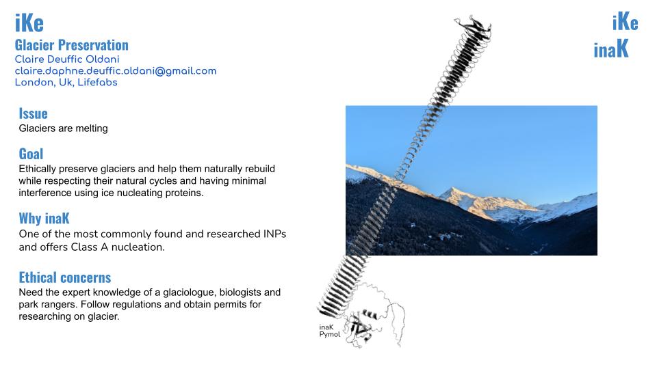
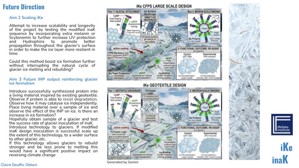
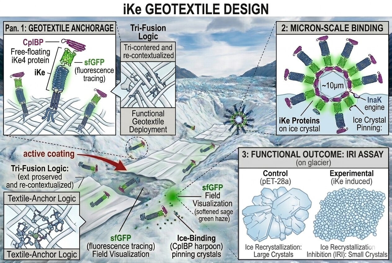
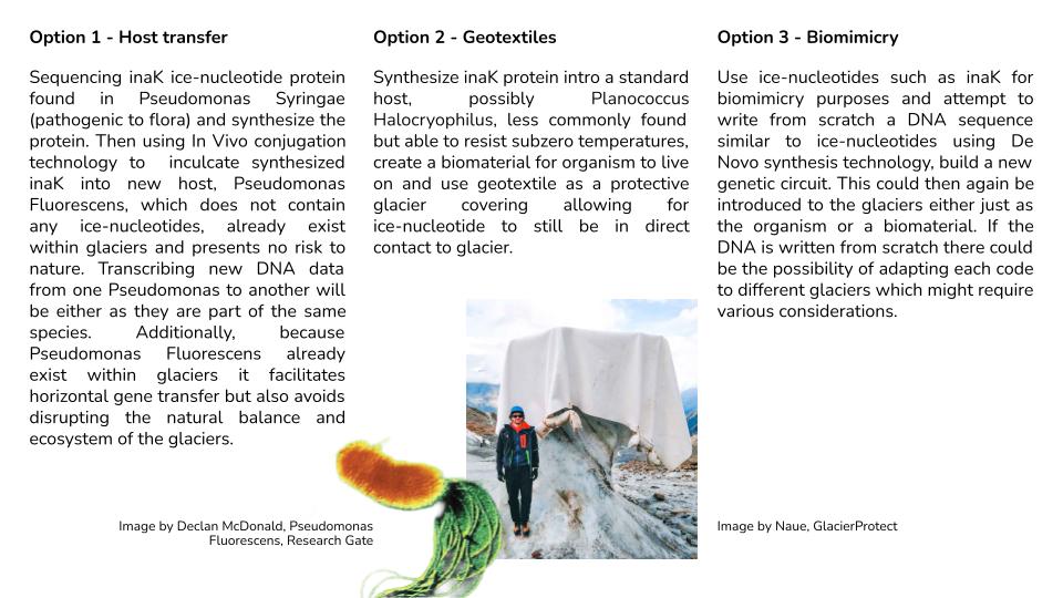
I could use cell free systems adapted to producing inaK in order to directly inoculate glaciers with the aim to preserve and boost ice formation, it would help glaciers rebuild and be more resistant to increasing temperatures caused by climate change. However, I would be interested in pushing the idea of geotextiles already helping preserve glaciers and design a living material, with inaK with a boosted ice nucleation function to create proactive glacier covering actively working to rebuild and preserve glacier ice.
2.How will the idea work, in more detail? Write 3-4 sentences or more.
The inaK would be synthesized through a cell free model, using an alternative to E. coli which could resist and be active in sub-zero conditions (to stay active in glaciers), such as Oleispira antarctica a psychrophiles bacteria which has evolved to have specialized ribosomes and enzymes able to remain flexible and functional in a frozen environment. Oleispira antarctica contains unique chaperone (Cpn60 and Cpn10) preventing protein misfolds in frozen temperatures. This cell design would include pores in the membrane so it can stay alive in the textile by having an ATP source of input.
These then freeze dried cells would be put into a textile (inoculated during the making of the textile), the textile can be brought to location and installed on the glacier and then be rehydrated to allow ice nucleation of the glacier to begin. This would permit me to create a live material that would be dormant in production and transportation and control its freezing function (preventing the textile from accidentally freezing its surrounding).
Note that because I am working with ice nucleation there might be challenges in freeze drying these cells.
Reference List
Ferrer, M. et al. (2003) ‘Low temperature-induced systems failure in Escherichia coli: Insights from rescue by cold-adapted chaperones’, Journal of Biological Chemistry.
Cui, Y. et al. (2022) ‘Cell-free PURE system: evolution and achievements’, Biodesign Research.
D’Amico, S. et al. (2006) ‘Psychrophilic microorganisms: challenges for life’, EMBO reports.
3.What societal challenge or market need will this address?
This addresses the environmental, social and political issue of melting glaciers caused by climate change, which only increases the power of climate change as glaciers are key factors in slowing climate change. We are actively losing biodiversities and ecosystems and doing very little about it. It is not seen as a profitable income so little motivation is inputted. However, in the longterm, this irreversible damage done to our nature will actively make climate change worse, and there will be many destructive environmental, social and economic consequences driven by this overlooked issue.
4.How do you envision addressing the limitation of cell-free reactions (e.g., activation with water, stability, one-time use)?
Working at very large scale, scale of the glacier, it can be a challenge to efficiently rehydrate the living material as it would be very energy consuming and costly to do it manually, but, if the living geotextile is strategically implemented at the right time of the year (early spring, already when they glacier coverings are usually installed) then nature itself through rain could activate the material naturally. The aim is to limit the human labor impact and simply give nature a tool to reinforce what it already knows how to do.
Considering the one time use issue, geotextile coverings which are already used to protect glaciers are removed and installed yearly according to their natural ice melting and forming cycles. The next step of my research would be to find a way to keep the textile created and reabsorb it with new inaK cell free protein systems when it is needed next. The goal is to create a regenerative textile and closed loop system to avoid waste through one time solutions.
Homework question from Ally Huang
1.Provide background information that describes the space biology question or challenge you propose to address. Explain why this topic is significant for humanity, relevant for space exploration, and scientifically interesting. (Maximum 100 words)
I am interested in exploring the purpose of ice nucleation in cell free design, freeze dried or not, taking the shape of a multipurpose textile which can be used as an alternative to current voluminous refrigeration tools or a freezing textile to activate. In space stations like the ISS a lot of research relies on lab samples being preserved in sub zero temperatures, from human research samples to organisms or protein crystals. Within research some experiments need cold induced phase changes to be activated or triggered. This technology could also be used for food or medical supplies.
2.Name the molecular or genetic target that you propose to study. Examples of molecular targets include individual genes and proteins, DNA and RNA sequences, or broader -omics approaches. (Maximum 30 words)
The genetic target of this project would be the inaK ice nucleating protein, commonly found in Pseudomonas syringae, with a wide potential of freezing functions.
3.Describe how your molecular or genetic target relates to the space biology question or challenge your proposal addresses. (Maximum 100 words)
The challenge is optimizing cold packaging and storage systems, a material which would require less space or a material able to be activated once in space again having less constraints in terms of space while travelling. The inaK offers a variety of possibilities in a cell free system whether freeze dried or not as it has a focused and controlled function to freeze. According to the development of the product it can be chosen at what temperature it freezes or activates and how resistant it can be to external temperatures. Creating a highly controlled and bespoke design for certain use in space allows for better control on the research done in space, every aspect of the research can be tailored in hopes to improve success rates of experiments. InaK is a relatively easy INP to work with.
4.Clearly state your hypothesis or research goal and explain the reasoning behind it. (Maximum 150 words)
I am interested in creating polyvalent designs with multiple usages and applications, this project aims to find an optimal alternative refrigerating system which can have bespoke qualities specific to in space research. As small of a detail it might seem every aspect and tool of experiments impacts the result of research and can lead to better efficiency, results or unexpected breakthroughs. During a space mission all equipment has to be optimized due to lack of space and need for many items and a polyvalent tool that can respond to a wide range of uses can help with the space optimization.
5.Outline your experimental plan - identify the sample(s) you will test in your experiment, including any necessary controls, the type of data or measurements that will be collected, etc. (Maximum 100 words)
I would design a cell free system for the inaK ice nucleating protein, freeze dry some and then create living textiles, some active and some dormant. The practicality of a textile is that it can be molded, cut, sewn, layered to adapt to any existing object which would then need a freezing function.
I can control the amount of inaK for the freezing rate needed, experiment with the different temperatures it can freeze at and the different temperatures it can stay frozen at, I can explore the threshold of the inaK.
I would then test the reactivation rates, how much water is needed and how long it would take.
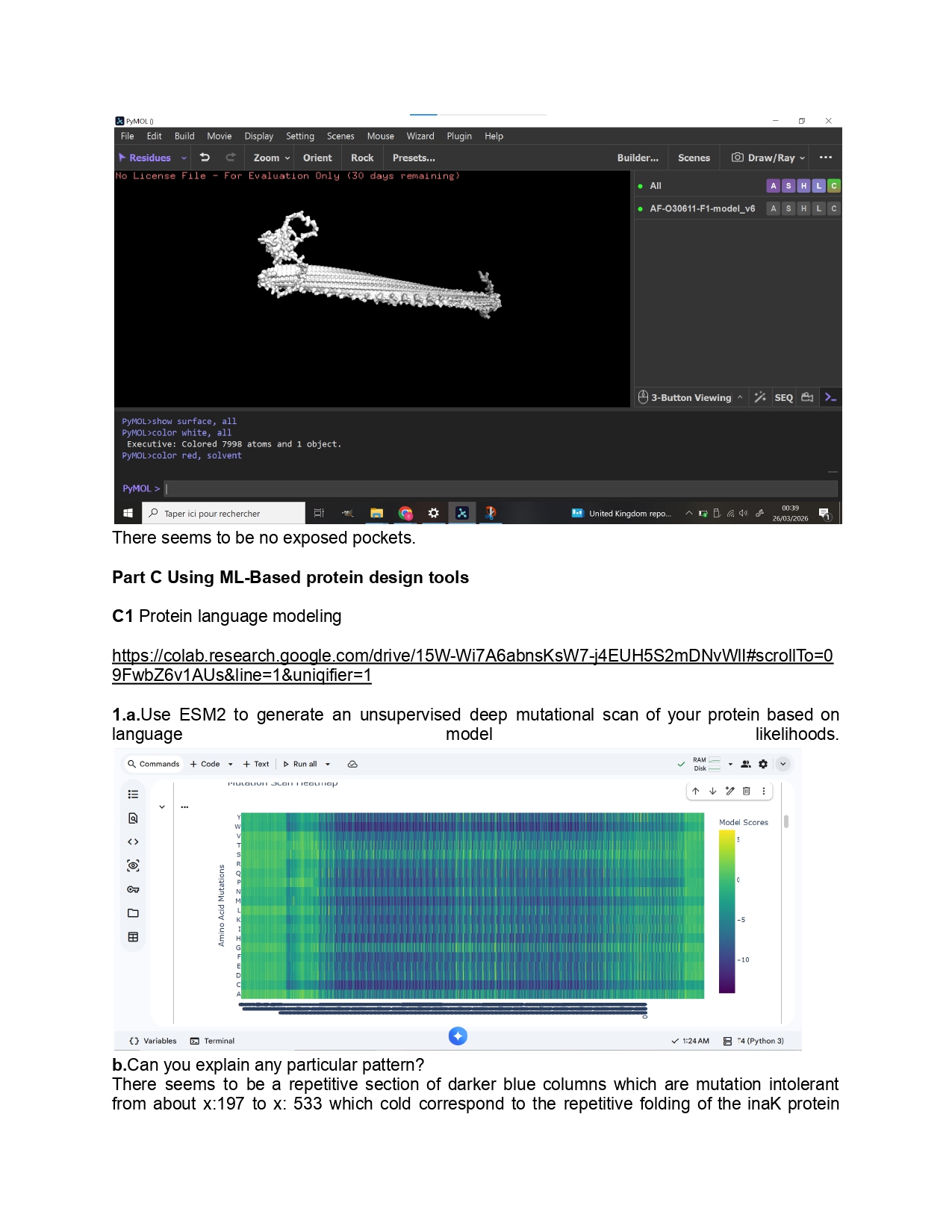
1.Please identify at least one (ideally many) aspect(s) of your project that you will measure. It could be the mass or sequence of a protein, the presence, absence, or quantity of a biomarker, etc.
As for this project I aim to use inaK for ice production I would like to measure the ice nucleation ratio and efficiency of the inaK protein. Additionally, I would like to measure the temperatures inaK can resist to, on its own and as a supplement to an ice sample.
If my initial experiments are successful I would like to measure the inaK ratio innoculated into ice to find the most optimal inaK quantity needed.
2.Please describe all of the elements you would like to measure, and furthermore describe how you will perform these measurements.
To measure the ice production ratio and efficiency of inaK I could use differential scanning calorimetry (DSC) which measures the difference in the amount of heat required to increase the temperature in the sample compared to a reference. It measures the ice nucleation ratio by calculating the enthalpy which is the area below the peak, allowing me to understand precisely how much of the water in the cell is being converted to ice. It measures the efficiency of ice nucleation by creating an exothermic peak ( release of a burst of energy) and analysing how high of a temperature the inaK can still function. This should give me information on thermodynamic efficiency.
I can also measure nucleation temperature through a droplet freezing assay for smaller samples allowing me to test a multitude of potential solution mixes. Here a high speed camera paired with a cooling plate ( a Linkam for example) can allow me to assess how fast a droplet of a solution containing inaK can freeze. Testing this on multiple samples containing different amounts of inaK will give me a spectrum of freezing capacity to find the most optimal ratio of inaK. This experiment could be coupled with an infrared thermography technology which will capture the heat spike and nucleation rate of an inaK and understand how fast the ice nucleation spreads through the cell membrane.
Reference List
Schmid, D. et al. (2016) ‘A high-throughput assay for the characterization of ice-nucleating proteins’, Biophysical Journal. This study outlines the specific use of droplet assays to quantify InaK efficiency.
3.What are the technologies you will use (e.g., gel electrophoresis, DNA sequencing, mass spectrometry, etc.)? Describe in detail.
For these experiments I will use a Differential Scanning Calorimeter for the DSC, a high precision camera, a cooling plate and IR thermography.
Waters Part 1 - Molecular Weight
For this section I used a combination of the tools provided, my knowledge and AI assistance as I have trouble understanding math related work.
Based on the predicted amino acid sequence of eGFP and any known modifications, what is the calculated molecular weight ?
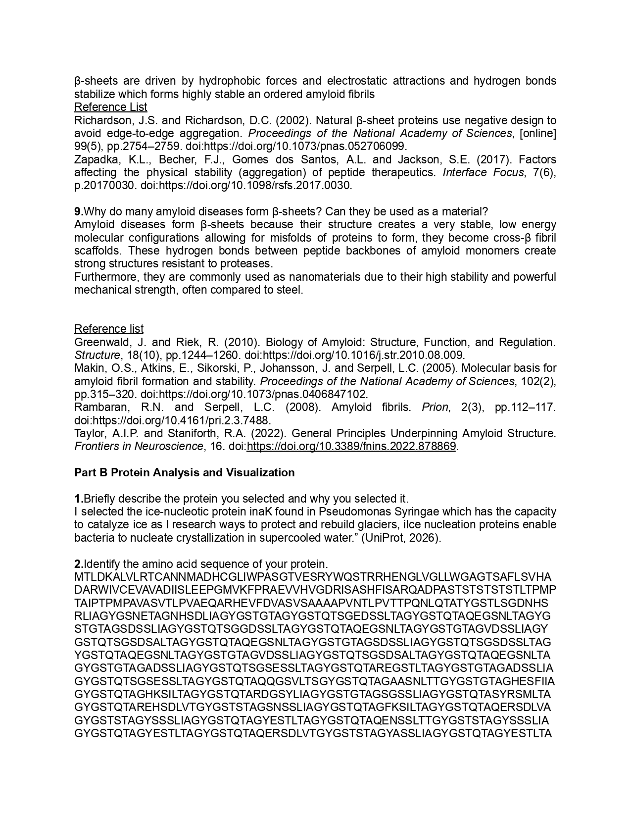
According to Expasy I found that this sequence has a theoretical pI/Mw of 5.90 / 28006.60.
To calculate the molecular weight of this sequence I referred to the standard isotopic mass of amino acids and subtracting H²O for each peptide bond. For this amino acid sequence I found that :
-eGFP of 238 AA = 26.735.6 Da
-LE Linker of 2 AA = 242.3 Da
-x-His Tag of 6 AA = 822.8 Da
Resulting in a total molecular weight of 27800.7 Da
Calculate the molecular weight of the eGFP using the adjacent charge state approach described in the recitation. Select two charge states from the intact LC-MS data (figure 1) and:
MW : molecular weight in Daltons
m/z : value of the peak on the x-axis of the spectrum
z : integer charge state of said peak
H+ : mass of a proton
a.Determine z for each adjacent pair of peaks $(n, n+1)$ using: $$ {\large z} = {\Large \frac{\frac{m}{z_{n+1}}}{\frac{m}{z_n} - \frac{m}{z_{n+1}}}} $$
Here I am using the adjacent peaks at 875.4421 and 903.7148.
m:zn = 875.4421
m:zn+1 = 903.7148
The lower the m/z the higher the charge versus the higher the m/z the lower the charge.
Using the provided formula
z = 903.7148 : (903.7148 – 875.4421) = 903.7148 : 28.2727 = 31.96
Charge states must be integers and the charge state for the peak at 903.7148 is z= 31
So, the peak at 875.4421 has a charge state of z+1=32
b.Determine the MW of the protein using the relationship between $\frac{m}{z_n}$, $MW$, and $z$
The base equation for the peaks in this figure is
m:z = (MW + (z x H+)) : z
Here I rearrange the formula to find MW
MW = z x (m:z) - (z x H+)
If z=31, then,
MW = 31 x(903.7148) - (31x1.008)
MW = 28 015.1588 – 31.248
MW = 27 983.91 Da
c.Calculate the accuracy of the measurement using the deconvoluted MW from 2.2 and the predicted weight of the protein from 2.1 using: $$ \text{Accuracy} = \frac{|MW_{\text{experiment}} - MW_{\text{theory}}|}{MW_{\text{theory}}} $$
Using the provided formula and substituting the values accordingly,
If,
Accuracy = (MW experimental - MWtheory) : MW theory
Then,
Accuracy = (27 982.90 - 27 782.70) : 27 782.70
Accuracy = 200.20 : 27 782.70
Accuracy = 0.0072
=0.72%
3.Can you observe the charge state for the zoomed-in peak in the mass spectrum for the intact eGFP? If yes, what is it? If no, why not?
One can observe the charge state in the zoomed in peak but not using the previous calculation method using the adjacent peak method used for the entire spectrum.
The zoomed in area presents an isotopic cluster of one charge state, therefore, the observation is focused on isotopic resolution. The other peaks represent different amounts of protons.
So, here, yes I can see the charge state using isotopic resolution because the peaks are distinct and separate and the instrument has high enough of a resolution to expose the isotopes separately. It is because the instrument used here is of high precision that we are able to have high resolution, if a lesser precise instrument were used then it would be very difficult to see the charge state.
The formula to calculate isotopic spacing here would be
z = 1 : Δm/z
Waters Part 2 - secondary & tertiary structure
1.Please explain the difference between native and denatured protein conformations. For example, what happens when a protein unfolds? How is that determined with a mass spectrometer? What changes do you see in the mass spectrum between the native and denatured protein analyses (figure 2)?
There is a structural difference between the native and the denatured protein conformations, the native protein conformation is a tightly folded protein, a unique three dimensional structure unique to its biological environment. The structure is compact but is held by weak non-covalent bonds. In contrast, a denatured protein conformation is the unfolded structure once the weak bonds are broken, the structure is flexible and unpredictable resulting in a random coil shape. The denatured protein no longer has a function compared to the native protein conformation.
A mass spectrometer scans the protein shape by measuring its mass and charge rather than measuring the shape directly.
The top green graph corresponds to the denatured confirmation and the bottom red chart corresponds to the native conformation.
The denatured conformation graph shows crowded peaks in the 500 to 1200 m/z range, these are low m/z values. A lower m/z equals a higher z charge and when a protein is unfolded into a random coil then the basic residues are exposed to a proton rich environment and can easily be protonated creating a protein with a very high net positive charge.
The native conformation has larger m/z values with peaks at 2545 and 2799, the higher the m/z value the lower the z charge. When the protein is still tightly folded the basic residues are buried in the hydrophobic core protecting it from its environment meaning protons cannot impact them so the protein is less protonated.
2.Zooming into the native mass spectrum of the eGFP from the Waters Xevo G3 QTof or MS (figure 3), can you discern the charge state of the peak at ~2800? What is the charge state? How can you tell?
It is possible to discern the charge state of the peak at ~2800 m/z because instrument provides a high resolution even if the image focuses on the 2525 m/z peak, we are still able to see the isotopic distribution by measuring the distance between the individual isotopes and we would be able to calculate the charge state.
Measuring the charge state of the peak at ~2800 m/z using isotopic spacing,
In a mass spectrum the individual isotopes of the same molecule differs by approximately 1 Dalton,
The distance between the isotopes here on the x-axis Δm/z can be calculated using the following formula,
Δm/z = 1: z
Zooming in at the 2799.4199 peak same as for the 2545 peak (with a peak separation of precisely 0.1 m/z) then,
z = 1: 0.1 = 10
So the charge state of the peak at ~2800 m/z is +10
Waters Part 3 - Peptide mapping, primary structure
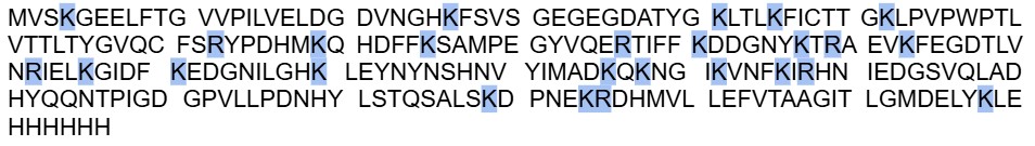
1.How many Lysines (K) and Arginines (R) are in eGFP? Please circle or highlight them in the eGFP sequence given in Waters Part I question 1 above. (Note: adding the sequence to Benchling as an amino acid file and clicking the biochemical properties tab will show you a count for each amino acid).
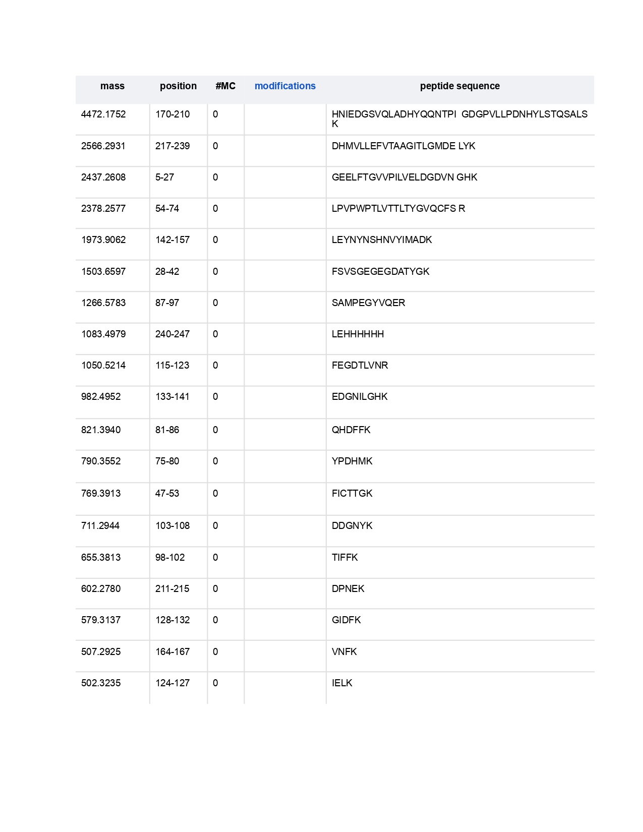
2.How many peptides will be generated from tryptic digestion of eGFP?
I found 19 peptides were generated from this sequence using the trypsin.
3.Based on the LC-MS data for the Peptide Map data generated in the lab ( please use Figure 5a as a reference) how many chromatographic peaks do you see in the eGFP peptide map between 0.5 and 6 minutes? You may count all peaks that are>10% relative abundance.
Between 0.5 and 6 minutes there are 14 distinct peaks above 10% relative abundance in this figure.
4.Assuming all the peaks are peptides, does the number of peaks match the number of peptides predicted from question 2 above? Are there more peaks in the chromatogram or fewer?
In comparison to the amount of predicted peptides, 19, there are fewer peptides in the chromatogram.
5.Identify the mass-to-charge (m:z) of the peptide shown in Figure 5b. What is the charge (z) of the most abundant charge state of the peptide (use the separation of the isotopes to determine the charge state). Calculate the mass of the singly charged form of the peptide ([M+H]+) based on its m:z and z.
The z charge of the most abundant peak in this peptide is m/z = 525.76712
To calculate the charge state,
First isotope peak 525.76712
Second isotope peak 526.25918
Calculating the spacing,
(Δm/z):526.25918 - 525.76712 = 0.49206
Using the formula,
z = 1: 0.49206≈2.03
So the charge state z of the most abundant peak of the peptide is of +2
Calculating the mass of the singly charged form of the peptide ([M+H]+),
First I need to calculate the neutral mass M using M=zx(m/z)-(zx1.00727),
1.00727 Da is the mass of a proton,
So,
M = 2 x (525.76712) - (2x1.00727)
M = 1051.53424 - 2.01454 = 1049.5197 Da
([M+H]+) can be calculated by adding one proton mass back to the neutral mass,
[M+H]+ = 1049.5197 + 1.00727 = 1050.5270 Da
6.Identify the peptide based on comparison to expected masses in the PeptideMass tool. What is mass accuracy of measurement? Please calculate the error in ppm. (Recall that Accuracy = (MW experimental - MWtheory) : MW theory)
To identify the peptide I consider that the experimental neutral molecular sight (MWexperimental) was calculated for the peak 525.76712 m/z and equaled MW: 1049.5197 Da, I will use the following peptide sequence as a theoretical tryptic digest as a comparison, LPDNHYLSTQSALSK, and considering theoretical MW (MWtheory) : 1049.5393 Da. This peptide corresponds to the residues 139–153 of the eGFP protein.
To calculate the mass accuracy, error in ppm (parts per million) I will use the following formula,
Accuracy (ppm) = ((MWexperimental - MW theory) : MWtheory) x 106
Now adding the values,
MW experimental = 1049.5197
MW theory = 1049.5393
Accuracy (ppm) = ((1049.5197 - 1049.5393) : 1049.5393) x 106
Accuracy (ppm) = (-0.0196) : 1049.5393) x 106
Accuracy (ppm) = –18.67ppm
7.What is the percentage of the sequence that is confirmed by peptide mapping ? (see figure 6)
The percentage of the sequence that is confirmed by peptide mapping seems to be indicated at 88%, the blue highlighted areas are confirmed amino acids in the sequence.
Waters Part 4 - Oligomers
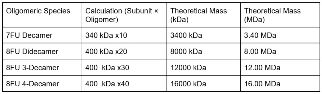
We will determine Keyhole Limpet Hemocyanin (KLH)’s oligomeric states using charge detection mass spectrometry (CDMS). CDMS single-particle measurements of KLH allow us to make direct mass measurements to determine what oligomeric states (that is, how many protein subunits combine) are present in solution. Using the known masses of the polypeptide subunits (Table 1) for KLH, identify where the following oligomeric species are on the spectrum shown below from the CDMS (Figure 7):
-7FU Decamer
-8FU Didecamer
-8FU 3-Decamer
-8FU 4-Decamer
I will first calculate the theoretical mass for the species using the given measures 7FU = 340 kDa and 8FU = 400 kDa. The axis of the spectrum is in MDa (megadaltons) where 1MDa = 1000 kDa.
I can then identify the peaks on the spectrum
-7FU Decamer ≈ 3.40 MDa is the peak labeled 3.4 on the spectrum
-8FU Didecamer ≈ 8.00 MDa correspond to the peak labeled 8.33
-8FU 3-Decamer ≈ 12.00 MDa matches the peak labeled 12.67
-8FU 4-Decamer ≈ 16.00 MDa would appear in the cluster of small unlabeled peaks around 16 to 17 MDa on the far right side of the chart
Waters part 5 - Did I make GFP?
Please fill out this table with the data you acquired from the lab work done at the Waters Immerse Lab in Cambridge, or else the data screenshots in this document if you were unable to have lab work done at Waters.
Week 11 HW: Building genomes
Week 11 Bioproduction & Cloud Lab
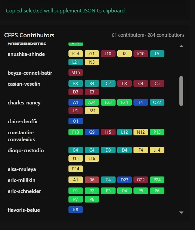
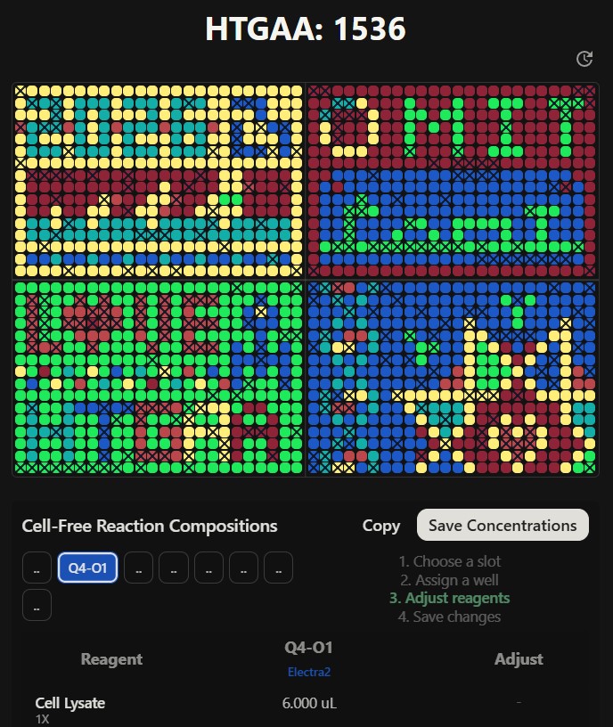
Part A - The 1.536 pixel art work canvas, collective artwork
1.Contribute at least one pixel to the global artwork
I added early on a pixel towards the top left corner. I do not have much to say about this section of the work except maybe understanding the full purpose of this exercise.
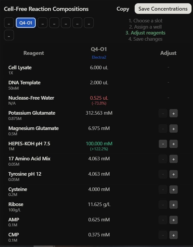
Part B - Cell Free protein synthesis, cell free reagents
Referencing the cell-free protein synthesis reaction composition (the middle box outlined in yellow on the image above, also listed below), provide a 1-2 sentence description of what each component’s role is in the cell-free reaction.
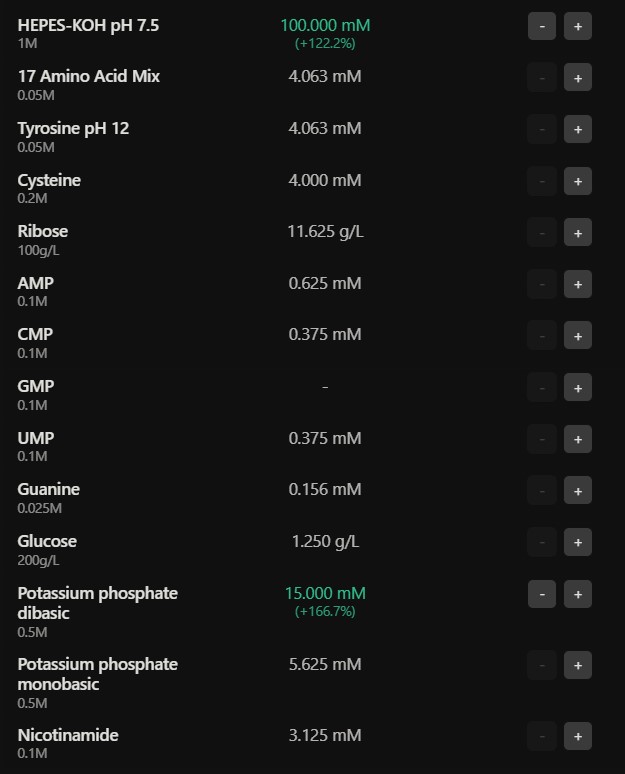
E. coli Lysate
BL21 (DE3) Star Lysate (includes T7 RNA Polymerase) : offers the base molecular machinery such as ribosomes, tRNAs and enzymes for translation, the Star Lysate strain reduces mRNA degradation, and, the T7 Polymerase drives high level transcription from T7 promoters
Salts / Buffer
Potassium Glutamate : primary salt that maintains ionic strength and provides potassium ions essential to ribosomal function and protein to nucleic acid exchange
HEPES-KOH pH 7.5 : chemical buffer which helps maintain a stable physiological pH which affects enzymatic function of the transcription and translation machinery
Magnesium Glutamate : magnesium ions are vital contributors to stabilizing the ribosome structure and enabling catalytic activity of the polymerases kinases
Potassium phosphate, monobasic and dibasic : functions as a secondary pH buffer and a source of inorganic phosphate essential for the regeneration of high energy molecules such as ATP
Energy / Nucleotide system
Ribose : serve as a carbon backbone precursor for the synthesis of nucleotides, allowing for regeneration of NTPs essential for transcription and energy transfer
Glucose : primary metabolic energy source fueled through glycolysis allowing to regenerate the ATP and GTP essential to the good functioning of protein synthesis
AMP / CMP / UMP : offers nucleotide building blocks for RNA synthesis and can be converted into triphosphate such as ATP, CTP, UTP needed in transcription
GMP : from the lack of GMP might demonstrate a dependency on salvage pathways to generate GTP essential to translation
Guanine : precursor for GMP/GTP synthesis through salvage pathways helpful to RNA synthesis and ribosomal function
Translation Mix (Amino acids)
17 Amino Acid Mix : provide the base building blocks to synthesize the polypeptide chain
Tyrosine : supplied separately because of its solubility limitations, becomes an essential building block for protein synthesis once it is adapted into a usable form
Cysteine : added separately due to its oxidation limitations, it is an essential compound in forming disulfide bonds in proteins
Additives
Nicotinamide : serves as a precursor for NAD+ / NADH synthesis reinforcing redox balance and metabolic reactions occurring in energy regeneration
Backfill
Nuclease Free Water : is used to adjust all the components to the desired the final reaction volume while it avoids degradation of DNA / RNA by nuclease and ensures stable transcription and translation processes
Describe the main differences between the 1-hour optimized PEP-NTP master mix and the 20-hour NMP-Ribose-Glucose master mix shown in the slide.
The main difference between the two master mix results in found in the energy and nucleotide sourcing a the 1-hour mix makes use of the PEP and pre-synthesized NTPs for instant and high burst protein synthesis compared to the 20-hour mix uses the ribose, glucose and NMPs as precursors to regenerate energy and nucleotides throughout time. Therefore, the 1-hour mix is designed for speed and rapid prototyping in contrast to the 20-hour mix allows to better optimize the cost for effectiveness by using the Lysate’s metabolic pathway to support the reaction for an extended period of time.
Part C - Planning the global experiment, cell-free master mix design
1.Given the 6 fluorescent proteins we used for our collaborative painting, identify and explain at least one biophysical or functional property of each protein that affects expression or readout in cell-free systems. (Hint: options include maturation time, acid sensitivity, folding, oxygen dependence, etc) (1-2 sentences each)
sfGFP : provides robust and rapid protein folding therefore the protein is less likely to aggregate enabling it to offer a strong fluorescent readout even if fused to complex proteins
mRFP1 : is a protein with a longer maturation time signifying the fluorescence develops slower after translation and might have a delayed signaling time in shorter experiments, it has a low acidity tolerance
mKO2 : is fast maturing and has a relative acidity tolerance, meaning the fluorescence will be less visible in a lower pH context but the fluorescence could increase in longer cell free reactions
mTurqoise2 : is a cyan protein with for a high quantum yield and high photostability making the fluorescence outread a great signal no matter the length of the reaction, it is very sensitive to pH
mScarlet_I : engineered for fast maturing and high brightness allowing for stronger fluorescence signals compared to older red proteins
Electra2 : protein engineered for very fast maturation making it very useful for fast consuming energy systems in an experiment where a rapid output is needed before the mix’s energy is used up
2.Create a hypothesis for how adjusting one or more reagents in the cell-free mastermix could improve a specific biophysical or functional property you identified above, in order to maximize fluorescence over a 36-hour incubation. Clearly state the protein, the reagent(s), and the expected effect.
Could the mTurquoise2 yield be increased or accelerated through pH stabilisation.
The reagents HEPES-KOH would be increased to 100mM and Potassium Phosphate would be increased to 15mM.
This adjustment should increase the capacity of the buffer within the mater mix and should neutralize organic acid by products such as lactate and acetate generated during the 36-hour metabolism of glucose and ribosome. Because mTurquoise2 is very reactive to pH, preserving the pH environment at 7.5 would prevent the typically occurring rapid cooling of the cyan signal which usually occurs as the mix acidifies over time. Therefore, the high quantum of yield of mTurquoise2 is complete and optimized leading to a bright and stable cyan readout which won’t dim as the energy levels decrease.
3.The second phase of this lab will be to define the precise reagent concentrations for your cell-free experiment. You will be assigned artwork wells with specific fluorescent proteins and receive an email with instructions this week (by April 24). You can begin composing master mix compositions here.
)
)
)
)
)
Copy of reagent doses
Reagent Preset:
Variant 1 Variant 2 (O1) Variant 3 Variant 4 Variant 5 Variant 6 Variant 7 Variant 8
Cell Lysate 6.000 uL 6.000 uL (-) 6.000 uL (-) 6.000 uL (-) 6.000 uL (-) 6.000 uL (-) 6.000 uL (-) 6.000 uL (-) 6.000 uL (-)
DNA Template 0.000 mM 2.000 uL (-) 2.000 uL (-) 2.000 uL (-) 2.000 uL (-) 2.000 uL (-) 2.000 uL (-) 2.000 uL (-) 2.000 uL (-)
Nuclease-Free Water 2.000 uL 2.000 uL (-) 0.525 uL (-73.8%) 2.000 uL (-) 2.000 uL (-) 2.000 uL (-) 2.000 uL (-) 2.000 uL (-) 2.000 uL (-)
Potassium Glutamate 312.563 mM 312.563 mM (-) 312.563 mM (-) 312.563 mM (-) 312.563 mM (-) 312.563 mM (-) 312.563 mM (-) 312.563 mM (-) 312.563 mM (-)
Magnesium Glutamate 6.975 mM 6.975 mM (-) 6.975 mM (-) 6.975 mM (-) 6.975 mM (-) 6.975 mM (-) 6.975 mM (-) 6.975 mM (-) 6.975 mM (-)
HEPES-KOH pH 7.5 45.000 mM 45.000 mM (-) 100.000 mM (+122.2%) 45.000 mM (-) 45.000 mM (-) 45.000 mM (-) 45.000 mM (-) 45.000 mM (-) 45.000 mM (-)
17 Amino Acid Mix 4.063 mM 4.063 mM (-) 4.063 mM (-) 4.063 mM (-) 4.063 mM (-) 4.063 mM (-) 4.063 mM (-) 4.063 mM (-) 4.063 mM (-)
Tyrosine pH 12 4.063 mM 4.063 mM (-) 4.063 mM (-) 4.063 mM (-) 4.063 mM (-) 4.063 mM (-) 4.063 mM (-) 4.063 mM (-) 4.063 mM (-)
Cysteine 4.000 mM 4.000 mM (-) 4.000 mM (-) 4.000 mM (-) 4.000 mM (-) 4.000 mM (-) 4.000 mM (-) 4.000 mM (-) 4.000 mM (-)
Ribose 11.625 g/L 11.625 g/L (-) 11.625 g/L (-) 11.625 g/L (-) 11.625 g/L (-) 11.625 g/L (-) 11.625 g/L (-) 11.625 g/L (-) 11.625 g/L (-)
AMP 0.625 mM 0.625 mM (-) 0.625 mM (-) 0.625 mM (-) 0.625 mM (-) 0.625 mM (-) 0.625 mM (-) 0.625 mM (-) 0.625 mM (-)
CMP 0.375 mM 0.375 mM (-) 0.375 mM (-) 0.375 mM (-) 0.375 mM (-) 0.375 mM (-) 0.375 mM (-) 0.375 mM (-) 0.375 mM (-)
GMP - - (-) - (-) - (-) - (-) - (-) - (-) - (-) - (-)
UMP 0.375 mM 0.375 mM (-) 0.375 mM (-) 0.375 mM (-) 0.375 mM (-) 0.375 mM (-) 0.375 mM (-) 0.375 mM (-) 0.375 mM (-)
Guanine 0.156 mM 0.156 mM (-) 0.156 mM (-) 0.156 mM (-) 0.156 mM (-) 0.156 mM (-) 0.156 mM (-) 0.156 mM (-) 0.156 mM (-)
Glucose 1.250 g/L 1.250 g/L (-) 1.250 g/L (-) 1.250 g/L (-) 1.250 g/L (-) 1.250 g/L (-) 1.250 g/L (-) 1.250 g/L (-) 1.250 g/L (-)
Potassium phosphate dibasic 5.625 mM 5.625 mM (-) 15.000 mM (+166.7%) 5.625 mM (-) 5.625 mM (-) 5.625 mM (-) 5.625 mM (-) 5.625 mM (-) 5.625 mM (-)
Potassium phosphate monobasic 5.625 mM 5.625 mM (-) 5.625 mM (-) 5.625 mM (-) 5.625 mM (-) 5.625 mM (-) 5.625 mM (-) 5.625 mM (-) 5.625 mM (-)
Nicotinamide 3.125 mM 3.125 mM (-) 3.125 mM (-) 3.125 mM (-) 3.125 mM (-) 3.125 mM (-) 3.125 mM (-) 3.125 mM (-) 3.125 mM (-)
The final phase of this lab will be analyzing the fluorescence data we collect to determine whether we can draw any conclusions about favorable reagent compositions for our fluorescent proteins. This will be due a week after the data is returned (date TBD!). The reaction composition for each well will be as follows:
6 μL of Lysate
10 μL of 2X Optimized Master Mix from above
2 μL of assigned fluorescent protein DNA template
2 μL of your custom reagent supplements
Total : 20 μL reaction
iKe Glacier Preservation Abstract Mountain glaciers are melting progressively due to climate change and human activity. I am inspired by the glaciers of the Italian Alps where my family is from and throughout generations have seen firsthand the glaciers progressively disappear. Glaciers are vital ecosystems which contribute to protecting nature and human existence.
Mountain glaciers are melting progressively due to climate change and human activity. I am inspired by the glaciers of the Italian Alps where my family is from and throughout generations have seen firsthand the glaciers progressively disappear. Glaciers are vital ecosystems which contribute to protecting nature and human existence.
I aim to use my knowledge in textiles, biology and biodesign to help preserve and rebuild glaciers using ice nucleating proteins (INPs).
I theorise that inoculating glaciers with modified INPs using cell free synthesis would help improve ice catalyzation and make glacier ice more resilient to face increasing temperatures. This method will work as a defensive tool helping restore natural and healthy glacier cycles benefiting a wider ecosystem and battling climate change.
Project overview
Growing up I spent a lot of time in the Italian Alps where part of my family originates from, within only 23 years of life I have seen first hand the glaciers of mountains surrounding me disappear progressively, my mother sees an even bigger decrease and my grandfather a shocking decrease. With my background in textiles, my current studies in Biodesign, my curiosity for biology and now partaking in HTGAAA I will conceptualise a project combining textiles and biology as a means to create a tool which could help prevent the fast disappearance of glaciers.
Glaciers are vital elements to regulating the earth’s temperature, they are ecosystems of their own across the world. As pointed out by Glacier Preservation, “glaciers are essential for sustaining millions of people by providing fresh water, supporting hydropower generation, and playing a key role in environmental stability. However, as climate change accelerates, glaciers are retreating at an unprecedented rate, threatening water security, energy infrastructure, and increasing natural hazards like flooding and avalanches.” (Un-glaciers.org. (2026). Glacier Preservation is the Key to Ensuring the Security of Water, Energy, and Environmental Resources. [online] Available at: https://www.un-glaciers.org/en/articles/glacier-preservation-key-ensuring-security-water-energy-and-environmental-resources.).
Glaciers are rapidly melting and disappearing but it is continuously overlooked, as explained by the Geneva Environment Network “Ice mass covers 10% of the Earth land surface (Antarctic ice sheet 8.3%, Greenland ice sheet 1.2%, glaciers and ice caps 0.5%), and its loss is a primary marker of climate change. The decrease has accelerated in recent decades and is now reaching concerning levels”. This issue will lead to (and has already started) ecosystem health, biodiversity health, human health and social cultural repercussions as well as a global increase of climate change consequences. Mountain glaciers are first most affected by glaciers disappearance, Alpine glaciers are expected to lose 90% of their mass by 2100 according to Harry Zekollari, 2019 and a third of global glaciers of World Heritage are predicted to have disappeared by 2050 according to the UNESCO in 2022, we are in 2026. (Please find below more complementary information)
My project aims to ethically preserve glaciers and help them naturally rebuild while respecting their natural cycles and having minimal interference.
I want to work with ice nucleating proteins, specifically the inaK strands, they are commonly found in nature and have the function to catalyze ice. These INPs should be used to improve ice formation in glaciers and sustain ice levels while they face a rise in temperatures.
I aim to work using a cell free synthesis method, potentially improve the inaK function through gene mutation and through different levels of test innoculate glaciers with inaK.
The following graphs and analysis by Geneva Environment Network depict the intense loss of mass of glaciers.
“Total annual loss 2022 of Swiss glaciers related to the current ice volume 2002–2022. The vertical bars indicate the percentage change in ice volume relative to the previous year. Red and purple bars are the 10 largest relative mass losses on record. The purple bar is the relative mass loss for 2022. The blue-shaded area in the background represents the overall ice volume. Source: Matthias Huss based on Glacier Monitoring Switzerland, 2022: Swiss Glacier Mass Balance (2022).
The summer of 2023 was the second-most negative year in history of Swiss glaciers retreat, with glaciers clearly below the average of the last 10 years. With 4% of ice volume destroyed in 2023, the Swiss Commission for Cryosphere Observation of the Swiss Academy of Sciences reported that a total of 10% of the ice volume disappeared in only two years.”
“Glaciers in Switzerland lost half their volume between 1931 and 2016 and another 12% between 2016 and 2021. This ice loss was described in a study published in the scientific journal The Cryosphere in August 2022 by a team of researchers from ETH Zurich and the Swiss Federal Institute for Forest, Snow and Landscape Research (WSL). The World Meteorological Organization 2022 State of Climate Report, signals that between 2021 and 2022, an additional 6% of glacier ice volume was lost.” (“Unprecedented Rates of Mountain Glacier Melting | Glaciers and the Role of Geneva”)
Source: GLAMOS – Glacier Monitoring in Switzerland
“Measurations undertaken by GLAMOS at the end of the winter season of 2024 found strongly above-average snow cover on glaciers in all regions of Switzerland with average snow depths of 3 to 6 meters. Extrapolated to all Swiss glaciers, a surplus of 31% more winter snow compared to the period 2010-2020 is found. After the very dry winters 2022 and 2023, with corresponding extreme ice loss in summer, the abundant snow falls during winter 2024 represent a blessing for glaciers. Despite exceptionally large volumes of snow last winter, during the summer of 2024, Swiss glaciers have lost 2.5% of their volume, with data recorded by the Glacier Monitoring in Switzerland (GLAMOS) network finding that the retreat of the glacier tongues and their disintegration, a clear symptom of climate change, has caused in 2022 and 2023 a total of 10% of Swiss glacier volume to disappear, with losses in 2024 exceeding the mean value of the last decade.” (“Unprecedented Rates of Mountain Glacier Melting | Glaciers and the Role of Geneva”)
Modify existing inaK construct to make it stronger, more efficient and more stable by combining to the initial sequence an sfGFP tag and an ice binding domain. The sfGFP reporter protein will increase stability and UV resistance making inaK more resilient to higher temperatures, external or at nucleation point. The ice binding domain will facilitate the INP to adhere to the surface of the glacier.
Use Cell Free Protein Synthesis and add a polyol to the master mix to help organize the water molecules around the inaK repeats.
Aim 2 Scaling inaK
Attempt to increase scalability and longevity of the project by testing the modified inaK sequence by incorporating extra melanin or Scytonemin to further increase UV protection and Hydrophins to promote better propagation throughout the glacier’s surface in order to make the ice layer more resilient in time.
Could this method boost ice formation further without interrupting the natural cycle of glacier ice melting and rebuilding?
Methods to apply iKe technology to glaciers at this point of research
Similarly to cloud seeding one could disperse the iKe technology using a bio-aerosol spraying technique or a pellet method. These applications would allow to cover a large surface of the glaciers efficiently with low impact.
Introduce successfully synthesized protein into a living material inspired by existing geotextile. Observe if protein is able to resist degredation. Observe how it may catalyse ice independently. Place living material over a sample of ice and observe the effect of the INP on ice. Is there an increase in ice formation?
Hopefully obtain sample of a glacier and test the success rate of glacier inoculation of inaK.
Introduce technology to glaciers. If modified inaK design inoculation is successful scale up the extent of this technology, to a wider surface to other glacier, etc.
If this technology allows glaciers to rebuild stronger and be less prone to melting this would have a significant positive impact on reversing climate change
Future direction
If I were to select a material to use for my aim three I would choose mycelium.
I could work with “deactivated” mycelium which has been pressed with heat and stunts its growth offering better control of the material, if working with living mycelium it would require nutrition but mostly would simply enter a dormant state in freezing conditions.
Mycelium is a wonderful alternative to regular synthetic geotextiles, it would limit impact on local ecosystems and is biodegradable if needed.
Mycelium also offers excellent insulation as it forms many air pockets which could further help protect ice mass of glaciers and the new provided ice crystallization as they face warm temperatures.
On one hand, I could inoculate my material fully with my protein, the mycelium is a beneficial material to ice nucleation as it is very porous, allowing water to enter deep in its layers, reaching all ice nucleating proteins and allowing them to execute their function.
On the other hand, I could inoculate my ike technology using a surface loading method like a textile anchor logic working with material affinity binding, in this case it could involve adding a cellulose binding domain for the proteins to “stick” to my material.
Additionally, mycelium could potentially provide a strong scaffold for ice crystallization as it is a very fibrous organism, offering many anchoring points for aggregation and ice lattice increasing the density of ice formation compared to regular flat surfaced synthetic fibers.
Note that if CFPS fails, then attempt in vivo synthesis and introduce inaK to new host, Pseudomonas Fluorescens, which does not contain any ice-nucleotides, already exist within glaciers and presents no risk to nature. Transcribing new DNA data from one Pseudomonas to another will be either as they are part of the same species. Additionally, because Pseudomonas Fluorescens already exist within glaciers it facilitates horizontal gene transfer but also avoids disrupting the natural balance and ecosystem of the glaciers.
Within this project I will also focus a section of my research in ethics, analyzing the impact my work could have on glaciers and their ecosystems. I aim to be educated about environmental laws and bioethics.
I recognise that within the context of this project I would need the expert knowledge of a glaciologue able to provide me with specific and reliable data to use within my research which would allow me to work as ethically, sustainably and durably as possible.
Background & Literature Context
Peer Review Research Citations
“Ice-nucleation active (INA) bacteria can promote the growth of ice more effectively than any other known material. Using specialized ice-nucleating proteins (INPs), they obtain nutrients from plants by inducing frost damage and, when airborne in the atmosphere, they drive ice nucleation within clouds, which may affect global precipitation patterns”
Roeters, S.J., Golbek, T.W., Bregnhøj, M., et al. (2021) ‘Ice-nucleating proteins are activated by low temperatures to control the structure of interfacial water’, Nature Communications
The opening line from Roeters in ‘Ice-nucleating proteins are activated by low temperatures to control the structure of interfacial water’ (2021), briefly shows the potential of ice nucleating proteins, it depicts very clearly that ice nucleating proteins (INPs) can “create” ice, or rather increase the catalyzation of ice. These INPs are commonly found in bacterias such as Pseudomona Synrigae which is classified as pathogenic as it contains INPs providing it with the capacity to freeze crops. INPs can act on a small scale in ice nucleating active bacteria as well as on a larger scale (as I will explore further in throughout my project) demonstrating potential of application as INPs affect natural systems on a whole. Overall, this citation and article explains how INPs increase the production of ice “more effectively than any other known material” to science as they have a unique way of organizing water molecules.
“Modeling of Pseudomonas borealis INP by AlphaFold suggests that the central domain of 65 tandem sixteen-residue repeats forms a beta-solenoid with arrays of outwardpointing threonines and tyrosines, which may organize water molecules into an ice-like pattern.”
Forbes, J., Bissoyi, A., Eickhoff, L., et al. (2022) ‘Water-organizing motif continuity is critical for potent ice nucleation protein activity’, Nature Communications
This citation and study from Forbes explains that the long continuous and repetitive nature of an INP domain is key to effective ice nucleation, it provides a surface to organize water molecules and form ice lattice. Furthermore, it demonstrates that disrupting the continuous motif by modifying the sequence, adding a construct with the sequence or changing the size of the sequence can lead to reducing nucleation function.
Literature review
There are a few ongoing projects which aim to protect glaciers. Mainly they appear in the form of geotextile covering.
GlacierProtect by Naue is a glacier protection project through geotextiles, it uses sustainable raw materials to reflect up to 75% of sunlight. Glaciers already have the capacity to reflect sunlight but this new textile supports this natural ability which preserves the glaciers and by preserving glaciers will enable them better form and themselves better reflect the sunlight. This project inspires me as a way of demonstrating these innovative projects are scalable but also that true regenerative design can be achieved with a mindset of supporting glaciers to promote their rebuild and growth where in turn they could thrive and function as they were meant to.
Ponte di Legno Tonale glacier project with a similar technology to Naue, the Presena glacier has been protected since 2008 after observing significant damage to the glacier in 2000 by “covering Presena glacier with geotextile fabric covers, which are able to reduce by 50% the melting of snow and ice during summer months” (Ponte di Legno Tonale, n.d.). This project demonstrating chronological progress shows the real potential and benefit of protecting glaciers and assisting them to rebuild naturally.
Furthermore, many companies are innovating and making use of ice nucleating proteins in snow canons to increase production of snow. Snowmax International has made great progress in this direction by using INP to make water free at higher temperatures because of a higher concentration of nuclides, increase ice catalyzation speed as it attaches to nuclides, and be more resistant in time as it makes use of less water therefore reducing evaporation capacity and due to its smaller crystal structures from stronger iconic bonds. According to GoldBio, “P. syringae’s ice-nucleating activities have long been used to make artificial snow. Products including Snomax® use the proteins derived from the outside of bacteria to enhance the snow generated by snow blowers. One study showed that Snomax® increases the amount of snow made by a snow blower by as much as 90% (Snomax® International, 2013)” (Christner et al., 2008) leading to my next point of research.
While this is mostly destined to ski track upkeep and not oriented to ice production it demonstrates that INPs are being used on a larger scale and prove to be effective. These technologies are also proving that they have no negative repercussions on the environment as they make use of all natural compounds.
Pseudomonas syringae is a bacteria which contains ice nucleating protein strands which have the ability to catalyze the formation of ice in sub zero temperatures. The main issue with pseudomonas syringae is that it is a pathogen in agriculture as it infects crops. However, would it be possible to extract the ice nucleating protein strands from the bacteria, use pseudomonas syringae as a model to synthesize ice nucleating protein or genetically modify the bacteria to neutralize its harmful effect to plants in order to use this genetically modified or synthesised bacteria as an ice forming agent to help preserve glaciers.
Instant ice packs are innovations of modern medicine where a sealed pouch of water is activated through an endothermic chemical reaction transforming surrounding heat into ice. Would it be possible to upscale this chemical reaction or let it inspire some biomimicry which through textiles and biology could use the increasingly hot temperature of the earth to transform it into ice on the surface of glaciers to help preserve them.
Snow Seeds by Tanay Wadokar is a project presented by a 2025 graduate of the MA Materials Future at Central Saint Martins (UAL, London). Tanay Wadokar created snow board stickers with cloud seeding technology. This would allow the deposit of ice nuclei while snowboarding which would enable locals and tourists to enjoy the inter sport while preserving mountains and glaciers by regenerating snowfalls. It is not about limiting the life we know today but rather by shaping it in a sustainable proactive way.
Ice nucleation is a mechanism where water molecules transform from a liquid state to a solid crystalline lattice, an existing nucleus or template needs to surpass the energy barrier of crystallization.
In a pure water sample, containing no foreign particles, water molecules can spontaneously collide and form stable crystals, this is homogeneous nucleation. In contrast a heterogeneous nucleation is when a foreign compound, an ice nucleator, forms a physical surface or template for the water molecules to attach themselves to which significantly lowers the energy needed for freezing. In the case of Pseudomonas syringae the INPs provide a heterogeneous surface for ice to form itself on.
Ice nucleation temperature will depend on the context in which ice nucleation is occurring, typically for pure water samples can stay liquid till -40°C in a supercooled state before freezing. With common nucleators such as minerals the water sample can freeze at about -10°C to -15°C. In the case of the inaK INP which I am studying, the ice catalyzation can be triggered at -2°C to -5°C, demonstrating the potential of using inaK compared to other INP. Being able to catalyze ice at higher subzero temperatures shows potential for environmental protection, as seen in the Snowmax project, helping to produce snow at higher temperatures, essential for protecting glaciers and reducing the ice loss. Additionally, they can have an atmospheric impact, because INPs are so effective they can truly affect weather patterns, similarly to cloud seeding technology they can trigger cloud formation and influence precipitation at warmer altitudes. For companies such as Snowmax the ice nucleation technologies allow them to produce the same amount of snow using less energy than they would need at regular ice nucleation temperature of -10°C or -15°C as machines don’t need to compensate for the temperature.
Different types of ice nucleation particles exist, they can be organized in categories.
The mineral particles which are mostly inorganic atmospheric aerosols like minerals and soot. They are the most common INPs in the atmosphere but are not considered very efficient as they require the very low temperatures of -10°C to -15°C to initiate heterogeneous nucleation.
The biological ice nucleators which consist of bacteria such as the Pseudomonas syringae containing inaK. They are the most effective ice nucleators as they can trigger ice at higher temperatures, between -2°C and -5°C. Like the inaK protein they are usually very efficient due to their repetitive genetic code improving crystal lattice capability.
Organic and macromolecular compounds which refer to non-living organic substances. Polyols are an example of macromolecular compounds which are not always ice nucleators but can improve the efficiency of existing biological nucleators by up to 100 fold.
Engineered materials, however, are synthetic substances designed to imitate natural ice nucleation. Silver iodide for instance is a synthetic protein used in cloud seeding.
Biological nucleators such as inaK are the most relevant for my work as, on one hand, they prove to be more efficient for ice nucleation at higher sub zero temperatures which will be essential when working on glaciers confronted with rising temperatures. On the other hand, they have a better chance of non disruptive inoculation into glaciers as they are more likely to assimilate naturally to the existing biological nucleators already present in glaciers.
Ice nucleating proteins are classified according to biological origin, physical assembly of the cell membrane and specific genetic variants.
Source class classification identifies INPs found across organisms and how they utilize their protein for different ecological advantages. The bacterial class is the most studied and their main function is to facilitate precipitation or cause frost damage on plants causing various nutrients to release, the most common bacteria is the Pseudomonas syringae which is recognized as pathogenic to nature. The fungal class is a newly recognized class usually sharing bacterial ancestry but can have different structural arrangements. The plant source is used to facilitate water uptake or environmental interaction at sub zero temperatures. The insect class refers to certain freeze tolerant insects which naturally produce INPs in order to control where and when ice forms on in their bodies to prevent lethal intracellular freezing.
INPs can also be classified according to activity type, or functional classes, categorized A, B and C according to the temperature at which ice nucleation is activated, this is directly tied to the size o the protein aggregate. Class A corresponds to the highly efficient INPs which nucleate ice between -2°C and -5°C, such as inaK, this type of nucleation requires very large and ordered protein aggregates and is usually connected to the presence of specific membrane lipids. Class B refers to moderately efficient INPs catalyzing ice between -7°C and -9°C, they appear as intermediate sized protein clusters. Class C identifies the less efficient INPs nucleating ice below -10°C, the activity is linked to smaller protein clusters or individual INP monomers.
Finally, INPs can be classified according to the protein family and various species of bacteria contain different genetic orthologs of the ice nucleation genes. There is the Pseudomonas family, including inaZ, inaK, inaV, inaQ, they often share similar genetic structure such as N-terminal anchors, repetitive fragments and C-terminals. There is the Erwinia and Pantoea families originating from Gram-negative bacteria found in plants, including inaA, inaU and inaE. Additionally, as aforementioned there is the non-bacterial families such as the fungal or the insect families.
Choosing an INP for a project should depend on functional performance (its nucleation temperature capacity), the ease of expression and genetic stability, the membrane and lipid dependence, the stability of the INP and its environmental robustness, and, its safety and regulatory approval.
I am choosing to work with inaK as it offers more genetic stability, efficiency, reliability and it already has records of it being used in synthetic biology. Inak is frequently used as it has a superior compatibility with surface display, it is easier to anchor secondary protein to a cell surface using inaK over other INPs, the N-terminal of the inaK is highly optimized for integration into Gram-negative bacterial membranes and will be less likely to produce misfolds when anchoring. Additionally, inaK offers better fusion stability as it preserves its own folding and ice nucleating properties even if fused with larger fluorescent reporters such as sfGFP. Furthermore, inaK is a Class A INP with the ability to freeze at higher subzero temperatures it also offers better predictability and consistency as it has been more studied. According to Jung H.C. in his study ‘Expression of Candida antarctica lipase B on the surface of Escherichia coli using InaK anchoring motif’ , Enzyme and Microbial Technology, (1998), inaK’s N-terminal domain is a superior anchor for displaying functional enzymes on the surface of bacteria. Shi H. also demonstrates in his study ‘A novel surface display system using the ice nucleation protein InaK-N as an anchor for the directed evolution of a highly active organophosphorus hydrolase’ , Applied and Environmental Microbiology, (2015), that inaK’s anchor is very stable even in harsh environmental conditions, reinforcing the idea of using them for glacier blanket technologies. Moreover, Li Q. also highlights in his research ‘Surface display of Vitreoscilla hemoglobin on Escherichia coli using InaK-N and its effects on cell growth’, Letters in Applied Microbiology, (2009), that inaK can be combined with complex proteins while maintaining its ice nucleation functionality and the physiological health of the host cell. The codon optimized inaK sequence has already been well characterized in E. coli systems making it easy to use in synthetic biology. Furthermore, the study by Roeters in 2024 in The Journal of Physical Chemistry C shows inaK has a sensitivity to enhancers when maximum efficiency is needed, inaK can have the ability to undergo a 100-fold enhancement in the presence of compounds such as polyols as they can improve stability of the hydration order in the highly repetitive segments of inaK.
Overall, inaK is the second INP to freeze at higher subzero temperatures after inaZ, both inaK and inaZ have been well characterized but inaK is more compatible with synthetic biology and it could easily be fused with an sfGFP, the inaK has extensively been studied. Inak is potentially the best choice amongst INPs as it responds very well to being modified and can be easily tailored according to what the end use is.
In terms of working with safety regulations, working with Pseudomonas syringae would involve more difficulties as it is a classified pathogenic bacteria, however, Snowmax makes use of it in its inactive form which its safety has been EPA regulated. Additionally, the DNA sequence has already been recorded on UniProt meaning I can directly synthesize it using Benchling and Twist avoiding safety issues as the inaK protein is not pathogenic.
There are motifs necessary to INPs activity and function of ice nucleation
INPs share general features, here I will focus on the bacterial INPs. Bacterial INPs have a three domain structure and repetitive motifs. All bacterial INPs have a 𝞫-helix fold shaped by tandem repeats. INPs also all have exact spacing of Threonine and Serine residues matching ice lattice, this geometric spacing quality is a requirement for the protein to function as a template. The domain structure is always comprises a non repetitive N-terminal domain, (hydrophobic region which can anchor the protein to the outer membrane), a non repetitive C-terminal domain (hydrophilic tail assisting in protein stability and folding) and a central repetitive domain CRD ( the engine of the protein formed of tandem repeats which organize water molecules). The CRD is structured in hierarchy repeats composed of 16 amino acids which are then grouped in larger 48 residue periodicities. The entirety of the CRD is essential for ice nucleation at higher temperatures. Additionally, nucleation activity cannot occur without the 𝞫-helix fold meaning any mutations brought to the CRD has to be done in a way that it does not affect the 𝞫-helix fold outcome, this structurally essential. Moreover, the Threonine rich motif is essential to creating a water binding surface, if Threonine is replaced with non-polar residues then the protein might still fold correctly but risks losing nucleation capabilities, this is essential for functionality. Finally, the amount of repetition is essential as for instance a single 16 residue motif cannot nucleate ice, a certain mass of residue motif is required, explaining why inaK for instance has such a long repeat and demonstrates how it correlates to it being a very effective INP, this is essential for efficiency.
The effect of motif repetition should be considered and how repeat number affects nucleation activity.
Indeed, the idea that more repeats involve better or more nucleation is not so linear, the number of repeats defines which class the INP belongs to and how much nucleation it can produce.
As aforementioned a 16 residue motif cannot produce any ice nucleation, there is a threshold of 15 to 20 repeats, which is the length of a typical CRD, to achieve the most basic level of ice nucleation at temperatures lower than -10°C. Below this number of repeats it is likely the protein could fold into a 𝞫-helix but it won’t catalyze ice as it will not have enough of a surface area to hold water molecules against temperature fluctuations. In order for the INP to function it needs an area large enough to stabilize ice catalyzation, this is referred to as the critical ice nucleus which is the smallest cluster of water molecules possible able to transform into a crystal rather than melt. The optimal number depends fully on the temperature the INP is supposed to be activated at or resist to. There is a correlation between the length of the sequence with which class the INP belongs to, the longer sequence belong to higher classes and vice versa. But, as much as longer repeats improve the capacity to nucleate at higher temperatures, too long of a sequence is likely to cause instability and become prone to genetic recombination and misfolding which can lead to failure to function. The activity does in fact reach a plateau as once the protein is long enough to stabilize a critical ice nucleus adding more repeats will not improve its function but rather might diminish its effectiveness, within each A B C class the INPs will plateau at their maximum capacity of repeats. Overcoming a plateau would depend more on aggregation capacity rather than length. The amount of repeats can impact aggregation, folding and membrane presentation. For class A the repeat number is essential to aggregation, longer repetitive sequences offer a better surface for proteins to stack on, without enough repeats proteins will not stack properly and won’t be very effective. The repeat number also impacts the folding process as the more the repeat number increases the larger the stress on the cell is for folding, the ribosome is challenged to reproduce with high fidelity a highly repetitive sequence. Finally, if the repetitive domain becomes too important it can become too heavy or too hydrophobic for the N-terminal anchor to be able to successfully connect the protein to the outer membrane, the protein might end up stuck in the cytoplasm where the INP function becomes useless.
INPs have specific mechanisms in order to organize water, template ice-like ordering and cluster ice nucleations at the membrane.
The molecular template and hierarchical assembly of INPs, especially inaK, able to organize water molecules into a structured solid crystal using only one protein is an incredible biological engineering mechanism of nature. An INP is able to actively organize water and direct its position through its hydrophobic and hydrophilic balanced motif. The 𝞫-helix structure provides a face with periodic motifs where the hydrophobic parts like Glycine prevent water molecules from attaching too strongly where in contrast the hydrophilic parts like Threonine groups form precise hydrogen bonds with the water molecules. This dual aspect of the surface offers a stable hydration layer as water molecules are linked to a 2D sheet imitating the surface of an ice crystal. The template ice-like ordering comes from the geometric matching of INPs’ structures with lattice matching, where spacing and distance of residues is very precisely organized in the repeat sections. Having an extremely organized and repetitive template allows the protein to use less energy and increase its nucleating ability as it facilitates the organization of the water molecules. Oligomerization or clustering affects activity by making it stronger or weaker. An individual INP is too weak to actively form ice whereas the class A INPs which are constituted of clusters have demonstrated to be more powerful, there is a correlation between the size of the repetitive section and the amount of ice the protein is able to produce. Additionally, a wider surface area can better stabilize a larger critical ice nucleus, which itself will be more resistant to higher temperatures making the INP more effective and stronger, this hierarchy is shown by Hudait in ‘Hierarchical assembly and environmental enhancement of bacterial ice nucleators’, Proceedings of the National Academy of Sciences, 2024.
Membrane localization matters because for INPs to function correctly and effectively they must be situated in the cell membrane, therefore, even through a cell free design one would still have to synthesize a cell membrane. If the INP doesn’t reach the cell membrane (thanks to the N-terminal) then it is rendered useless, for ice catalyzation to be used it needs to occur on the outside of the cell.
INP production has progressed from harvesting wild type bacteria to synthetically engineered ones.
The native microbial production involved cultivating naturally ice nucleating bacterias such as the Pseudomonas syringae. The bacterias would be cultivated in large scale fermenters, once the desired density was reached the cell often deactivated through UV or chemical treatment to prevent environment damage from their pathogenic nature. Companies like Snomax use pelletized inactive Pseudomonas syringae.
Recombinant in vivo bacterial expression is safer, non pathogenic, process of inserting the INP gene into a lab strain such as E.coli or B.subtilis, the protein will be expressed in the cytoplasm or led to the outer membrane.
Cell free protein synthesis is used to produce INPs from DNA templates and added to a cell free solution, this allows to bypass the need for living cells and only requires the mechanical components such as the ribosomes, enzymes and amino acids extracted from a cell. This method is faster and offers better control for longer, repetitive proteins.
Lastly, membrane based reconstitution or display systems are methods which combine recombinant expression with artificial membranes. For example, proteoliposomes is when INPs are retracted and reconstituted in a synthetic lipid vesicle, a liposome. This is the current method used to synthetically produce snow and allows researchers such as Hudait to study the different lipid types and understand the clustering required for high temperature activity.
INPs are now used in a wide range of applications from environmental and ecological focused uses and research to commercial sectors. These proteins are utilized in many industries as they can accurately control the ice nucleation phase.
As aforementioned, they are often used in artificial snow production, as demonstrated by Snomax International using native bacterial proteins for snow making at temperatures where traditional machines would not be effective. The use of INPs in snow production significantly reduces the energy and water cost for ski resorts and therefore reduces the commercial impact on the environment. This application is close to my area of research as it is used within a similar context of mountains and rebuilding and reinforcing skiing tracks against rising temperatures. However, the mindset and end use varies widely, the aim to protect and preserve glaciers does not have a commercial use as it focuses on an ecological solution to climate change rather than compensating for climate change consequences human activity does not want to face. Additionally, the technology would still vary as the aim for me is not to produce snow but ice which will have different temperature requirements and a different upkeep.
My research of INPs would come closer to the geo-engineering experimental approach of considering the use of INPs for cloud seeding, where silver iodide is commonly used and can have some toxic secondary effects but INPs are biodegradable and highly efficient making them a possible sustainable alternative. These approaches are still conceptual and theoretical projects as seen in the following projects; for atmospherical cloud seeding in the Walser 2024 project ‘Fungal ice nucleation proteins open new pathways for weather modification and biopreservation’, Science Advances, or, in idea of developing glacier blankets in the case study of Biotreks 2021, ‘Ice nucleation proteins – a synthetic pathway to alleviate ice loss’ .
INPs are also commonly used in the food industry as freezing structuring and preservation technologies allowing to control the size and distribution of ice crystals. Large ice crystals can damage the texture of frozen food whereas precise ice nucleation can preserve it, the INPs are used as freeze structuring agents to form many small ice crystals simultaneously. Similarly to the artificial snow making technologies using INPs to freeze food also allows to reduce energy waste as the process is controlled and optimized.
Furthermore, INPs are used in the biotechnology or medical field for biopreservation. INPs have the ability to prevent supercooling and are used to better preserve sensitive biological samples. For example it can be used for cryopreservation where samples are frozen in a homogeneous and controlled way preventing cells from being damaged or dying. There are case studies where this technology is being experimented with in organ preservation, with controlled nucleation in liver preservation as discussed in ‘Controlled ice nucleation by ice-nucleating proteins for the cryopreservation of complex biological systems’, Biomaterials (38, pp. 11–21. doi: 10.1016/j. ), 2015, by Lee, C.Y., et al..
For this section I made use of Gemini assistant by inputting my information collected with the comparative elements needed so I could create a clear comparative table.
There are clearly very strong advantages to using CFPS compared to an in vivo method.
My main challenge when working with inaK, a class A INP, will be the membrane dependence issue, as class A INPs require a membrane in order to form larger ice clusters using the lipid bilayer. Class C INPs would be able to function without a membrane but would be significantly less useful for the needed output working on glacier preservation faced with the rise of temperatures as they will produce much less ice and much lower sub zero temperature. An in vivo method using bacteria naturally provides a base for the protein to attach to, however, large amounts of inaK can become toxic to the host and might just become ineffective. Moreover, CFPS offers better optimization and enhancement enabling a superior platform of the 100 fold provided by polyols. In a cell free design the polyol concentration can be precisely adjusted to avoid killing the host which can enable better maximization to template inaK. The other main issue I would be faced with using CFPS is the limited scalability as it can be more costly, using an in vivo method the INP like inaK could be inoculated into a glacier in a new host and naturally form horizontal gene transfer, which would not be possible with CFPS. Finally, CFPS has much less regulatory and biosafety issues which will be extremely relevant for my project as my technology would be directly inoculated into nature and the natural consequence this could have must be very carefully considered.
Understanding inaK and INPs involves understanding whether using the whole protein or a fragment is the most effective. Interfering with the natural structure of an INP, here inaK, can completely shift its ability to correctly nucleate ice.
Expressing the full protein allows for the hierarchical architecture which focuses on geometry. The N-terminal and C-terminal cannot be taken out of the structure and the first plays the essential role of an anchor and the second is essential in the folding process, with these the central repetitive domain would likely fail to nucleate ice, its primary function. Additionally, the large size of the inaK gives it stability and ensures the protein can withstand the mechanical stress it undergoes in a synthetic cell surface or within a Glacier Blanket concept for instance, it is “important to strike a balance between adequately mutating conserved residues and avoiding large-scale disruptions to the overall fold of the repetitive region”, (Forbes, 2022).
As aforementioned a single nucleation motif cannot function alone, a single motif would be too small to overcome the thermal energy of liquid water, the long continuous and repetitive surface of the protein is essential to stabilize the critical ice nucleus. Additionally the 𝜷-helix is a critical part of the structure as this is what fold the individual strings of amino acid into the 𝜷-helix shape again essential to stability and the nucleation of ice.
A truncate construct would on one hand reduce the genetic burden during PRC or translation as the sequence would be shorter and could offer higher reliability in data recovery. However, on the other hand, as aforementioned this method would simply reduce the ice nucleation capability and efficiency and essentially would downgrade the protein, likely reducing an inaK protein from a class A to a class C.
Again, removing the scaffold would render the INP useless as without the N-terminal anchor it will not be able to orient itself to the membrane and might be able to template a few water molecules but won’t be able to form crystal lattice as it will not have a flat 2D surface and the motif will randomly float within the cell. Additionally, the N-terminal anchor is also what enables clustering, without it the protein will not efficiently nucleate ice.
Thus, the motifs provide the chemical code of the hydrogen bonding patterns, the spacing and repetition provides the physical geometrical template allowing the ice to form, and, the supramolecular assembly provides the scale neede for a class A INP like inaK to nucleate ice at a higher temperature.
The membrane is an essential part of an INPs ability to express ice nucleation.
The membrane is not simply a container for the protein, in this case the membrane becomes a functional factor or tool. Ice nucleation produced by an INP like inaK forms on the surface of the cell at the membrane for better more effective ice catalyzation. Without a membrane imitating scaffold like a liposome or nanodisc the INP, inaK, would fail to reach its class A efficiency. The membrane is essential for clustering, a lipid bilayer would act as a fluid 2D scaffold and the inaK would be restricted to a 2D plane which increases concentration. Orientation is not possible without a membrane, the protein would simply tumble in the space and not align to each other preventing ice lattice, the N-terminal ensures proper orientation with the membrane. On a mechanical aspect the membrane offers a resilient yet flexible scaffold preventing the template from collapsing. A membrane facilitates multivalent display as it becomes a hub for ice binding sites to form simultaneously. The membrane also plays a critical role in the protein folding as the lipid tales help the N-terminal domain to work properly in a hydrophobic environment. In CFPS system the lipid membrane is vital to avoid dead aggregates, inclusion bodies, and give the protein a direction upon translation.
Overall, a membrane of membrane like material is essential for ice nucleation and makes it more stable and efficient. For my project I will need to create a synthetic cell membrane.
Understanding the literature gaps allows to better see the potential and limitations within research and development .
While we know how to identify INPs and how their general mechanism works there are still many existing gaps within literature. For instance, while there is an understanding of the different classes and that 8.33 MDa didecamers correlates with high temperature nucleation there is still uncertainty around the tipping point of minimal clusters, of how many proteins are actually needed. Additionally, there is a lack of high resolution structural data mapping the transition from a single 𝞫-helix monomer to a supramolecular assembly and without understanding what is the minimal viable cluster it makes it difficult to optimize synthetic cells the best efficiency with the least protein expression. On top of it, the relationship between repetitive sequence length and the freezing temperature is still to some extent speculative as INPs are usually tested on within similar context using the same amount of repeats and working within a membrane like environment, as explorations have been limited it is likely a lot of potential has not yet been explored. Moreover, the main evident gap in literature is the lack of knowledge on how to achieve class A function without relying on a membrane or membrane like environment. There is also limited information on the comparative data between in vivo and CFPS systems, currently it appears to be more of an overall idea. Finally, the potential of INPs has not been explored much further from what we know already, the idea of tailored design through the exploration of specific mutations has not been explored. In conclusion, in the context of my glacier preservation project using inaK I am faced with a few literature gaps regarding clustering requirements, precise manufacturing comparison and varied environmental application.
I believe from this research that the best current option to explore relies around protein engineering and chimeric design. Structural modifications appear less likely to be successful from research.
My personal interrogation to redefine and push my aims:
Can I increase the efficiency of inaK by combining it with another compound?
sfGFP tag : increased stability and UV protection with both aspects increase ability to produce ice nucleation and be more resilient to higher temperatures (whether increasing the nucleation point or afterwards resisting warmer external temperatures)
Polyol : (Sorbitol, Glycerol, Xylitol) a hydroxyl rich molecule which can behave as a partner template, it cannot nucleate ice itself but can help organize the water molecules around the inaK repeats reducing potential entropy
Ions : adding specific ions to a CFPS design can stabilize the 𝞫-helix and inaK repeats improving nucleation
Ice Binding Domains (IBDs): added to the C-terminal help the INP adhere to the surface of another layer of ice (such as the glacier) preventing INPs to be naturally washed away by meltwater
Hydrophins : small surface active protein enable INP to spread evenly across a surface ( such as the ice layer of a glacier)
Melanin / Scytonemin : pigments which could serve as a sunscreen to the INP, protecting it from UVs would make it more resistant to melting
Lipid nanodiscs/liposomes: in CFPS combining inaK with a synthetic scaffold will increase the productivity of the N-terminal anchoring the INP with a stronger bond to the membrane
Protein cage: is a method where the INP is fused to self assembling protein cage (such as Ferritin or Encapsulin) combining inaK proteins into a larger single molecule creating a super-cluster by design rather than relying on the membrane activity
Christner, B.C., Morris, C.E., Foreman, C.M., Cai, R. and Sands, D.C. (2008). Ubiquity of Biological Ice Nucleators in Snowfall. Science, 319(5867), pp.1214–1214. doi:https://doi.org/10.1126/science.1149757.
Davies, P.L. (2014) ‘Ice-binding proteins: a remarkable capacity to adapt for life at cold temperatures’, Biochemical Journal, 458(1), pp. 9–20. doi: 10.1042/BJ20131291.
Dr. Tobias Weidner, Dr. Janine Fröhlich-Nowoisky (2016). The effect of bacterial ice nuclei. [online] Www.mpg.de. Available at: https://www.mpg.de/10470442/ice-formation-bacteria-syringae.
Experimental Data Figure (2026) Intact Mass Spectra: Native vs. Denatured sfGFP Analysis.
Experimental Data Figure (2026) Sequence Coverage Report: 88% Confirmation of sfGFP.
Experimental Data Figure (2026) CDMS Spectrum of KLH Oligomeric States.
Forbes, J., Bissoyi, A., Eickhoff, L., et al. (2022) ‘Water-organizing motif continuity is critical for potent ice nucleation protein activity’, Nature Communications
Garnham, C.P. et al. (2011) ‘A conserved water-organizing motif in ice-nucleating proteins’, Molecular Microbiology, 79(6), pp. 1419–1427. doi: 10.1111/j.1365-2958.2011.07546.x.
Govindarajan, A.G. and Lindow, S.E. (1988) ‘Size of bacterial ice-nucleation sites measured in situ by radiation inactivation’, Proceedings of the National Academy of Sciences (PNAS), 85(5), pp. 1334–1338.
Hudait, A. et al. (2024) ‘Hierarchical assembly and environmental enhancement of bacterial ice nucleators’, Proceedings of the National Academy of Sciences (PNAS), 121(18), p. E2409283121.
James Dalton, Global Head, Water and Wetlands Team, IUCN (2025). Protecting glaciers – our most effective natural water manager. [online] IUCN. Available at: https://iucn.org/blog/202503/protecting-glaciers-our-most-effective-natural-water-manager.
Jung, H.C. et al. (1998) ‘Expression of Candida antarctica lipase B on the surface of Escherichia coli using InaK anchoring motif’, Enzyme and Microbial Technology, 22(5), pp. 348–354.
Lee, C.Y., et al. (2015) ‘Controlled ice nucleation by ice-nucleating proteins for the cryopreservation of complex biological systems’, Biomaterials, 38, pp. 11–21. doi: 10.1016/j.biomaterials.2014.10.050.
Li, Q. et al. (2009) ‘Surface display of Vitreoscilla hemoglobin on Escherichia coli using InaK-N and its effects on cell growth’, Letters in Applied Microbiology, 49(1), pp. 71–76.
Ling, M. L. et al. (2018) ‘The constructive role of protein repeats in ice nucleation’, Nature Communications, 9, p. 3314.
Lukas, M. et al. (2025) ‘A New Class of Fungal Ice-Nucleating Proteins with Bacterial Ancestry’, ChemRxiv. doi: 10.26434/chemrxiv-2025-73058.
Lindow, S.E. et al. (1989) ‘Relationship between Ice Nucleation Frequency and inaZ Protein Content in Escherichia coli’, Molecular Plant-Microbe Interactions, 2(5), pp. 262–272.
O’Sullivan, D. et al. (2016) ‘The influence of pH, ionic strength and soluble organics on the ice nucleating ability of Pseudomonas syringae’, Atmospheric Chemistry and Physics, 16(11), pp. 7443–7454
Pandey, R. et al. (2016) ‘Ice-nucleating bacteria control the order and dynamics of interfacial water’, Science Advances, 2(4), p. e1501630. doi: 10.1126/sciadv.1501630.
Roeters, S.J., Golbek, T.W., Bregnhøj, M., Drace, T., Alamdari, S., Roseboom, W., Kramer, G., Šantl-Temkiv, T., Finster, K., Pfaendtner, J., Woutersen, S., Boesen, T. and Weidner, T. (2021). Ice-nucleating proteins are activated by low temperatures to control the structure of interfacial water. Nature Communications, [online] 12(1), p.1183. doi:https://doi.org/10.1038/s41467-021-21349-3.
Schmid, D. (2026) Glacier Blankets Could Help Prevent Melting. [Online Video]. 25 April. Available at: https://www.youtube.com/watch?v=hKT_SGK2qtY (Accessed: 27 April 2026).
Schoborg, J.A. et al. (2014) ‘Aqueous two-phase system (ATPS) for direct fractionation of proteins from cell-free protein synthesis’, Biotechnology and Bioengineering, 111(12), pp. 2405–2415.
Silverman, A.D. et al. (2020) ‘Cell-free gene expression: an expanded repertoire of applications’, Nature Reviews Genetics, 21(3), pp. 151–170. doi: 10.1038/s41576-019-0186-3.
Shi, H. et al. (2015) ‘A novel surface display system using the ice nucleation protein InaK-N as an anchor for the directed evolution of a highly active organophosphorus hydrolase’, Applied and Environmental Microbiology, 81(15), pp. 5128–5135.
Snomax.com. (2015). FAQ - Snomax. [online] Available at: https://www.snomax.com/faq.html.
Snomax International (2026) The Science of Snomax: Maximizing Snow Production Efficiency. [Online Technical Bulletin].
Walser, A. et al. (2024) ‘Fungal ice nucleation proteins open new pathways for weather modification and biopreservation’, Science Advances, 10(12), p. eadl1234. doi: 10.1126/sciadv.adl1234.
Addressing the issue of glaciers melting means directly confronting climate change. It might seem like it but glaciers protect the Earth and its ecosystems which we are an active part of by helping to regulate the Earth’s temperature, with the deregulation of temperature comes a rise of temperatures and a disruption of the natural cycle of melting and reforming of glaciers. With increasing temperatures glaciers melt more intensely in spring and summer, less snow is produced during winter and winters are not cold enough to correctly and efficiently rebuild enough layers of ice. The weaker glaciers grow as years pass the less resistant they become to warmer temperatures and as their surface diminishes the less they are able to protect ecosystems.
While some attempts are done to protect glaciers I argue that not nearly enough is being done most likely because protecting glaciers does not directly create a financial profit, less investments are being made in research projects aiming to protect nature (which directly benefits the health of human and non human species) compared to human centric research.
This project demonstrates how we could make use of existing research and projects studying and using INPs and optimize and push further this knowledge to shape an innovative solution to glaciers melting. The aim of this project and the DNA construct outcome is to experiment with how to increase the activity and function of natural mechanisms such as ice nucleation without distorting completely how nature already performs it. This design offers a supportive system respectful of what nature has already created rather than a core modification into something unnatural.
This project can highlight an issue too often overlooked as well as providing more data about glaciers, as they would have to be thoroughly studied in order to work with them, and the inaK INP which my project focuses on, an INP studied only to an extent. This research would add a large amount of qualitative and quantitative data, observations and explore in depth the potential of a class A INP which might open up new exploratory routes and inspire more bioremediation projects.
Ethics Context
Working and researching on a delicate, threatened ecosystem is no mundane task, it requires care and vigilance. No synthetic biology project which may involve (genetic) modifications of an environment should be done lightly, synthetic biology is a powerful tool and has the capacity to be very beneficial or detrimental to a system. Ethical considerations of this project are vital to the well being of natural ecosystems.
Geopolitics play a large role in how glaciers are treated. As the Geneva Environment Network states we “ require targeted policy and a geopolitics of ice” and “this environmental issue is directly linked to human rights” (“Unprecedented Rates of Mountain Glacier Melting | Glaciers and the Role of Geneva”). Laws and objectives have been progressively put in place with a goal of glacier protection and tracking. Firstly, the UN has consistently raised awareness for the need of national initiative to protect glaciers, such as during the coming seventh United Nations Environmental Assembly in December of 2026. The UNEP is in agreement with the UN’s Decade on Ecosystem Restoration calling to prevent and reverse any harm brought to mountain ecosystems. This was taken further under the Adaptation at Altitude programme where UNEP is a partner to the Swiss Agency for Development and Cooperation where the goal was to find innovative mountain solutions around the world in order to regroup policy representatives from a variety of countries in order to share plans surrounding mountain and glacier preservation solutions. Moreover, the High Mountain Summit of October 2019 held in Geneva called for actions with the goal “to support more sustainable development, disaster risk reduction and climate change adaptation” (“Unprecedented Rates of Mountain Glacier Melting | Glaciers and the Role of Geneva”). Additionally, global data collection about glacier evolution started in August of 1894 with the foundation of the International Glacier Commission during the sixth International Geological Congress in Zurich. On an Italian scale, for the purpose of this case study, there is a lack of a singular law protecting glaciers, an aspect within this project which could aim to change through petitioning and lobbying for policy change, synthetic biology needs to extend further than the research and development. However, Italy has made mountain glaciers public property preventing any private appropriation of natural resources such as water, plus regional committees in conjunction with the Italian Alpine Club and other specialized committees oversee high altitude areas in order to prevent unsustainable development. Overall, there are wider legal and policy considerations to understand and respect in order to research and work in a protected natural environment.
Permits, working and researching on glaciers requires strict permits which I would obtain in order to further this project. Permits (here in the case of my local inspiration of the Italian Alps) include authorizations from National Protected Parks, the Stelvio National Park. As well as regional authorisations to access and research local glaciers, this includes permits for samples and equipment. Action would require a declaration to the Italian Glaciologue Committee (IGC). Ethical clearance would be needed by global institutions such as the Royal Geographical Society and local institutions like the Italian National Research Council ( Consiglio Nazionale delle Ricerche)
Furthermore, I would aim for transparency and clear communication, while specific technical knowledge might not be shared the overall impact of this projects, in how it is situated, its purpose, the way it is being done, the final output should always be shared globally and with the local communities as it directly affects nature and human health and lifestyle.
Team and expert knowledge, in the scope of this project and ensuring to the best of my abilities the ethical preservation of (mountain) glaciers I would work with a team of varied experts which I could rely on for knowledge and feedback. I am not a scientist, I am a designer and a local seeing first hand the disappearance of glaciers. Attempting to problem solve on my own would not be effective or successful. I would build a team of : glaciologists (as they have the knowledge of glaciers), synthetic biologists (as they have the required knowledge and skill to see this project to term), conservation biologist ( as they have the aim and knowledge to protect biodiversities, they need to be familiar with the local biodiversity), a botanical biologist (as they would have a wider perspective of the impact of this work on the surrounding flora), a geologist (familiar with the local environment, again to better understand the area and the effects of this project), a textile engineer (with the practical design knowledge for aim 3), a biodesigner (myself with a systems thinking and innovative design approach) and last but not least, the park rangers (locals with direct knowledge of the environments, usually protected, with a social cultural historical attachment to the land). Moreover, I would aim to collaborate with local university departments and public institutions such as the Consiglio Nazionale delle Ricerche also specialized in local biodiversity or glaciology in order to expand further input knowledge and resources and reinforce the success of outcome. The aim of building a team with varied profiles is to anticipate any harmful consequences this project could have on the environment. Any small change or addition to a functioning ecosystem can have drastic consequences even if a project, technology or species wouldn’t appear harmful at first. This project should not be put to use on glaciers until it has been verified thoroughly that it could only benefit glaciers and the surrounding ecosystems.
Reference List
Anacona, P.I., Kinney, J., Schaefer, M., Harrison, S., Wilson, R., Segovia, A., Mazzorana, B., Guerra, F., Farías, D., Reynolds, J.M. and Glasser, N.F. (2018). Glacier protection laws: Potential conflicts in managing glacial hazards and adapting to climate change. Ambio, 47(8), pp.835–845. doi:https://doi.org/10.1007/s13280-018-1043-x.
NANGERONI, G. and VANNI, M. (1963). THE ACTIVITIES OF THE ITALIAN GLACIOLOGICAL COMMITTEE ON THE OCCASION OF THE INTERNATIONAL GEOPHYSICAL YEAR (IGY) / L’activité du Comité Glaciologique Italien à l’occasion de l’Année Géophysique Internationale (A. G. I.). International Association of Scientific Hydrology. Bulletin, 8(3), pp.97–101. doi:https://doi.org/10.1080/02626666309493342.
Codon optimized and scrambled coding sequence of inaK: offers class A nucleation (scrambling the sequene allowed me to limit repetitive segments in the sequence)
Reporter protein sfGFP: increased stability and UV protection with both aspects increase ability to produce ice nucleation and be more resilient to higher temperatures (whether increasing the nucleation point or afterwards resisting warmer external temperatures)
Glycine serine linker (GGGGSGGGGS): A flexible tether that allows the IBD to move freely and “find” the ice surface.
IBD codon optimized: added to the C-terminal help the INP adhere to the surface of another layer of ice (such as the glacier) preventing INPs to be naturally washed away by meltwater
with CpIBP: The anchor that locks the entire complex to the glacier.
7His-tag : protein purification very suitable with the sfGFP
Stop codon
Terminator
I used Pymol to visualize the inaK protein (unmodified strain) I worked on
I used Alpha Fold 3 to visualize the sfGFP and IBD I chose to add to my protein sequence
sfGFP protein reporter
Ice binding domain
How my iKe construct works:
In order to visualize what was happening on a molecular level I generated the following illustration using Gemini
How is the construct interacting with its environment?
How is the protein organizing water molecules?
How is the ice binding domain IBD connecting to the ice surface of the glacier?
As you will see from the translation of my sequence and the explanation in the design development process using Twist to order my sequence appears challenging, my construct is and needs to be long and repetitive which presents too much complexity for Twist even after scrambling my sequence.
I opted to see if using GenScript a more technically powerful syntethis platform if my construct would be validated, it was. I was warned by GenScript that my construct could face some difficulties due to repitition as expected but I was able to purpose my order. From my research and examples like Snowmax syntethising inaK to use on a big scale in snow canons I know thi construct can be fesable.
GenScript order
Twist order
As I faced complexity issues with Twist and choose to order my sequence using Genscript I still simulated an order using Twist with a simpler structure using only the sfGFP protein as an example of what a successful Twist order would appear as and to demonstrate that a main domain of my construct can be synthesized using my design.
Final sfGFP construct using pTwist amphigh copy
Full iKe construct sequence with translation:
First Design Attempt
Initial project proposal choice
Initial project proposal
Found limitation
It appears a project is already making use of the technology I was planning on innovating on, Snowmax International is already making use of ice nucleating proteins to improve ice catalyzation at higher temperatures in snow cannons to replenish skiing tracks.
Therefore, I will keep going with my second aim of modifying certain amino acids of my DNA sequence to improve the function of inaK and find an additional way of pushing further the innovation for this project.
Second Design Attempt & Experimentation Process
Following the limitations found throughout my first design attempts I adjusted my aims accordingly.
Redefining my aims
Aim 1 - modify my inaK sequence to be stronger, more efficient and more stable by combining to the initial design structure an sfGFP tag (contains some UV protective benefits)and an ice binding domain, additionally, add to the master mix a polyol which will help the ice binding domain stick to the surface glacier better.
Aim 2 - increase the scalability and longevity of the project by testing the modified inaK sequence by adding extra Melanin or Scytonemin to increase UV protection and hydrophins to increase better propagation on glaciers and making the ice layer more persistent in time.
According to my aim 1 I designed a protein construct which would boost stability and efficiency of the inaK INP to be used on glaciers.
DNA sequencing
I followed the base DNA sequencing structure shown to us in week two and adapted it accordingly to my project.
T7 promoter
RBS
Start codon
6His-tag : most common protein purificator
Codon optimized coding sequence : offers class A nucleation
Reporter protein sfGFP : increased stability and UV protection with both aspects increase ability to produce ice nucleation and be more resilient to higher temperatures (whether increasing the nucleation point or afterwards resisting warmer external temperatures)
Glycine serine linker : forms a flexible tether allowing the IBD to move freely and locate the ice surface.
IBD codon optimized : added to the C-terminal help the INP adhere to the surface of another layer of ice (such as the glacier) preventing INPs to be naturally washed away by meltwater
CpIBP: compound anchoring the entire complex to the glacier surface
7His-tag : protein purification
Stop codon
Terminator
Add Sorbitol polyol to inaK master mix which will increase inaK function
Attempt at Twist order
I was consistently faced with Twist issues, at first simple nuclotide sequence errors which I rapidly corrected but then hierarchal design issues where I added linkers to clarify my sequence although and finally I always had the issue of using a repetititive sequence which Twist would not accept its complexity, I did try slpitting my construct in two plasmid simply to see if it would solve the issue but even then it was too complex. I cannot reduce, cut or use only a fragment of my sequence as effective and strong nucleation require a long continuous repetitive motif to organize water molecules and offer a surface for ice crystalization. I attempted to scramble my sequence as much as possible to reduce repitition as much as possible but it still did not work.
I did 19 construct designs on Benchling and 18 attempts at a Twist order.
From a basic construct simply with my base protein inaK,
to my most promosing construct,
I tried optimizing my construct through Twist, however, it remained to complex
Attempt at seperating construct in two plasmid
As one can notice the second plasmid is marked as complex but is still not accepted by Twist, the second plasmid still presents too much complexity even as it is much shorter and sipler than the first plasmid
I attempted using a different ice nucleating protein,here the inaQ with less repetitive sections than an inaK. The inaQ sequence on its own was still too complex for Twist to synthesize it.
iKe Design Protocol
Ike Aim 1 experiment and timeline:
Pre, research and development
In depth research and successful DNA construct design, 4 weeks
Ordering construct from Twist or Genscript, 10 business days
Phase 1 cloning and growing
Preparation and transformation : reconstitute ordered plasmid (pET-28a-iKe) and transform into a chemically competent E. coli BL21(DE3) cell via heat shock at 42°C, the pores of the bacteria open and the plasmid can enter and the bacteria have the instructions to produce the protein, 3 days.
Bacterias are grown on LB-kanamycin plates, only the cells which have accepted the plasmid will survive.
Grow the successful cells at varying temperatures, single colonies on 5mL LB media, 18°C - 25°C - 37°C to find optimal growth condition and yield, if it is too warm the protein will clump up and if it is too cold it will grow too slowly, 2 days.
Phase 2 making the protein
Induce IPTG chemical to create a reaction with the T7 promoter to force mass production in iKe protein construct
Expose construct to fluorescence as a green glow should be seen from the sfGFP, in each cell a bright green halo should be observed as proof of the protein reaching the membrane of the cell. Use a Confocal Laser Scanning Microscope (CLSM), 1 day.
Cell fractionation, using physical pressure like osmotic shock to break the cells open, centrifuge at high speed to separate the cell content (cytoplasm and periplasm) from the membrane, 3 days.
Phase 3 testing function and efficiency
Use Western Blot to sort the proteins by size and use antibody to detect 7His-tag and confirm it is found in the outer membrane fraction. Run fractions on a 10% SDS-PAGE gel for Western Blot targeting C-terminal 7His-tag, results should appear at about 85 kDa in the membrane, 2 days.
Test ice binding by dropping the cells onto a thin layer of ice, the CpIBP should link onto the surface of the ice and prevent ice crystals from growing larger, 2 to 3 days.
Droplet freezing assay, test the boosted function of the nucleation of inaK by placing droplets of the cells onto a cold plate and progressively lower the temperature, if the inaK nucleates as intended it should force water to freeze at higher sub-zero temperature, 3 days .
Use a flow cytometer to run cells individually through the laser in order to count precisely how many cells present green fluorescence and provide data on how successful the protein growth was, 1 to 2 days.
Post, quality control ensuring plausibility and reproducibility
Verify iKe sequence by performing Sanger sequencing on the transformed plasmid using universal T7 primers to confirm the repetitive domain of the inaK has not been affected and recombined in the host.
Sterility and negative controls will allow to control the difference between the iKe construct ice binding and the background cellular protein behaviour, run an empty vector pET-28a without insert.
Check proper folding of sfGFP by measuring the excitation spectra (485\nm / 510nm) using a plate reader. A low signal to noise ratio would indicate that the inaK is forcing the sfGFP to misfold into inclusion bodies.
Reference list
Chen, X., Zaro, J.L. and Shen, W.C. (2013) ‘Fusion protein linkers: property, design and functionality’, Advanced Drug Delivery Reviews, 65(10), pp.1357–1369.
Forbes, J., Bissoyi, A., Eickhoff, L., et al. (2022) ‘Water-organizing motif continuity is critical for potent ice nucleation protein activity’, Nature Communications
Gibson, D.G., Young, L., Chuang, R.Y., Venter, J.C., Hutchison, C.A. and Smith, H.O. (2009) ‘Enzymatic assembly of DNA molecules up to several hundred kilobases’, Nature Methods, 6(5), pp.343–345.
Hudait, A., Odendahl, N., Qiu, Y., Paesani, F. and Molinero, V. (2018) ‘Ice nucleation by bacterial proteins: how larger aggregates initiate freezing at warmer temperatures’, Journal of the American Chemical Society, 140(14), pp.4905–4912.
Kim, E.J. and Kim, S. (2010) ‘A versatile microbial cell surface display system using InaK anchor protein’, Methods in Molecular Biology, 605, pp.353–365.
Li, Q., Yan, Q., Chen, J., He, Y., Wang, J., Zhang, H., Yu, Z. and Li, L. (2012) ‘Molecular characterization of an ice nucleation protein from the ice nucleating bacterium Pseudomonas syringae’, Gene, 502(1), pp.1–7.
Pédelacq, J.D., Cabantous, S., Tran, T., Terwilliger, T.C. and Waldo, G.S. (2006) ‘Engineering and characterization of a superfolder green fluorescent protein’, Nature Biotechnology, 24(1), pp.79–88.
Schmid, M.A., Jensen, G.J. and Grimm, R. (2010) ‘A new mechanism for bacterial ice nucleation’, Journal of Molecular Biology, 397(3), pp.802–813.
Vance, T.D., Graham, L.A. and Davies, P.L. (2019) ‘Developing a surface-display system for the evaluation of ice-binding proteins’, Journal of Applied Microbiology, 126(3), pp.812–824.
Check List
As a committed listener working in silico I validated the aim for designing a DNA protein construct and tested the order in Twist and Genscript, please refer to project development documentation.
Throughout my final project I made use of a variety of synthetic biology techniques and tools such as DNA sequencing, making a DNA construct using Benchling, genetic databases (NCBI, Uniprot, ENA), , Alpha Fold 3, Pymol, designing a Twist and Genscript order. In order to design my DNA construct I used genetic data bases to source sequences for each element of my construct, I used reverse translation tool, I codon optimized and scrambled my coding sequence, I learn how to build a construct using Benchling where I also annotated each element and exported a circular plasmid, I learned how each piece of the puzzle affects the outcome and success of a construct and guides my choices. Additionally I used Pymol and AlphaFold 3 to visualize and better understand the main domains of my structure. With all the challenges I was faced with using Twist I learn in depth how to use Twist, its limitations and a variety of issues one can be faced with when designing a construct, this really pushed by design and project overall.
Expected results and data
By inputting my research, sequence and protocol into Gemini Assistant I collected mock data and predicted results of my in silico project.
Protein expression and localization
Using Western Blot targeting the 7His-tag at the C-terminal I expect to see a clear band at approximately 85 kDa
Generated by Gemini
Generated by Gemini
The chart shows a high intensity in the outer membrane area and minimal intensity in the cytoplasm area. This confirms that the inaK is correctly expressing its function in the membrane, as it should for effective ice nucleation.
Fluorescence tracking
Run cells through a flow cytometer to find the correct green fluorescence.
Generated by Gemini
Negative control (pET-28a empty) with a single peak at 10(1). Experimental design with a shift at 10(4) or 10(5). The theoretical result shows >85% of the group should demonstrate high fluorescence confirming the sfGFP domain is folding correctly on the surface of the majority of the cells.
Ice nucleation activity
Using the droplet freezing method to test the increased function of the inaK engine.
Comparing freezing temperatures of 50 droplets of my culture compared to a control.
Generated by Gemini
The control in pure water freezes from -18°C to -20°C. The iKe inoculated cells droplets should initiate freezing at -3°C t -5°C. The inaK is creating the necessary ice templates to trigger crystallization on the surface of the cell at high sub zero temperatures.
Ice binding activity
Testing the cpIBP domain to measure the size of ice crystals (mean grain size MGS) over time at -6°C
Generated by Gemini
Generated by Gemini
The theoretical data depicts that the iKe inoculated cells should express an MGS reduction of about 75% compared to the control, proving that the cpIBP domain is exposed to the water and successfully linking to the ice crystal borders. The cpIBP is functioning correctly as an anchor between the protein and the ice crystal boundaries.
Theoretically my project appears successful as it presents a positive triple positive logic with successful green fluorescence, water droplets freezing at high sub zero temperatures and a cluster of small ice crystals rather than large plates.
Overall, this data is speculative as my project was run in silico, however, if I were to perform my experiment I would overlay my real data with my predicted results and observe if these align. If my real data matches my theoretical curves then my hypothesis would be supported.
Project cost estimate
As a committed listener working fully in silico I used Gemini by inputting my protocol and project information to create a cost estimate of my project.
Production of my iKe construct
Genscript order - 717.64£
Expression Vectors & Bacterial Hosts: Reagents, growth media (LB or TB broth), and competent E. coli or Bacillus subtilis strains for bulk production - 1500£ – 3700£
Bioreactor / Fermentation Equipment: Access to a pilot-scale bioreactor (10L to 100L) to cultivate high densities of the engineered protein - 11000£ – 30 000£ (or 4 000£ – 7 500£ if outsourced to a contract manufacturing organization).
Protein Purification Supplies: Centrifuges, lysis buffers, and chromatography columns (FPLC, Ni-NTA resins for His-tagged constructs) to isolate pure iKe proteins from cell debris – 6 000£ – 11 000£
Testing of the construct
Western Blot (7His-tag detection) via 10% SDS-PAGE to verify expression in the outer membrane fraction (~85 kDa).
Custom Anti-7His (or Anti-6His) Primary Antibody: To specifically target your C-terminal tag - 250£ – 350£ (per 100 $\mu$L vial).
Secondary Antibody (HRP-conjugated anti-mouse/rabbit): For chemiluminescent visualization- 100£ – 200£.
Outer Membrane Fractionation Kit (or Lysozyme + Polyethylene Glycol/Ultracentrifugation reagents): Reagents to selectively isolate the bacterial outer membrane from the cytoplasm and inner membrane - 150£ – 300£.
SDS-PAGE & Western Blot Consumables: Precast 10% polyacrylamide gels, PVDF or Nitrocellulose membranes, transfer sandwiches, protein ladders (covering 85 kDa explicitly), and ECL chemiluminescence substrate - 200£ – 400£.
Specialized Cold Stage or Controlled Temperature Enclosure: To maintain a precise sub-zero microscopic field without ambient room heat melting the thin ice sheet - 1800£ – 5 000£ (to purchase a basic thermal stage) OR 150£ – 400£ instrument rental/core facility access.
Assay Consumables: High-purity coverslips, cryo-protectants (like sucrose or PBS buffers for background comparison), and a digital camera attachment for an existing microscope to record crystal sizes over 2–3 days - 100£ – 250£.
Droplet freezing assay
Automated Micro-Droplet Freezing Plate/Rig: A cooling block capable of precise, linear temperature ramp downs (e.g., down to -20°C at 0.5°C/min) coupled with a video system to catch the exact moment a droplet turns opaque (freezes) - 2 000£ – 6 000£ to buy/build, or 400£ in custom parts using a thermoelectric Peltier cooling module if built DIY.
Assay Surface Modifiers: Hydrophobic glass slides or silicone oil matrix to prevent the droplets from prematurely nucleating on the plate surface itself rather than via the InaK engine - 80£ – 150£.
Flow cytometry quantification
Flow Cytometer Core Facility Fees: Standard charge for using a shared university analyzer machine (e.g., BD FACSCanto or similar), typically billed hourly - 60£ – 100£ per hour (Estimated 4 hours total for replicates: 250£ – 450£).
Cytometry Consumables: 96-well round-bottom plates or specialized polystyrene tubes, calibration beads, and cell-staining/dilution buffers - 100£ – 200£.
Quality control
Sanger Sequencing (with Universal T7 Primers): Forward and reverse reads to verify that the unstable, repetitive domains of InaK haven’t recombined out of your plasmid sequence - 5£ – 10£ per reaction (Assuming 5 clone validations = 40£ – 60£).
Empty Vector Control Culture: Reagents to maintain and express the control group containing the blank pET-28a plasmid (kanamycin selection antibiotics, cell growth media) - 40£ – 80£.
Fluorescence Microplate Reader Access/Consumables: Black-walled 96-well plates (essential to prevent signal bleed-through when measuring 485nm excitation / 510nm emission spectrums) and instrument time - 100£ – 200£.
The price would also vary if working with university labs or public institution labs.
Bibliography
Anacona, P.I., Kinney, J., Schaefer, M., Harrison, S., Wilson, R., Segovia, A., Mazzorana, B., Guerra, F., Farías, D., Reynolds, J.M. and Glasser, N.F. (2018). Glacier protection laws: Potential conflicts in managing glacial hazards and adapting to climate change. Ambio, 47(8), pp.835–845. doi:https://doi.org/10.1007/s13280-018-1043-x.
Arnold, D.L. and Preston, G.M. (2019). Pseudomonas syringae: enterprising epiphyte and stealthy parasite. Microbiology, 165(3), pp.251–253. doi:https://doi.org/10.1099/mic.0.000715.
Biotreks (2021) ‘Ice nucleation proteins – a synthetic pathway to alleviate ice loss’, Biotreks, (e202111).
Christner, B.C., Morris, C.E., Foreman, C.M., Cai, R. and Sands, D.C. (2008). Ubiquity of Biological Ice Nucleators in Snowfall. Science, 319(5867), pp.1214–1214. doi:https://doi.org/10.1126/science.1149757.
Davies, P.L. (2014) ‘Ice-binding proteins: a remarkable capacity to adapt for life at cold temperatures’, Biochemical Journal, 458(1), pp. 9–20. doi: 10.1042/BJ20131291.
Experimental Data Figure (2026) Intact Mass Spectra: Native vs. Denatured sfGFP Analysis.
Experimental Data Figure (2026) Sequence Coverage Report: 88% Confirmation of sfGFP.
Experimental Data Figure (2026) CDMS Spectrum of KLH Oligomeric States.
Forbes, J., Bissoyi, A., Eickhoff, L., et al. (2022) ‘Water-organizing motif continuity is critical for potent ice nucleation protein activity’, Nature Communications
Garnham, C.P. et al. (2011) ‘A conserved water-organizing motif in ice-nucleating proteins’, Molecular Microbiology, 79(6), pp. 1419–1427. doi: 10.1111/j.1365-2958.2011.07546.x.
Govindarajan, A.G. and Lindow, S.E. (1988) ‘Size of bacterial ice-nucleation sites measured in situ by radiation inactivation’, Proceedings of the National Academy of Sciences (PNAS), 85(5), pp. 1334–1338.
Hudait, A. et al. (2024) ‘Hierarchical assembly and environmental enhancement of bacterial ice nucleators’, Proceedings of the National Academy of Sciences (PNAS), 121(18), p. E2409283121.
Jung, H.C. et al. (1998) ‘Expression of Candida antarctica lipase B on the surface of Escherichia coli using InaK anchoring motif’, Enzyme and Microbial Technology, 22(5), pp. 348–354.
Lee, C.Y., et al. (2015) ‘Controlled ice nucleation by ice-nucleating proteins for the cryopreservation of complex biological systems’, Biomaterials, 38, pp. 11–21. doi: 10.1016/j.biomaterials.2014.10.050.
Li, Q. et al. (2009) ‘Surface display of Vitreoscilla hemoglobin on Escherichia coli using InaK-N and its effects on cell growth’, Letters in Applied Microbiology, 49(1), pp. 71–76.
Ling, M. L. et al. (2018) ‘The constructive role of protein repeats in ice nucleation’, Nature Communications, 9, p. 3314.
Lukas, M. et al. (2025) ‘A New Class of Fungal Ice-Nucleating Proteins with Bacterial Ancestry’, ChemRxiv. doi: 10.26434/chemrxiv-2025-73058.
Lindow, S.E. et al. (1989) ‘Relationship between Ice Nucleation Frequency and inaZ Protein Content in Escherichia coli’, Molecular Plant-Microbe Interactions, 2(5), pp. 262–272.
NANGERONI, G. and VANNI, M. (1963). THE ACTIVITIES OF THE ITALIAN GLACIOLOGICAL COMMITTEE ON THE OCCASION OF THE INTERNATIONAL GEOPHYSICAL YEAR (IGY) / L’activité du Comité Glaciologique Italien à l’occasion de l’Année Géophysique Internationale (A. G. I.). International Association of Scientific Hydrology. Bulletin, 8(3), pp.97–101. doi:https://doi.org/10.1080/02626666309493342.
O’Sullivan, D. et al. (2016) ‘The influence of pH, ionic strength and soluble organics on the ice nucleating ability of Pseudomonas syringae’, Atmospheric Chemistry and Physics, 16(11), pp. 7443–7454
Pandey, R. et al. (2016) ‘Ice-nucleating bacteria control the order and dynamics of interfacial water’, Science Advances, 2(4), p. e1501630. doi: 10.1126/sciadv.1501630.
Pédelacq, J.D., Cabantous, S., Tran, T., Terwilliger, T.C. and Waldo, G.S., 2006. Engineering and characterization of a superfolder green fluorescent protein. Nature Biotechnology, 24(1), pp.79-88. (Note: This reference validates the use of sfGFP for your visual tracing and quality control folding assay).
Roeters, S. J. et al. (2024) ‘Polyol-Induced 100-Fold Enhancement of Bacterial Ice Nucleation Efficiency’, The Journal of Physical Chemistry C, 128(15).
Roeters, S.J., Golbek, T.W., Bregnhøj, M., Drace, T., Alamdari, S., Roseboom, W., Kramer, G., Šantl-Temkiv, T., Finster, K., Pfaendtner, J., Woutersen, S., Boesen, T. and Weidner, T. (2021). Ice-nucleating proteins are activated by low temperatures to control the structure of interfacial water. Nature Communications, [online] 12(1), p.1183. doi:https://doi.org/10.1038/s41467-021-21349-3.
Schmid, D. (2026) Glacier Blankets Could Help Prevent Melting. [Online Video]. 25 April. Available at: https://www.youtube.com/watch?v=hKT_SGK2qtY (Accessed: 27 April 2026).
Schmid, J., Sperl, J.M. and Sieber, V., 2015. Cell-free protein synthesis: an emerging technology for the production of tailored proteins and biopolymers. Applied Microbiology and Biotechnology, 99(21), pp.8893-8907.
Schoborg, J.A. et al. (2014) ‘Aqueous two-phase system (ATPS) for direct fractionation of proteins from cell-free protein synthesis’, Biotechnology and Bioengineering, 111(12), pp. 2405–2415.
Silverman, A.D. et al. (2020) ‘Cell-free gene expression: an expanded repertoire of applications’, Nature Reviews Genetics, 21(3), pp. 151–170. doi: 10.1038/s41576-019-0186-3.
Shi, H. et al. (2015) ‘A novel surface display system using the ice nucleation protein InaK-N as an anchor for the directed evolution of a highly active organophosphorus hydrolase’, Applied and Environmental Microbiology, 81(15), pp. 5128–5135.
Snomax.com. (2015). FAQ - Snomax. [online] Available at: https://www.snomax.com/faq.html.
Snomax International (2026) The Science of Snomax: Maximizing Snow Production Efficiency. [Online Technical Bulletin].
Turner, M.A., Arellano, F. and Kozloff, L.M., 1991. Components of ice nucleation structures of Pseudomonas syringae, Erwinia herbicola, and Escherichia coli in plasma membranes. Journal of Bacteriology, 173(21), pp.6874-6880. (Note: This supports your Phase 3 protocol verifying the fractionated outer-membrane localization of the InaK engine).
Walser, A. et al. (2024) ‘Fungal ice nucleation proteins open new pathways for weather modification and biopreservation’, Science Advances, 10(12), p. eadl1234. doi: 10.1126/sciadv.adl1234.
Warren, G.J. and Wolber, P.K., 1991. Molecular biology of microbial ice nucleation. Biomedical and Life Sciences, 55(3), pp.1043-1048.
Zachariassen, K.E. and Kristiansen, E., 2000. Ice-nucleating agents: definitions, methods of structural assay and mechanisms of action. Cryobiology, 41(4), pp.257-279. (Note: This research provides the methodological framework for your droplet freezing and Ice Recrystallization Inhibition assays).