Week 9 HW:Cell-Free Systems
Homework Part A: General and Lecturer-Specific Questions
General homework questions
Explain the main advantages of cell-free protein synthesis over traditional in vivo methods, specifically in terms of flexibility and control over experimental variables. Name at least two cases where cell-free expression is more beneficial than cell production.
Cell-free protein synthesis is an open system that enables you to directly add, remove or titrate any reaction without the constraints of using a living system. Reaction conditions can be freely manipulated and this is special because a living system with cellular homeostasis resists such changes. CFPS provides advantages over cell-based production in scenarios like toxic protein production. Many protein that are lethal to the host cell when expressed in vivo. In a cell-free system, there is no host, so the reaction can pump out higher yields. Another case where CFPS fares better than cell-based approach is experimentation, CFPS can be used to test gene variants, enzymes, tweak pathways without waiting for or managing a living system.
Describe the main components of a cell-free expression system and explain the role of each component.
A CFPS system contains following components:
- Cell extract
- The central component of the system. It is prepared by lysing cells and removing cell debris. It contains ribosomes, Translation factos, tRNA synthesases, chaperones, RNA Polymerase and tRNAs. It is the entire transcription and translation machinery.
- DNA Template
- This is the gene of interest with a promoter that is compatible with the machinery in the cell extract.
- Amino Acids
- These are the substrates/building blocks for the translation. All amino acids are provided at saturating concentrations.
- Energy Regeneration Systems
- The system provides and recycles ATP and GTP. common systems include phoshpocreatine/creatine kinase, PEP etc. This system ensure that the translation doesn’t stall.
- Buffer and Salts
- Usually Tris buffer at neutral pH with Mg2+ and K+ concentrations optimized for the reaction. Mg2+ is critical for ribosome stability.
- Cofactors
- The cofactors like NAD, CoA etc. support enzyme activity, protein folding and reaction stablity.
- Cell extract
Why is energy provision regeneration critical in cell-free systems? Describe a method you could use to ensure continuous ATP supply in your cell-free experiment.
A cell-free extract is not a living system, therefore it doesn’t actively generate ATP through processes like oxidative phosphorylation/glycolysis. A cell-free extract is a closed system in which ATP is consumed irreversibly unless actively replenished. ATP is important to the entire system as ATP drives AA activation by aminoacyl-tRNA synthases and fuels the GTPase driven translocation on the ribosome. So a continuous regen system becomes crucial to the process. A method for ensuring a continuous supply of ATP is using Phosphocreatine/Creatine Kinase system. Phosphocreatine donates its phosphate group to ADP, regenerating ATP via the enzyme creatine kinase. This is a standard system used in E.coli based CFPS systems.
Compare prokaryotic versus eukaryotic cell-free expression systems. Choose a protein to produce in each system and explain why.
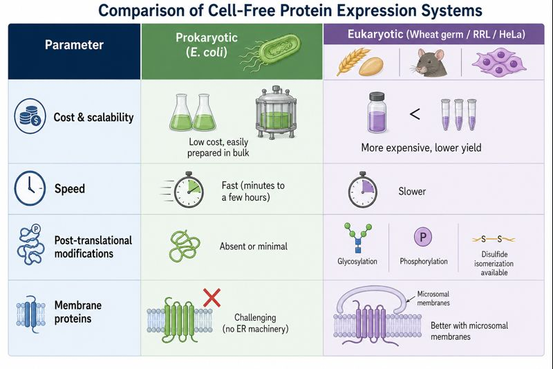 Diagram created Via ChatGPT-Image2 Model.
A protein for the prokaryotic system would be GFP (Green Fluoroscent Protein) GFP is a simple protein that doesn’t need any post-translational modification for fluorescence, therefore it folds correctly in an E.coli system.2 It is an ideal candidate for rapid prokaryotic cell-free expression. It’s is frequently used in E.coli as a reporter.
A protein for the eukaryotes would be erythropoietin, which is a human glycoprotein hormone, it is a complex protein. Producing it in E.coli would produce a non-glycosylated product as E.Coli lacks the machinery for post-translational modifications. Using a eukaryotic system like HeLa with the necessary ER machinery to glycosylate the protein will produce a better product.
How would you design a cell-free experiment to optimize the expression of a membrane protein? Discuss the challenges and how you would address them in your setup.
Membrane proteins are amphipathic in nature and due to the transmembrane domains being hydrophobic, they will aggregate in aqueous conditions. This will lead to precipitation of the target protein before it can fold properly in a standard CFPS reaction, reducing the amount of protein product. One approach to solve this would be to incorporate the reaction with liposomes or some type of a scaffold that bypasses aggregation of the product. In order to supplement this approach, reducing the Mg2+ concentration and thus reducing the translation rate would slow the reaction, giving the product time to properly engage with the scaffold/liposome.
Imagine you observe a low yield of your target protein in a cell-free system. Describe three possible reasons for this and suggest a troubleshooting strategy for each.
The cell-free system hinges mainly on three main components; The template, the energy system and the product. The problems could be in any of these three components. One of the reasons for a low yield would be the template instability, the template might be getting degraded quickly or the design of the template makes it tough to be expressed. Another reason for low yield could be the energy system, if the energy system in non-functional or sub-optimal in function (there are inhibitory byproducts etc.) then the reaction will stall before the yield can peak. Aggregation of the product protein could make it invisible to the western blot, this could also be one of the reasons.
Homework question from Kate Adamala
1a. What would your synthetic cell do? What is the input and what is the output?
The synthetic cell is a low-cost arsenic contamination sensor for drinking water. Arsenic poisoning via contaminated groundwater harms a considerable amount of people every year. Working in an environmental testing lab, I know the level of contamination that exists in rural water bodies. The synthetic cell would have the arsenite dissolved in the water as the input. The output would be a visible blue color produced inside the vesicle by the chromoprotein amilCP. If the water sample turns the synthetic cell suspension blue, arsenic is present above the detection threshold.
1b. Could this function be realized by cell-free Tx/Tl alone, without encapsulation?
Yes, a bulk cell-free reaction with the same ArsR circuit and amilCP reporter would also produce color in the presence of arsenite. But encapsulation would provide benefits like protection of the cell machinery from other contaminants in the water that could possibly inhibit the reaction. Also encapsulation, adds stability to the entire system, making it possible to be dried, stored, shipped making it viable for low resource regions of the world.
1c. Could this function be realized by a genetically modified natural cell?
Yes, possibly E. coli engineered with the same ArsR-pArs-amilCP circuit would function as a whole-cell biosensor and has been demonstrated in the literature. (Chen, S., Zhang, Y., Li, R., Wang, B., & Ye, B. (2022). De Novo Design of the ArsR Regulated Pars Promoter Enables a Highly Sensitive Whole-Cell Biosensor for Arsenic Contamination. Analytical Chemistry, 94, 7210 - 7218. https://doi.org/10.1021/acs.analchem.2c00055.) However, using living GMO cells for field water testing carries biosafety concerns (containment, environmental release), requires special handling procedures and might invite ethical concerns. The synthetic cell is non-living, non-replicating, and contains no viable organism, which makes it simpler to use.
1d. Describe the desired outcome of your synthetic cell operation.
When a water sample containing arsenic above the threshold (~10–50 µg/L, the WHO guideline limit) is added to a vial of synthetic cells, arsenite diffuses across the membrane, binds the ArsR repressor inside, de-represses the arsenic-responsive promoter, and the cell-free system produces amilCP. The suspension turns visibly blue within 4–6 hours, readable by eye with no equipment. Clean water samples produce no color change.
2a. What would the membrane be made of?
POPC and cholesterol in a 4:1 molar ratio. POPC provides a stable, fluid bilayer at room temperature. No special modifications are needed. The key property required is that the membrane is permeable to small neutral molecules, which POPC:cholesterol bilayers naturally are. Arsenite at physiological-range pH exists primarily as arsenious acid (H₃AsO₃), an uncharged small molecule that crosses lipid bilayers by passive diffusion without any transporter.
2b. What would you encapsulate inside?
- Bacterial cell-free Tx/Tl system (E. coli S30 extract)
- DNA construct: amilCP (blue chromoprotein) under the arsenic-responsive promoter pArs, with ArsR binding operator
- ArsR protein: either purified and added directly, or encoded on the same DNA under a constitutive promoter so it is expressed first and accumulates before any arsenite arrives
- Phosphocreatine (40 mM) + creatine kinase for ATP regeneration
- Standard CFPS buffer: HEPES-KOH pH 7.4, Mg-glutamate, K-glutamate, NTPs, 20 amino acids
2c. Which organism will the Tx/Tl system come from?
Bacterial (E. coli S30 extract). ArsR is a native E. coli regulator, the pArs promoter functions in E. coli-based cell-free systems, and amilCP expresses well in prokaryotic extract.
2d. How will the synthetic cell communicate with the environment?
Only in one direction, and passively. Arsenite (H₃AsO₃) diffuses across the POPC membrane without any transporter, driven by its concentration gradient from the water sample into the vesicle interior. Once inside, it binds ArsR. There is no output molecule that needs to cross the membrane, amilCP stays inside the vesicle and is visible as a blue color through the transparent bilayer. The system requires zero membrane proteins.
3a. List all lipids and genes
Lipids:
- POPC (1-palmitoyl-2-oleoyl-sn-glycero-3-phosphocholine) `
- Cholesterol
Genes:
- arsR (E. coli K-12, UniProt P0AE18) — under constitutive promoter J23101; encodes the arsenic-responsive repressor that sits on the pArs operator and is displaced by As³⁺
- amilCP (blue chromoprotein from Acropora millepora, Addgene) — under pArs promoter with ArsR operator; produces visible blue color without UV light, no cofactor required
Additional CFPS components:
- E. coli S30 extract, NTP mix, amino acid mixture, phosphocreatine/creatine kinase, HEPES buffer
3b. How will you measure the function of your system?
Visual readout (primary): Add synthetic cells to water samples spiked with 0, 10, 50, 100, and 500 µg/L arsenite. Incubate at room temperature for 6 hours. Score color development by eye; blue = arsenic detected. This is the intended end-use readout requiring no instruments.
Controls:
- Positive control: synthetic cells + 100 µg/L arsenite → should turn blue
- Negative control: synthetic cells + clean water → no color
- Specificity control: synthetic cells + other heavy metals (lead, cadmium, mercury at equivalent concentrations) → should produce no color, confirming ArsR specificity for arsenic
Homework question from Peter Nguyen
Write a one-sentence summary pitch sentence describing your concept.
“Pollumin” A biosensor paint embedding freeze-dried cell-free reactions is applied to industrial smokestack exits, producing a vivid, indelible color change visible from a distance whenever emissions exceed a regulatory threshold. It creates a passive, tamper-evident public record of pollution events.
How will the idea work, in more detail? Write 3-4 sentences or more.
The paint would be a two-component formulation. The first component is a durable, microporous binder matrix — a silicone-based paint maybe with high surface porosity to allow gas and moisture exchange with the exhaust. We then embed trehalose-stabilized freeze-dried cell-free Tx/Tl pellets (E. coli S30 extract). Each pellet contains a DNA construct encoding a high-yield chromoprotein (e.g., amilCP for deep blue) under the control of a promoter that responds to the pollutant. In the case of SO₂ a commonly known industrial pollutant, we can use a sulfite-responsive promoter (e.g., derived from the sox regulon of Paracoccus pantotrophus) that drives chromoprotein expression above a defined concentration threshold. For NOₓ, a nitric-oxide-sensitive promoter (e.g., norV from E. coli, activated by the NorR transcription factor) could be used instead, or both encoded in a single construct for a multi-pollutant readout. The exhaust from a smokestack would carry significant moisture and heat. This moisture would be the activation trigger, it would rehydrate the freeze-dried cell-free pellets embedded in the paint surface. If the SO₂ or NOₓ concentration simultaneously exceeds the promoter activation threshold, gene expression initiates and chromoprotein accumulates within the paint matrix over several hours, developing a vivid color patch visible from hundreds of meters. Identifiable by satellite imaging or public observers. Once triggered, the chromoprotein is permanently fixed within the crosslinked polymer matrix and cannot be washed away. The facility must repaint with fresh biosensor paint to reset the monitoring cycle, and the repainting itself is a documentable event. (Controlling the access to the paint would make sure there’s no illicit cover-ups) also attempting to cover the color change with ordinary paint would be visibly obvious against the textured smokestack surface.
What societal challenge or market need will this address?
Working at a environmental testing lab, I know that a lot of industrial emission monitoring is currently self-reported or requires expensive on-site sensors that are rarely audited. Companies with facilities in low-oversight regions often exceed limits without consequence for extended periods. This paint turns every smokestack into a passive, publicly legible pollution record, one that cannot be selectively deleted, readable by anyone with eyes or a camera, requiring no infrastructure beyond the paint itself. The accountability mechanism derives from the indelible ink used in elections: the point is not just detection, but the social and legal weight of a visible, persistent, non-removable signal that was not there before the violation. Environmental regulators, satellite-monitoring NGOs, and local communities all become distributed inspectors without any specialized equipment.
How do you envision addressing the limitation of cell-free reactions (e.g., activation with water, stability, one-time use)?
The design tries to turn the limitations into a a feature, the activation via water property turns into an actual trigger feature that activates the paint naturally. In the case of dry stacks, maybe using a secondary layer can work as a latent trigger. In order to keep the system stable, lyophilizing the cell-free extract with the trehalose will provide desiccation protection and spraying the paint with a desiccant overcoat also provides more stability. Batch codes on each cartridge allow traceability of manufacture date, so facilities cannot apply expired paint and claim it as a functioning sensor. The one-time use limitation actually serves as a stellar feature as a triggered patch is a permanent record of a threshold, patterned boxes like 12 patches might serve as a yearly record that can be annually cleaned. The facility’s obligation to repaint with fresh certified biosensor paint (sourced from a controlled regulated supplier, with batch traceability) creates a documented compliance cycle.
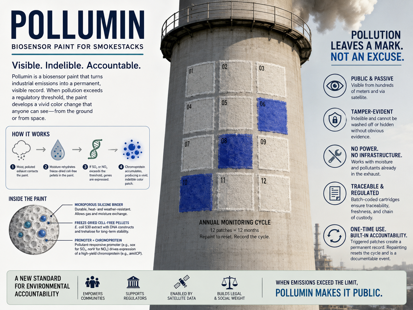
Poster-generated via ChatGPT Image-2 Model.
Homework question from Ally Huang
One of my final project ideas is the Astrobioreactor, a bioreactor redesigned to work in space but building for the absence of gravity as gravity is one of the major driving forces in earthly fermentation. One of the challenges that the project might face is the necessity of modifying strains for surviving physical forces as one of the approaches for the Astrobioreactor is using centrifugal force, the cells might not be able to withstand that and as a result might need to be modified but in space, microorganisms face cosmic radiation and it may mutate the organisms mid-mission in turn compromising their viability and performance. Studying how cosmic radiation affects microorganisms can help better plan the modifications.
Provide background information that describes the space biology question or challenge you propose to address. Explain why this topic is significant for humanity, relevant for space exploration, and scientifically interesting. (Maximum 100 words)
Prolonged spaceflight exposes microorganisms to cosmic radiation (galactic cosmic rays and solar energetic particles) at doses higher than on Earth. This radiation induces double-strand DNA breaks and oxidative lesions that can accumulate in fermentation cultures, causing loss of productivity, phenotypic instability, or outright culture collapse on long-duration missions. Monitoring microbial DNA integrity in real time is impossible with current ISS capabilities. A rapid, equipment-light diagnostic for radiation-induced genomic stress would enable astronauts to assess fermentation health and make timely decisions about culture maintenance or replacement.
Name the molecular or genetic target that you propose to study. Examples of molecular targets include individual genes and proteins, DNA and RNA sequences, or broader -omics approaches.
The SOS regulon transcriptional response, specifically the promoter activity of recA and sulA genes, as biomarkers of active DNA damage and repair stress in Saccharomyces cerevisiae or E. coli fermentation cultures.
Describe how your molecular or genetic target relates to the space biology question or challenge your proposal addresses.
The SOS response is the conserved bacterial stress-signaling network activated when DNA damage exceeds the cell’s baseline repair capacity. Elevated recA expression is a direct, real-time proxy for double-strand break frequency and replication fork collapse the precise damages caused by cosmic ionizing radiation. In yeast, the homologous RAD51 regulon serves the same function. By measuring SOS/RAD51 promoter-driven reporter protein expression in samples drawn from a space bioreactor culture, astronauts gain quantitative insight into the cumulative genomic stress state of their fermentation organism without requiring sequencing or microscopy.
Clearly state your hypothesis or research goal and explain the reasoning behind it.
Hypothesis: Freeze-dried BioBits cell-free reactions can be used as a quick, portable test for radiation stress in space-grown microbes or cells. The system (BIoBits Bright) uses a luciferase reporter linked to the DNA-damage response genes recA (in bacteria) or RAD51 (in yeast/eukaryotes). When these stress-response mRNAs are present in a culture sample, the reaction produces light, and brighter light means higher DNA damage stress. Reasoning: The BioBits system only needs water to activate. After adding a lysed culture sample, the reaction can produce a measurable glow within a few hours. Since recA and RAD51 are activated during DNA damage responses, higher radiation exposure should lead to more of their mRNA being present. This results in stronger luciferase production and a brighter signal, giving a simple readout of culture health in about 2–4 hours.
Outline your experimental plan - identify the sample(s) you will test in your experiment, including any necessary controls, the type of data or measurements that will be collected, etc.
Samples: (1) Unirradiated fermentation culture lysate (negative control), (2) UV-irradiated culture lysate (positive control, known SOS induction), (3) Simulated cosmic-radiation-exposed culture lysate (experimental condition, irradiated via ISS radiation phantom data-matched dose).Procedure: Pellet cells from 1 mL samples, lyse by freeze-thaw, add 5 µL lysate to rehydrated BioBits pellet containing PrecA-luciferase construct. Incubate 3 hours at 29°C. Read luminescence with P51 Viewer. Data collected:_ Luminescence intensity per sample, normalized to total protein concentration (Bradford assay). Comparison across dose conditions establishes a dose-response curve for SOS induction vs. radiation exposure.