Week 5 HW: Genetic circuits part 1
Assignment: DNA Assembly
- What are some components in the Phusion High-Fidelity PCR Master Mix and what is their purpose?
Phusion High-Fidelity DNA polymerase: enzyme responsible for synthesizing new DNA strands while possessing activity, which reduces errors during replication.
dNTPs (deoxynucleotide triphosphates): Nucleotide substrates incorporated by DNA polymerase into the elongating DNA strand during synthesis
reaction buffer: contain compounds as Tris-HCl that maintains the correct pH and salts like KCl which help stabilize primer binding and enzyme activity
MgCl₂: Essential cofactor required for DNA polymerase catalytic activity.
- What are some factors that determine primer annealing temperature during PCR?
Tm Range: reflects the temperature at which half of the primer–template duplex dissociates, it depends largely on the primer nucleotide composition, particularly the GC content.
Primer length: As longer primers higher melting temperatures due to more base-pair interactions with the template.
Secondary structures: May require adjustment of the annealing temperature.
Reaction conditions: can alter primer–template stability and thus influence the optimal annealing temperature.
- There are two methods from this class that create linear fragments of DNA: PCR, and restriction enzyme digests. Compare and contrast these two methods, both in terms of protocol as well as when one may be preferable to use over the other.
PCR and restriction digests serve different purposes: PCR amplifies a specific DNA fragment using primers and an enzyme like Taq polymerase, making it ideal when you need a lot of a precise sequence or start with very little DNA. In contrast, restriction enzymes such as EcoRI cut DNA at specific sites, which is useful for cloning or checking constructs. So basically, PCR = amplify, restriction digest = cut, and they’re often used together.
- How can you ensure that the DNA sequences that you have digested and PCR-ed will be appropriate for Gibson cloning?
Primers should be designed to add some bp of overlapping homologous sequences between the insert and the vector. After PCR or digestion, fragments are verified by gel electrophoresis to confirm the correct size and purity, and the overlap regions should be checked in silico, for example by calculating the predicted digest in Benchling to verify the expected band sizes.
- How does the plasmid DNA enter the E. coli cells during transformation?
Heat shock: Generate pores in bacterial cell wall with an abrupt temperature change
Electroporation: Generate pores in bacterial cell wall with high electrical voltage
In both methods the cells are shocked causing the cell membrane to “open up”
- Describe another assembly method in detail
Golden Gate Assembly
Allows the assembly of multiple DNA fragments in a single reaction, using a Type IIS restriction enzyme, which cuts DNA outside of its recognition sequence to create specific overhangs. DNA fragments and the vector are designed so that these overhangs are complementary, ensuring that the fragments assemble in the correct order. During the reaction, the restriction enzyme cuts the DNA while T4 DNA Ligase simultaneously ligates the compatible ends. Because the restriction sites are removed after ligation, the final assembled plasmid cannot be cut again, which improves assembly efficiency. This method is widely used for assembling multiple fragments quickly and accurately in a one-pot reaction.

Assignment: Asimov Kernel
Create a Repository for your work
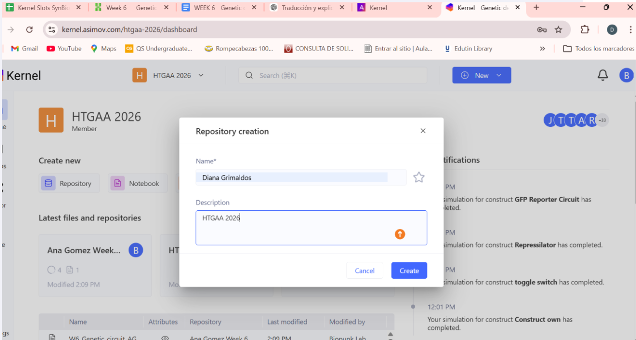
Create a blank Notebook entry to document the homework and save it to that Repository

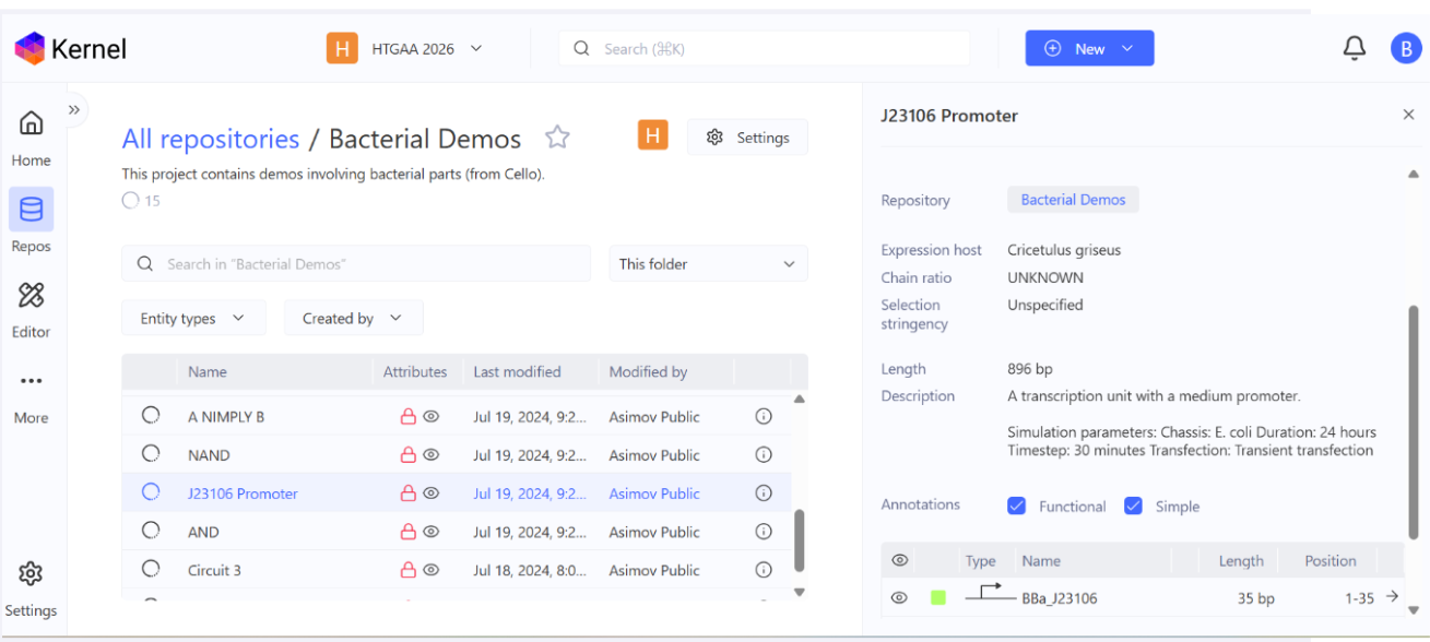
Explore the devices in the Bacterial Demos Repo to understand how the parts work together by running the Simulator on various examples, following the instructions for the simulator found in the “Info” panel (click the “i” icon on the right to open the Info panel)
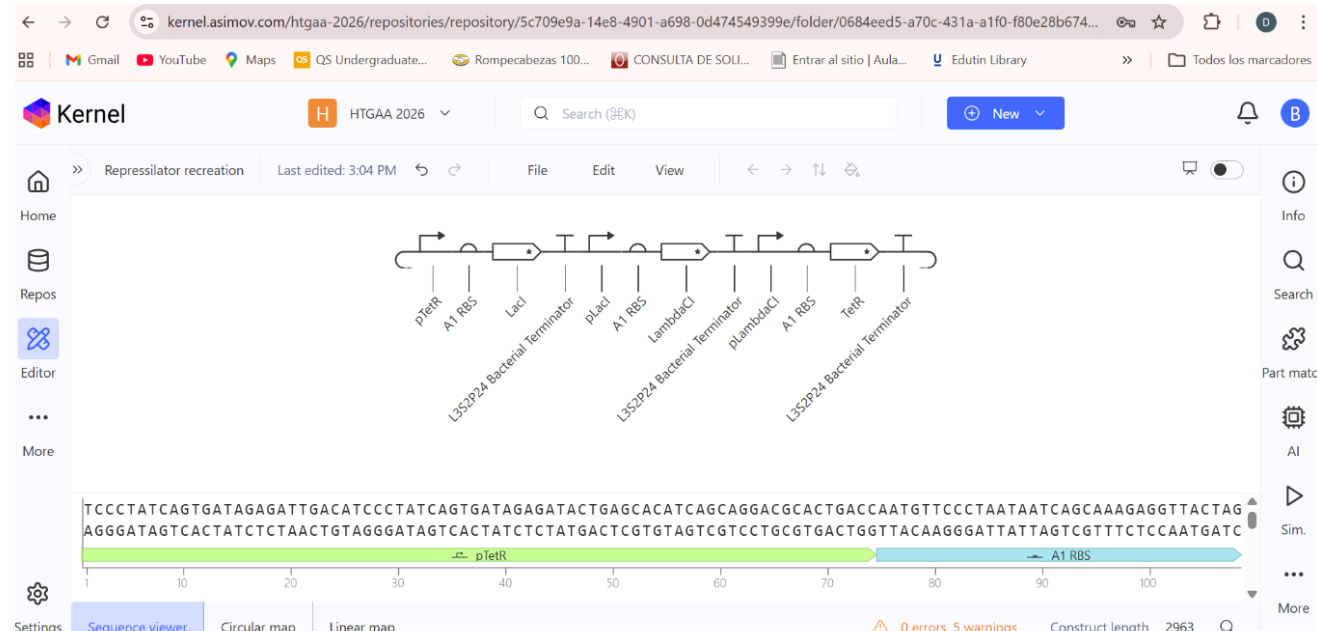
Create a blank Construct and save it to your Repository

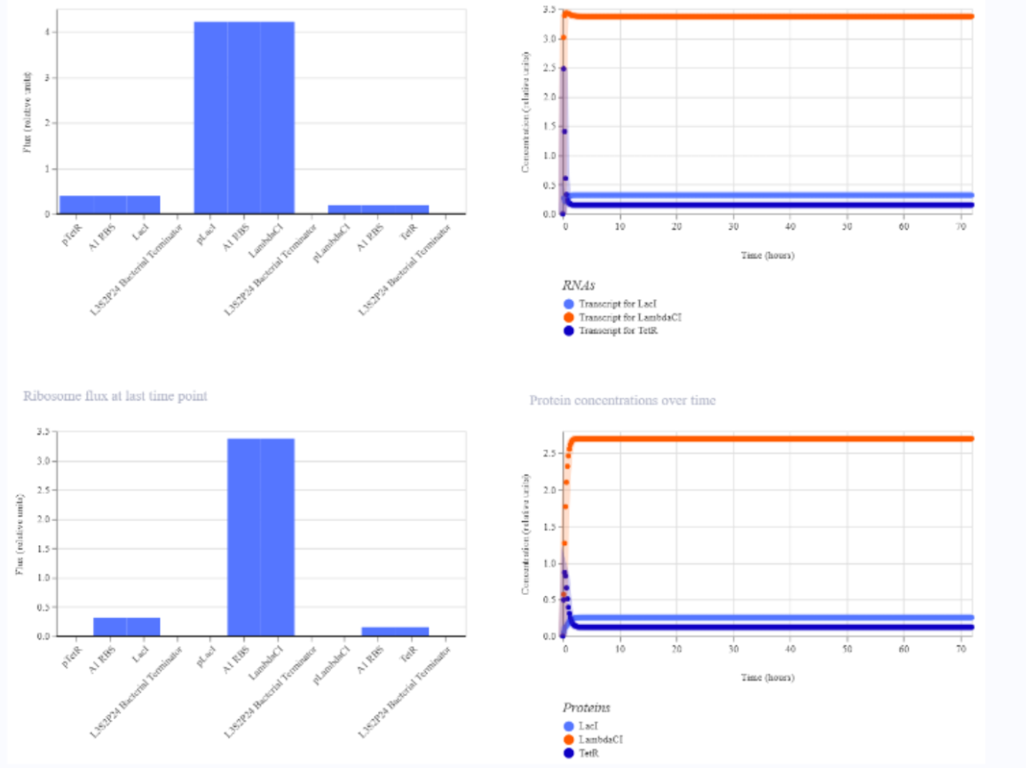
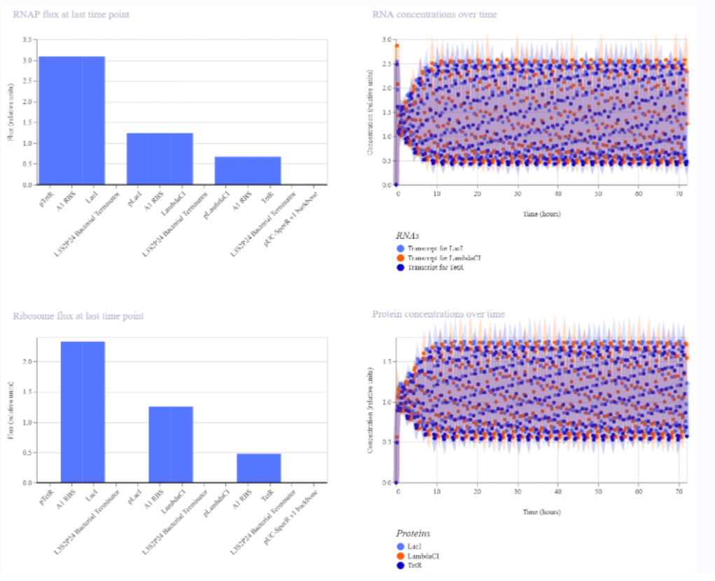
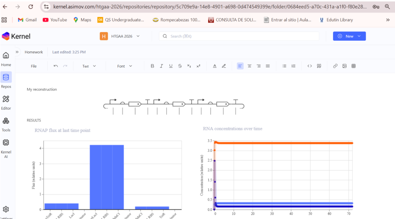
- Build three of your own Constructs using the parts in the Characterized Bacterials Parts Repo
a) A dengue-responsive genetic sensor based on a derepression mechanism. An inducible promoter continuously drives the expression of the repressor TetR, which binds to the Tet-regulated promoter (PTet) and blocks transcription of the reporter gene (sfGFP) under normal conditions, keeping fluorescence at basal levels. When dengue is present, its biomarker (E), represented as an inducible promotor, is recognized by an aptamer that functionally inhibits TetR. This inhibition prevents TetR from binding to PTet, thereby relieving repression and allowing transcription and translation of sfGFP. As a result, GFP fluorescence increases in the presence of NS1, meaning the construct converts dengue detection into a measurable fluorescent output, with low signal in the absence of the ligand and high signal when it is present.
b) Three expression cassettes assembled in a single construct. Together, these cassettes implement a threshold-detection circuit that produces GFP only when the dengue-derived ligand is present above a sufficient concentration.
The circuit integrates two constitutively expressed repressors (TetR and LacI) and a dual-repressed reporter cassette, allowing the system to behave as a biological logical gate sensitive to ligand concentration. Correct functionality relies on specifically chosen promoters for each cassette, as the use of pLtetO‑1 in Cassette 2.
C) Genetic construct designed as a ligand-responsive transcriptional amplifier based on TetR–pTet regulation. The system operates through double negation and feedback, such that an external ligand indirectly activates gene expression by inhibiting a transcriptional repressor.
Final project