Week 11 HW: Building Genomes


Part A: The 1,536 Pixel Artwork Canvas | Collective Artwork

image image
  • Colaborated in the stage where a single picture was being formed throughout the 4 plates
  • It was a fun premise to explore could lab automation collaboratively
  • Next year there could be the “constraint” of creating a single image/pattern throughout all the plates to really engage in the collective activity of trying to figure out how to expand on what other people started like a Cadavre Exquis logic

Part B: Cell-Free Protein Synthesis | Cell-Free Reagents

1. Referencing the cell-free protein synthesis reaction composition, provide a 1-2 sentence description of what each component’s role is in the cell-free reaction.

In a cell-free master mix, the lysate, in this case E. coli, has all the machinery needed to process DNA to RNA to protein. In order for this to work, there is a mix of salts such as Potassium Glutamate, Magnesium Glutamate and Potassium phosphate, which maintain proper ionic concentrations to allow for correct DNA/RNA folding, enzymes to bind, and protein synthesis to occur efficiently, and buffers (HEPES + Potassium phosphate monobasic/dibasic) which maintain the optimal ph conditions for enzymatic activity.

2. Describe the main differences between the 1-hour optimized PEP-NTP master mix and the 20-hour NMP-Ribose-Glucose master mix.

The main differences rest on how they supply energy and nucleotides, as well as in their intended performance (for fast expressions or for expressions that might need more time) The 1-hour PEP–NTP system, uses pre-supplied NTPs (ATP, GTP, CTP, UTP) and the energy comes from PEP (phosphoenolpyruvate)— which is a high-energy molecule that can donate a phosphate group to regenerate ATP—, designed for rapid expression, by including high-energy, ready-to-use components.

While the 20-hour NMP–Ribose–Glucose system uses NMPs (AMP, CMP, UMP) instead of full NTPs. Nucleotides and energy are generated in situ from Ribose and Glucose, by relying on native enzymatic pathways in the extract. It is designed for longer expressions, having fewer synthetic additives— includes nicotinamide— which supports metabolic pathways that convert glucose into usable energy— instead of multiple cofactors. Therefore, being more metabolically integrated and sustainable.


Part C: Planning the Global Experiment | Cell-Free Master Mix Design

1. Given the 6 fluorescent proteins we used for our collaborative painting, identify and explain at least one biophysical or functional property of each protein that affects expression or readout in cell-free systems.
  1. sfGFP — Engineered for robust folding, even under non-optimal conditions, making it highly reliable in cell-free systems
  2. mRFP1 — Has a relatively slow maturation time, meaning fluorescence appears later, which can delay readout despite successful expression.
  3. mKO2 — Exhibits pH sensitivity, so fluorescence intensity can decrease in more acidic environments typical of some cell-free reactions over time
  4. mTurquoise2 — Known for high quantum yield and brightness, but requires proper folding, making its performance sensitive to reaction conditions
  5. mScarlet_I — Designed for fast maturation and high brightness, allowing rapid and strong fluorescence readout in cell-free systems.
  6. Electra2 — Displays oxygen-dependent chromophore formation, so fluorescence requires sufficient oxygen availability, which can be limiting in dense or sealed reactions.
2. Create a hypothesis for how adjusting one or more reagents in the cell-free mastermix could improve a specific biophysical or functional property you identified above, in order to maximize fluorescence over a 36-hour incubation.

mKO2 — Exhibits pH sensitivity, so fluorescence intensity can decrease in more acidic environments typical of some cell-free reactions over time As mKO2 exhibits sensitivity to ph shifts which decrease fluorescence over-time as the reaction gets more acidic, one solution could be making a stronger buffer concentration, both with HEPES and the potassium phosphate (mono/dibasic) In order to validate and optimize this hypothesis the following experimental set could be performed:

Sample 1 — Control

  • HEPES-KOH pH 7.5: 45 mM
  • K-phosphate dibasic: 5.63 mM
  • K-phosphate monobasic: 5.63 mM

Sample 2 — Moderate buffer increase

  • HEPES-KOH pH 7.5: 60 mM
  • K-phosphate dibasic: 5.63 mM
  • K-phosphate monobasic: 5.63 mM

Sample 3 — Stronger buffer increase

  • HEPES-KOH pH 7.5: 75 mM
  • K-phosphate dibasic: 5.63 mM
  • K-phosphate monobasic: 5.63 mM

Sample 4 — HEPES + phosphate support

  • HEPES-KOH pH 7.5: 60 mM
  • K-phosphate dibasic: 7.5 mM
  • K-phosphate monobasic: 7.5 mM

mTurquoise2 — Exhibits high brightness and quantum yield but depends on efficient translation and proper protein folding, making its fluorescence sensitive to ionic conditions in cell-free reactions. As mTurquoise2 fluorescence depends on correct folding and efficient translation, one approach to improve its performance is to optimize magnesium concentration, since Mg²⁺ plays a key role in ribosome function and protein folding in cell-free systems. In order to validate and optimize this hypothesis, the following experimental set could be performed:

Sample 1 — Control Magnesium Glutamate: 6.975 mM

Condition 2 — Moderate Mg increase Magnesium Glutamate: 8.000 mM

Condition 3 — Strong Mg increase Magnesium Glutamate: 10.000 mM

Condition 4 — Very strong Mg increase Magnesium Glutamate: 12.000 mM

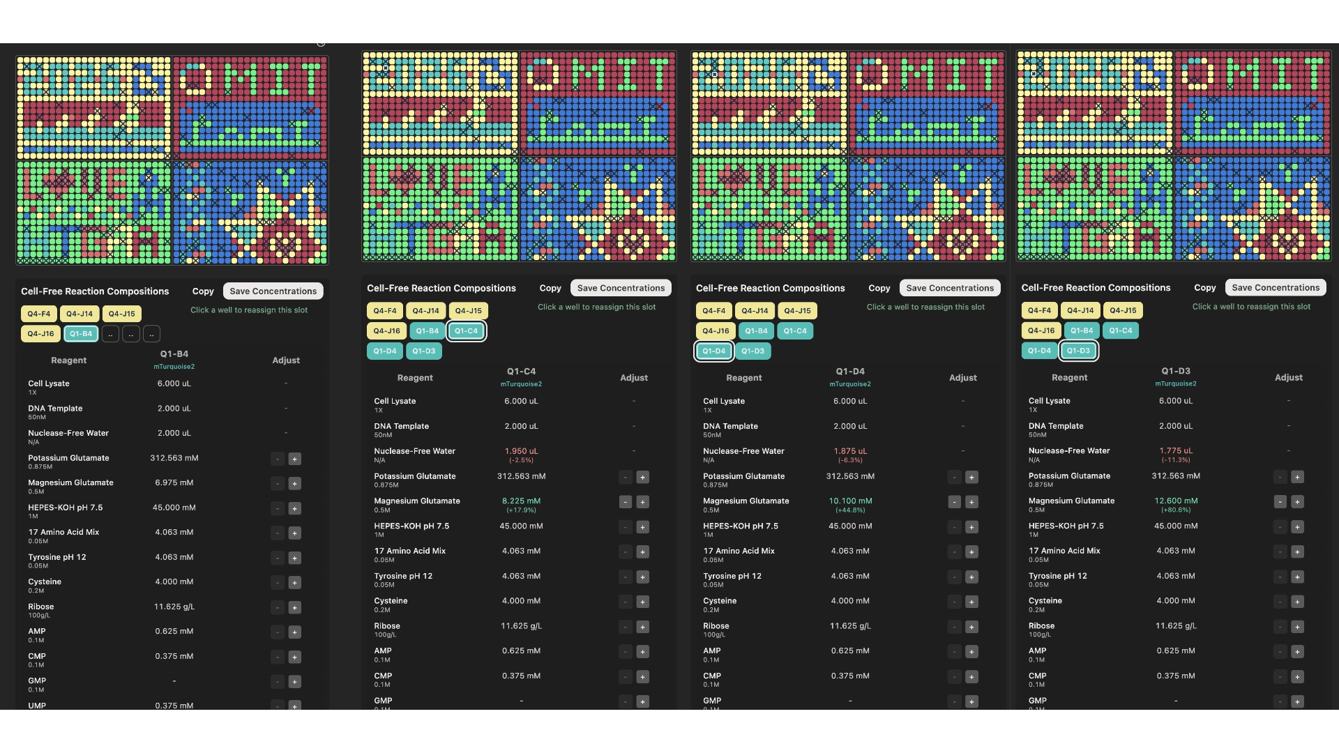
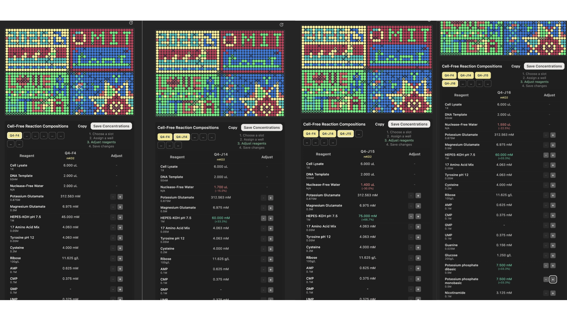
3. The second phase of this lab will be to define the precise reagent concentrations for your cell-free experiment.

According to the hypothesis presented in the previous step, I created these 8 wells, 4 for the mKO2 and 4 for the mTurquoise2.

mKO2
image image
mTurquoise2
image image