Week 8 HW: Cell-Free Systems
General and Lecturer-Specific Questions
1. Explain the main advantages of cell-free protein synthesis over traditional in vivo methods, specifically in terms of flexibility and control over experimental variables. Name at least two cases where cell-free expression is more beneficial than cell production.
There are many advantages of cell-free protein synthesis over traditional in vivo methods. Cell-free systems allow you to have more control over the conditions of your experiment (ie. DNA, proteins, small molecules, etc.). Unlike working in vivo methods, where things are a bit more of a “black box”.
Two cases where cell-free is more beneficial:
i. Cell-free is more beneficial when it comes to rapid prototyping of metabolic pathways. In the lecture it was mentioned that a four-enzyme pathway would have taken weeks of cloning and transformation with an in-vivo method could take about an hour if done in a cell-free system.
ii. Cell-free systems can be more affordable. For example, there are cell-free freeze dried systems that cost under a dollar and can therefore be used in low-resource communities to detect Ebola, Zika or other diseases.
2. Describe the main components of a cell-free expression system and explain the role of each component.
A cell-free expression system essentially mimics what is going on in a bacteria. You scoop out the ribosomes, polymerases, the tRNAs - and then you components like amino acids and an energy source.
Below are the main components in detail:
DNA template - the DNA for the protein you want to make.
Amino acids - the building blocks for protein synthesis.
tRNAs - small RNA molecule that decodes mRNA and delivers amino acids to ribosome.
Energy source - something that powers the transcription and translation (could be ATP regeneration)
Cell extract - This is what includes the ribosomes and RNA polymerases (PURE express is a version of this)
3. Why is energy provision regeneration critical in cell-free systems? Describe a method you could use to ensure continuous ATP supply in your cell-free experiment.
Transcription and translation cost energy. In a cell-free system the reaction quicky uses up either the ATP or GTP - it’s designed more for a one time use. To avoid this there are ways you can create a regenerative ATP supply. One of the ways to create this is to use the Phosphoketolase (PKT) Systems: these use a pathway of enzymes to turn phosphorylate ADP into usable ATP.
4. Compare prokaryotic versus eukaryotic cell-free expression systems. Choose a protein to produce in each system and explain why.
A prokaryotic cell-free expression system is fairly straightforward, it can be E.coli-based which is already highly understood. It is high-yield and cheaper. You could produce GFP in this system and it would be quick and efficient.
A eukaryotic cell-free system is useful if you need post-translational modifications. There are extra components in eukaryotic cells like the endoplasmic reticulum that attaches sugar chains to the proteins being expressed. There are also chaperones that help the protein fold correctly. It more expensive and has a lower yield than prokaryotic cell free system.
5. How would you design a cell-free experiment to optimize the expression of a membrane protein? Discuss the challenges and how you would address them in your setup.
Membrane proteins normally live inside cell membranes (of course), so the challenge when you try to make them in a cell-free system is that they can clump together because there’s no membrane for them to fold into. A way you can fix this is by adding lipids (like lipid vesicles) to create a sort of artificial membrane. This helps give the protein membranes somwhere to go.
6. Imagine you observe a low yield of your target protein in a cell-free system. Describe three possible reasons for this and suggest a troubleshooting strategy for each.
One of the issues could be degradation of mRNA by RNases. A way to get around this is to add more Murine RNase Inhibitor, as was mentioned in the lab. Another issue could be a deficiency in the energy supply. A way to fix this would be to increase energy like in the form of 3-PGA or PEP. A third issue could be plasmid quality, maybe there is an issue with the T7 promoter. It would be good in that case to verify the plasmid by running it in a gel.
Homework question from Kate Amadala
Design an example of a useful synthetic minimal cell as follows:
1. Pick a function and describe it.
a. What would your synthetic cell do? What is the input and what is the output?
I would want to make a synthetic cell that would detect heavy metals in the soil (inspired by my earlier homeworks about Schewanella and Geobacter bacterias). The input would be the heavy metal molecules that would diffuse into the cells. The output would be a chromoprotein or maybe GFP that would be expressed if the heavy metals were detected.
b. Could this function be realized by cell-free Tx/Tl alone, without encapsulation?
It could be. Without encapsulation the cell-free system is more one-shot (not reusable). So it would functionally still work, but it would be more useful if the cell-free system had a membrane to better protect it from the soil elements. Although technically it’s supposed to be used as more of a one shot because once the GFP proteins are expressed you can’t reuse it. But you might want the sensor to last a while until it does detect something.
c. Could this function be realized by genetically modified natural cell?
Totally. E.coli and Shewanella have been genetically modified with metal-detecting promoters to respond to heavy metals. Of course, when working with natural cells you have to make sure that the environmental conditions are perfect in order to keep the natural cell alive.
d. Describe the desired outcome of your synthetic cell operation.
The idea would be to mix these freeze-dried synthetic cells with soil and water. And then if heavy metals are detected the cells will change color. In the case of using GFP you will have to flash blue or UV light in order to detect the change in color. There could be a variety of synthetic cells that change different colors based on different toxic metals. Since there’s a very limited biosecurity risks with the natural cells, these detectors could be used by non-scientists (like farmers) to detect issues with their soil.
2. Design all components that would need to be part of your synthetic cell.
a. What would be the membrane made of?
The membrane would be a classic synthetic minimal cell membrane: so by that I mean that it would be a lipid membrane made with cholesterol and phospholipids. This would help protect the synthetic cell while still letting the heavy metal molecules enter.
b. What would you encapsulate inside? Enzymes, small molecules.
You would have to use all the mastermix components like ribosomes, enzymes, small molecules, tRNAs, etc. Then of course you would need a toxic metal responsive promoter and either the chromoprotein or a GFP.
c. Which organism your Tx/Tl system will come from? Is bacterial OK, or do you need a mammalian system for some reason? (hint: for example, if you want to use small molecule modulated promotors, like Tet-ON, you need mammalian)
Bacterial cell here will work fine! I don’t have any specific need for a mammalian system. Metal responsive promoters are very well understood in bacterials system, so no need to switch it up.
d. How will your synthetic cell communicate with the environment? (hint: are substrates permeable? or do you need to express the membrane channel?)
I won’t need any special membrane channel proteins, like the ones Kate Amadala mentioned in their lecture. For my design the heavy metal ions are small enough to diffuse through the synthetic cell membrane.
Experimental details a. List all lipids and genes. (bonus: find the specific genes; for example, instead of just saying “small molecule membrane channel” pick the actual gene.)
The setup is pretty straightforward, nothing crazy. As mentioned in the previous questions here are my lipids and genes: Lipids - phospholipids, cholesterol Genes - chromoprotein or GFP under a metal-responsive promoter (MerR for mercury, ArsR for arsenic) TX-TL system - a lyophilized E. coli cell extract with basic master mix
b. How will you measure the function of your system?
I would use a plate reader or a UV light in order to monitor the function of heavy metals. If they are bright and there is fluorescence then that would show that toxic metals were detected in the soil.
References:
Lentini, R. et al., 2014. Communicating with the outside world from within a lipid vesicle. Nature Communications, 5, p.4012. Available at: https://www.nature.com/articles/ncomms5012
Homework question from Peter Nguyen
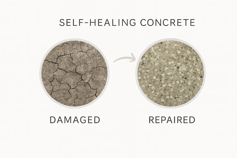
Summary Pitch: The development of freeze-dried cell-free systems that are embedded into building materials and are activated by water that seeps through the cracks in ceilings or wall, which in turn produces urease enzyme to start a biocementation process - to seal the crack before it spreads.
The freeze-dried cell-free systems will be mixed in with the concrete, so that there are cell-free systems a little everywhere in the mixture. When water touches the freeze-dried systems the production of the urease systems begins. This breaks down urea (also in the material) into ammonia and CO2. This raises the pH and provides carbonate which then bonds with calcium in the material to create limestone (calcium carbonate). Which seals the crack in the material.
This addresses a common contruction issue that affects many urban areas. Building materials are constantly degrading due to environmental issues so this will help the longevity of materials.
The cell-free solution for this issue does have a big limitation in that it is a one-time use. Using freeze-dried cell free systems to create self-healing concrete will not create a perpetually self-healing material. If there is a recurring water issue that causes a crack then the cell-free system will increase the longevity of the material but not solve the issue permanently.
Homework question from Ally Huang
1. Provide background information that describes the space biology question or challenge you propose to address. Explain why this topic is significant for humanity, relevant for space exploration, and scientifically interesting.
A challenge with space biology is the fact that DNA can degrade when exposed to the high levels of cosmic radiation that exist outside of Earth’s atmosphere. There is no easy way to detect how much radiation is affecting the DNA of astronauts. We could use the Biobits cell-free system to test DNA that has been exposed to radiation and compare it to regular control DNA to get a better understanding of how the astronauts are being impacted.
2. Name the molecular or genetic target that you propose to study. Examples of molecular targets include individual genes and proteins, DNA and RNA sequences, or broader -omics approaches. (Maximum 30 words)
The integrity of DNA templates will be studied by encoding for GFP after exposure to space radiation (and comparing with the control DNA), using fluorescence output from BioBits cell-free expression as the readout.
3. Describe how your molecular or genetic target relates to the space biology question or challenge your proposal addresses. (Maximum 100 words)
DNA damage caused by radiation in space would pose a big health risk to astronauts. By being able to track the amount of radiation damage is done over time we can better time space missions to protect the health of space travelers. A great next step would be understanding how to conduct DNA repair while in space, but at the moment I am working with the constraints of the Biobit.
4. Clearly state your hypothesis or research goal and explain the reasoning behind it. (Maximum 150 words)
The hypothesis is that the DNA exposed to space will produce less fluorescent proteins in the Biobit cell-free system. I also believe that the longer the DNA is in space the more radiation damage it will recieve and the fewer and fewer proteins it will be able to express over time. This knowldege of when radiation damage occurs to DNA could help researchers and scientists develop better preventative measures for astronauts’ protection.
5. Outline your experimental plan - identify the sample(s) you will test in your experiment, including any necessary controls, the type of data or measurements that will be collected, etc. (Maximum 100 words)
While on Earth the idea would be to test a GFP-encoding DNA template wusing the BioBits cell-free system. This will help to create a fluorescence baseline as the control for the experiment. The same DNA template will then be put onboard a spacecraft. Then, at regular intervals throughout the mission (weekly?), astronauts will run the same reaction again. They will rehydrate the freeze-dried BioBits kit with water, then incubate, and finally measure the fluorescence using the P51 Molecular Fluorescence Viewer. If there is a decrease in fluorescence compared to the Earth baseline one that will indicate a relative measure of DNA damage from cosmic radiation. It will help to give an idea of how DNA integrity changes the further and longer we travel from Earth.
Homework Part B: Individual Final Project
Updated on the Individual Final Project page