Week 11 HW: Building Genomes
Cloud laboratories are making science accessible, affordable, and reproducible. Our aim this semester is to showcase how they can enable human creativity at scale, and how they provide a platform for collaboration and community.
How To Grow (Almost) Anything is about synthetic biology, bioengineering, robotics, automation, art, and AI. But it is also about friendship, shared purpose, and the freedom to build beyond what we know and to be inspired by what can be. To that end, the goal with this cloud lab unit and homework assignment is to inspire collaboration and creativity while designing a scientifically rigorous cell-free fluorescent protein optimization experiment together.
Tip
As you plan for final projects, you may want to refer to the provided non-exhaustive list of common Nebula protocols and their parameters in the “Reading & Resources” section below.
Homework
Info
Note that this homework is due a week later than it ordinarily would due to its release a week later than normal.
Part A: The 1,536 Pixel Artwork Canvas | Collective Artwork
- Contribute at least one pixel to this global artwork experiment before the editing ends on Sunday 4/19 at 11:59 PM EST.
- A personalized URL was sent to the email address associated with your Discourse account, and you can discuss the artwork on the Discourse.
- If you did not have a chance to contribute, it’s okay, just make sure you become a TA this fall! 😉
So I contributed 29 pixels to the collective Artwork! Placing 39th on the dashboard (Not bad eh).

Essentially I wanted to leave a yellow heart since on the first day of going live there were three hearts that needed some color. So I first added yellow pixels mostly to try and make the heart more heart-shaped.
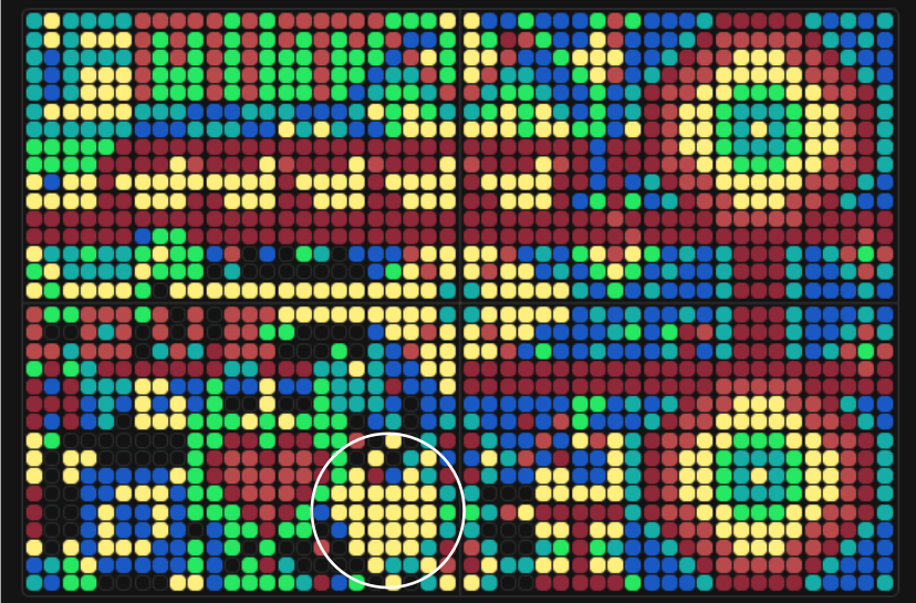
Then after a day or two I came back and noticed that the overall drawing completely changed, which was very fun to see. Adapting to that change, I filled another yellow heart, and it stayed like that for some time. Besides the yellow hearted adventure I added some cyan and red pixels to make some figures symmetrical.

Make a note on your HTGAA webpages including:
- what you contributed to the community bioart project (e.g., “I made part of the DNA on the bottom right plate”)
I contributed with yellow hearts on the bottom right plate and on the upper left plate. However, these hearts were later changed since the overall scheme of the collective artwork changed too! I also contributed with some cyan and red pixels on the bottom left plate to “even out” some corners and make the drawings more symmetrical.
- what you liked about the project, and
What I really liked about the collective artwork project is how community can change a blank slate (in this case an agar plate) and transform it into something memorable and beautiful in just some days! I remember very well how people chatted and discussed it over the classes and on the forum. I also loved the interface so much (Shoutout to Ronan!!!!), it was really well implemented (wish I could learn some of those interface creation abilities for more projects). I also really loved how much love and appreciation went into each pixel since it represented a person in a place in our world.
- what about this collaborative art experiment could be made better for next year.
I think the only thing that could be better for next year is to maybe reduce the time limit just a little bit more :D (although there was a secret technique to get unlimited time!). And also it would be nice to expand the artwork every year (that way we can have more space for initial exploration). It would be really impressive to see a collective 4x4 plate artwork fully completed, too.
Part B: Cell-Free Protein Synthesis | Cell-Free Reagents
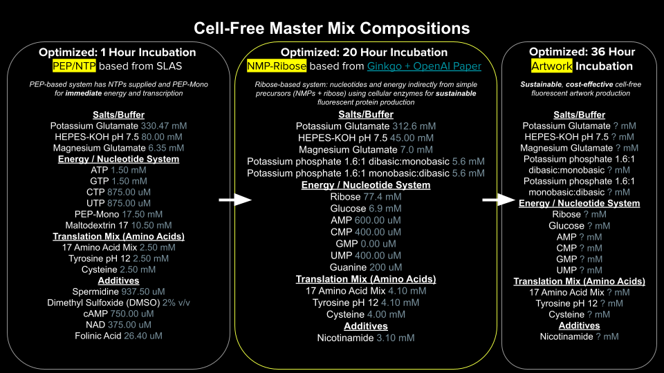
Referencing the cell-free protein synthesis reaction composition (the middle box outlined in yellow on the image above, also listed below), provide a 1-2 sentence description of what each component’s role is in the cell-free reaction.
E. coli Lysate
BL21 (DE3) Star Lysate (includes T7 RNA Polymerase)
Provides the essential molecular machinery, including ribosomes, tRNAs, and initiation factors, required to execute the translation of target proteins. The BL21 (DE3) Star Lysate specifically includes T7 RNA Polymerase to drive highly efficient, coupled transcription from plasmids containing a T7 promoter.
Salts/Buffer
Potassium Glutamate
HEPES-KOH pH 7.5
Magnesium Glutamate
Potassium phosphate monobasic
Potassium phosphate dibasic
Maintains the optimal physiological pH and osmotic balance required to preserve protein stability and enzymatic activity throughout the long incubation. Potassium Glutamate and Magnesium Glutamate supply indispensable cofactors that stabilize mRNA structures, ribosomal subunits, and tRNA-aminoacyl interactions, while HEPES-KOH pH 7.5, Potassium phosphate monobasic, and Potassium phosphate dibasic establish a robust buffering system to prevent pH drifts.
Energy / Nucleotide System
Ribose
Glucose
AMP
CMP
GMP
UMP
Guanine
Functions as a sustainable recycling system that slowly generates ATP and GTP locally from simple precursors, preventing rapid phosphate accumulation while continuously fueling transcription and translation. Within this system, Ribose and Glucose serve as primary carbohydrate energy sources, while AMP, CMP, GMP, UMP, and Guanine act as nucleotide precursors that endogenous enzymes progressively phosphorylate into active substrate building blocks.
Translation Mix (Amino Acids)
17 Amino Acid Mix
Tyrosine
Cysteine
Supplies the fundamental substrate building blocks necessary for the ribosome to polymerize and synthesize the primary polypeptide chain of the target protein. The 17 Amino Acid Mix provides the majority of the standard genetic code requirements, while Tyrosine and Cysteine are added individually to bypass the solubility limits typical of highly concentrated master stocks, ensuring complete dissolution.
Additives
Nicotinamide
Acts as a stabilizing metabolic precursor that sustains the functional lifetime of essential nicotinamide adenine dinucleotide (NAD+/NADH) cofactors within the lysate. Specifically, Nicotinamide prevents the premature degradation of these electron carriers, supporting the homeostatic core metabolic pathways that generate chemical energy for the cell-free system.
Backfill
Nuclease Free Water
Brings the overall master mix to its exact final required reaction volume without introducing external biological contamination. Nuclease Free Water ensures that no trace DNA or RNA degrading enzymes jeopardize the stability of the genetic template or the newly synthesized transcripts.
Describe the main differences between the 1-hour optimized PEP-NTP master mix and the 20-hour NMP-Ribose-Glucose master mix shown in the Google Slide above. (2-3 sentences)
So, the 1-hour PEP-NTP system relies on a high concentration of readily available nucleoside triphosphates (NTPs) paired with phosphoenolpyruvate (PEP) as a rapid, direct phosphate donor for immediate transcription and translation. In contrast, the 20-hour system is designed for long-term sustainability, utilizing cheap precursors like ribose, glucose, and nucleoside monophosphates (NMPs) that endogenous enzymes slowly convert into energy. This metabolic pacing keeps chemical energy generation active over nearly a day and avoids the toxic accumulation of inorganic phosphate that prematurely halts the PEP-based reaction.
- Bonus question: How can transcription occur if GMP is not included but Guanine is?
This one is interesting! From what I’ve read, transcription can still proceed effectively because the E. coli lysate contains active endogenous salvage pathway enzymes, such as purine nucleoside phosphorylase and phosphoribosyltransferases. These enzymes can salvage free Guanine by combining it with ribose-1-phosphate or PRPP to generate GMP directly within the reaction. Once GMP is produced, native adenylate and nucleoside diphosphate kinases sequentially phosphorylate it into GDP and functional GTP, successfully fueling T7 transcription.
Part C: Planning the Global Experiment | Cell-Free Master Mix Design
Given the 6 fluorescent proteins we used for our collaborative painting, identify and explain at least one biophysical or functional property of each protein that affects expression or readout in cell-free systems. (Hint: options include maturation time, acid sensitivity, folding, oxygen dependence, etc) (1-2 sentences each)
sfGFP: Superfolder GFP possesses an exceptionally robust folding kinetics network that prevents aggregation during rapid transcription-translation coupling, making it highly efficient in cell-free systems. However, its rapid expression can outpace the chemical maturation of its chromophore, which strictly requires molecular oxygen ($O_2$) to become fluorescent.
mRFP1: Monomeric RFP1 features a notoriously slow chromophore maturation time of over an hour, which delays real-time fluorescence readouts during the early phases of cell-free incubation. Additionally, it exhibits relatively low photostability and brightness compared to newer generation red fluorescent proteins, limiting its long-term detection sensitivity.
mKO2: Monomeric Kusabira Orange 2 is characterized by a high molar extinction coefficient and excellent brightness, but its unique azoline-containing chromophore exhibits significant acid sensitivity. As a cell-free reaction progresses and metabolic waste products accumulate, any decrease in pH can drastically quench mKO2 fluorescence.
mTurquoise2: This enhanced cyan fluorescent protein has a uniquely high quantum yield and fast maturation due to a stabilized tryptophan side chain within its beta-barrel core. A key functional constraint in cell-free platforms is its narrow excitation/emission gap, which requires precise optical filtering to avoid overlap with background autofluorescence from the lysate.
mScarlet_I: mScarlet_I is an engineered red fluorescent protein with a highly rigidified chromophore that yields record-breaking brightness and quantum efficiency. Its main operational challenge in extended reactions is its high susceptibility to photobleaching under continuous excitation, requiring carefully pulsed plate reader protocols over long timelines.
Electra2: Electra2 is a specialized, rapidly maturing yellow-green fluorescent protein specifically engineered for high-throughput visibility. Its primary biophysical limitation is its high sensitivity to chloride ion concentrations and ionic strength changes, meaning slight variations in the cell-free salt balance can destabilize its tertiary structure.
The amino acid sequences are shown in the HTGAA Cell-Free Benchling folder.
Create a hypothesis for how adjusting one or more reagents in the cell-free mastermix could improve a specific biophysical or functional property you identified above, in order to maximize fluorescence over a 36-hour incubation. Clearly state the protein, the reagent(s), and the expected effect.
High-Illumination and Metabolic Oscillator Hypotheses
To achieve high-illumination pixels and maximize absolute fluorescence over a 36-hour incubation, I hypothesize that supplementing high concentrations of HEPES-KOH pH 7.5 paired with a tightly metered dose of Magnesium glutamate will drive translation velocity while delaying system shutdown. The primary bottleneck for sustained cell-free brightness is the progressive acidification of the reaction combined with the accumulation of free inorganic phosphate from energy depletion, which typically kills translation within hours. By expanding the buffering capacity with HEPES-KOH, we actively neutralize the acid waste, preserving the unquenched state of mScarlet_I and sfGFP while magnesium glutamate supports ribosomal elongation stability to maximize the total number of synthesized protein copies before the system reaches exhaustion.
Alternatively, to explore the feasibility of an intermittent light effect or a metabolic blinking pixel, I hypothesize that a transient, self-limiting loop can be established by intentionally overloading the L-tyrosine substrate concentration against a minimal HEPES-KOH baseline using sfGFP. In this metabolic context, the cell-free machinery will initially synthesize highly fluorescent sfGFP, causing a rapid spike in light emission. Concurrently, the active MelA tyrosinase enzyme will consume the abundant L-tyrosine to produce dark melanin pigment while the unbuffered system undergoes natural metabolic acidification. As the reaction darkens and the pH drops, the optical interference of the melanin combined with the acid-induced quenching of the green reporter will systematically extinguish the visible light, creating a distinct, self-terminating pulse of fluorescence that mimics a delayed metabolic countdown.
These flowcharts illustrate the operational logic and downstream biological consequences of both experimental designs:
flowchart LR
    %% High-Illumination Pathway
    subgraph High_Illumination [High-Illumination Pixels]
        A1[High HEPES & Mg²⁺] --> B1[Neutralizes acid waste & stabilizes ribosomes]
        B1 --> C1[Sustained transcription & translation velocity]
        C1 --> D1[Maximum protein accumulation over 36h]
    end

    %% Blinking Pixel Pathway
    subgraph Blinking_Pixel [Metabolic Oscillator / Blinking Pixel]
        A2[High Tyrosine & Low Buffer] --> B2[Rapid MelA conversion & metabolic acidification]
        B2 --> C2[Melanin accumulation + Protonated chromophore]
        C2 --> D2[Optical masking + Acid quenching of sfGFP]
        D2 --> E2[Self-terminating fluorescence pulse]
end- The second phase of this lab will be to define the precise reagent concentrations for your cell-free experiment. You will be assigned artwork wells with specific fluorescent proteins and receive an email with instructions this week (by April 24). You can begin composing master mix compositions here.
Important
In order to be eligible for this, make sure that your final project slide is in the “2026 Committed Listener ONE FINAL PROJECT IDEA” slide deck.
To test the hypothesis, I have selected the following wells

Which are located on the left of the Q3 plate (forming a little snake!):
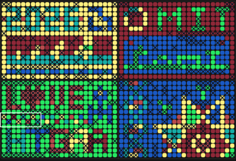
Custom Reagent Supplement Designs for Assigned Artwork Wells (2 μL Additions)
| Well Coordinate | Target Reporter | Operational Design | Reagent to Adjust | Concentration to Set in Interface | Expected Phenotype / Visual Outcome |
|---|---|---|---|---|---|
| Q3-I6 | mScarlet_I | High-Illumination Base | HEPES-KOH pH 7.5 Tyrosine pH 12 Magnesium Glutamate | 95.000 mM 5.938 mM 13.225 mM | Sustained, ultra-bright red pixel using the absolute maximum translation capacity allowed by the 2 µL limit. |
| Q3-H3 | mRFP1 | High-Illumination Buffer Alt | HEPES-KOH pH 7.5 | 120.000 mM | Steady red pixel fully protected against metabolic organic acid quenching. |
| Q3-I2 | Electra2 | High-Illumination Base | HEPES-KOH pH 7.5 Tyrosine pH 12 Magnesium Glutamate | 95.000 mM 5.938 mM 13.225 mM | Optimized translation kinetics yielding maximum initial green-yellow emission intensity within volume limits. |
| Q3-H5 | mTurquoise2 | High-Illumination Buffer Alt | HEPES-KOH pH 7.5 | 120.000 mM | Deep cyan pixel shielded from degradation with an extended functional translation lifetime. |
| Q3-I8 | mTurquoise2 | Blinking / Intermittent Pixel | HEPES-KOH pH 7.5 Tyrosine pH 12 | 57.500 mM 8.438 mM | Cyan pixel that hits an early fluorescence peak before rapidly dark-quenching due to maximal Tyrosine-to-melanin conversion. |
| Q3-H7 | mKO2 | High-Illumination Base | HEPES-KOH pH 7.5 Tyrosine pH 12 Magnesium Glutamate | 95.000 mM 5.938 mM 13.225 mM | High-intensity orange/yellow display that counteracts the default acid sensitivity of the reporter. |
| Q3-I4 | mKO2 | Blinking / Intermittent Pixel 1 | HEPES-KOH pH 7.5 Tyrosine pH 12 | 57.500 mM 8.438 mM | Yellow pixel engineered to shut down prematurely via high-velocity substrate-driven pigmentation under zero-water saturation. |
| Q3-H1 | mKO2 | Blinking / Intermittent Pixel 2 | HEPES-KOH pH 7.5 Tyrosine pH 12 | 57.500 mM 8.438 mM | Replicate yellow blinking node to validate the kinetic timing of the metabolic countdown loop using interface limits. |
The final phase of this lab will be analyzing the fluorescence data we collect to determine whether we can draw any conclusions about favorable reagent compositions for our fluorescent proteins. This will be due a week after the data is returned (date TBD!). The reaction composition for each well will be as follows:
- 6 μL of Lysate
- 10 μL of 2X Optimized Master Mix from above
- 2 μL of assigned fluorescent protein DNA template
- 2 μL of your custom reagent supplements
Total: 20 μL reaction
This section will be finished once we get the results back from the global Cell-Free experiment!
Part D: Build-A-Cloud-Lab | (optional) Bonus Assignment
Assignees for the following sections
| MIT/Harvard students | Optional |
| Committed Listeners | Optional |
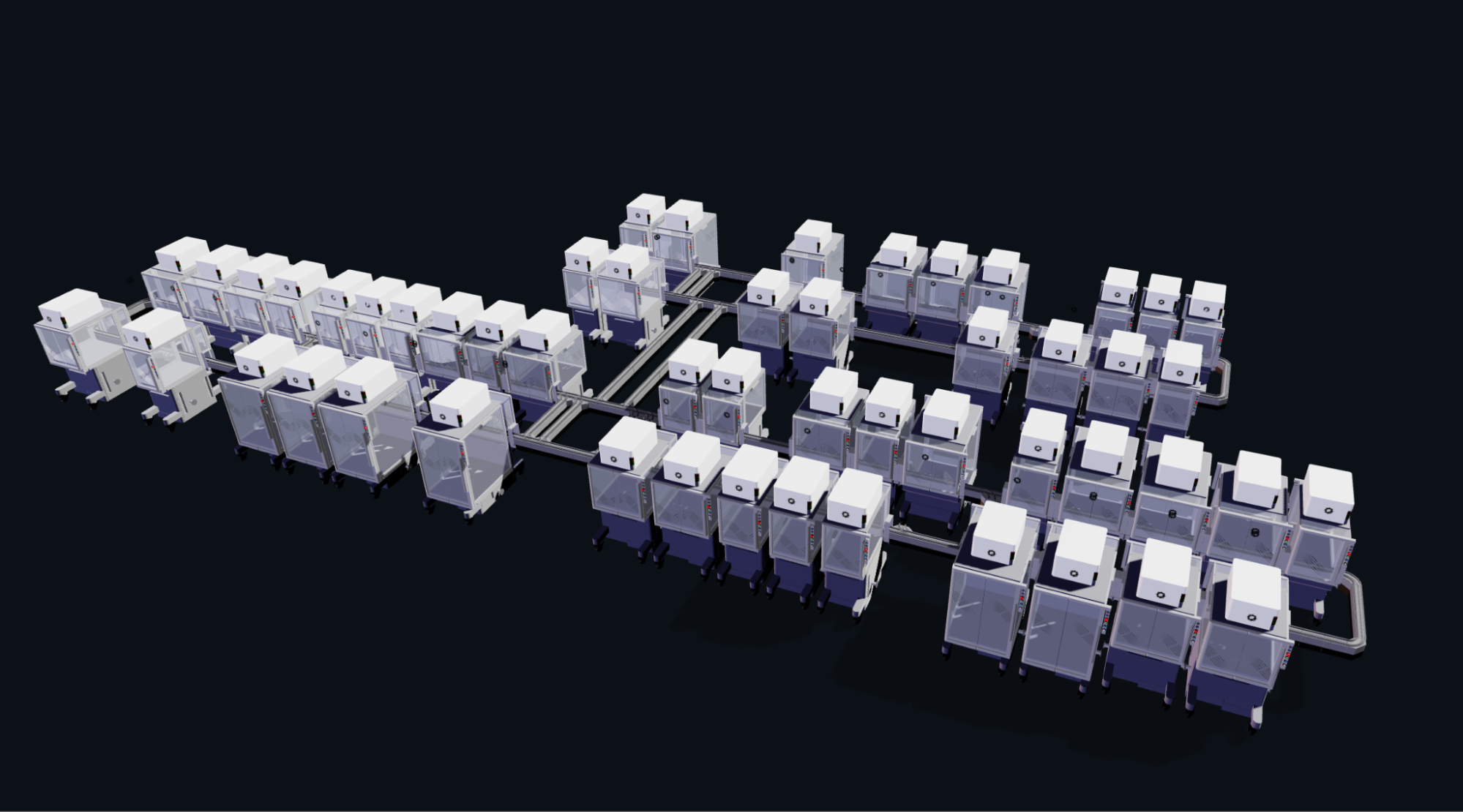
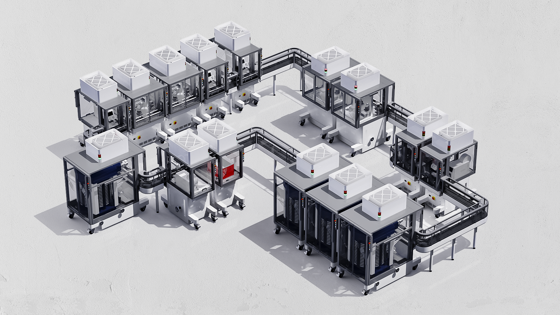
Ginkgo Nebula Cloud Laboratory Rendering, 2025
- Use this simulation tool to create an interesting looking cloud lab out of the Ginkgo Reconfigurable Automation Carts. This is just a minimal implementation so far, but I would love to see some fun designs!
Tip
Note from Ronan: If you are interested in helping me build out future HTGAA cloud lab software, please fill out this form!
Skipping this one for now.