Synthetic Cell Design
1. Oestradiol Biosensor with Bioluminescent Output
1a. Biosensor Description
I would like to design a synthetic cell that can continuously monitor the extracellular concentration in oestradiol such (e.g. 17 β-oestradiol) and emit a quantifiable bioluminescent signal whose intensity is proportional to the oestradiol concentration.
Input: oestradiol concentration
Output: bioluminescent signal
1b. Cell-Free System vs Encapsulation
The design may function in a cell-free system, but encapsulation would probably improve:
- The stability and overall duration of the experimental conditions, usually limited to 2-6 hours in cell-free systems
- The signal-to-noise ratio by inducing a stronger output signal
1c. Oestradiol Biosensor in Genetically Modified Natural Cell
The oestrogen biosensing function has already been realized in genetically modified natural cells: estradiol-inducible gene expression systems (see GEV example below) have been created in yeast.
GEV: special hybrid protein that can switch on the expression of selected genes in yeast when binding to estradiol. GEV is made of three parts: a GAL4 DNA-binding domain (from yeast, can attach to specific portions of the DNA), Human estrogen receptor domain (detects β-estradiol) and VP16 activation domain (from herpes virus, activates gene expression). References: Louvion et al. (1993) Ottoz et al. (20214)
1d. Desired Outcome of the Synthetic Cell Operation
Upon exposure to oestradiol, the synthetic cells produce a sustained, concentration-proportional bioluminescent signal via e.g. NanoLuc luciferase (NLuc) acting on its substrate furimazine.
NanoLuc offers >150-fold increase in luminescence compared to established luciferase systems, along with enhanced stability and a smaller size (19 kDa). Reference: NanoLuc: A Small Luciferase Is Brightening Up the Field of Bioluminescence
2. Components
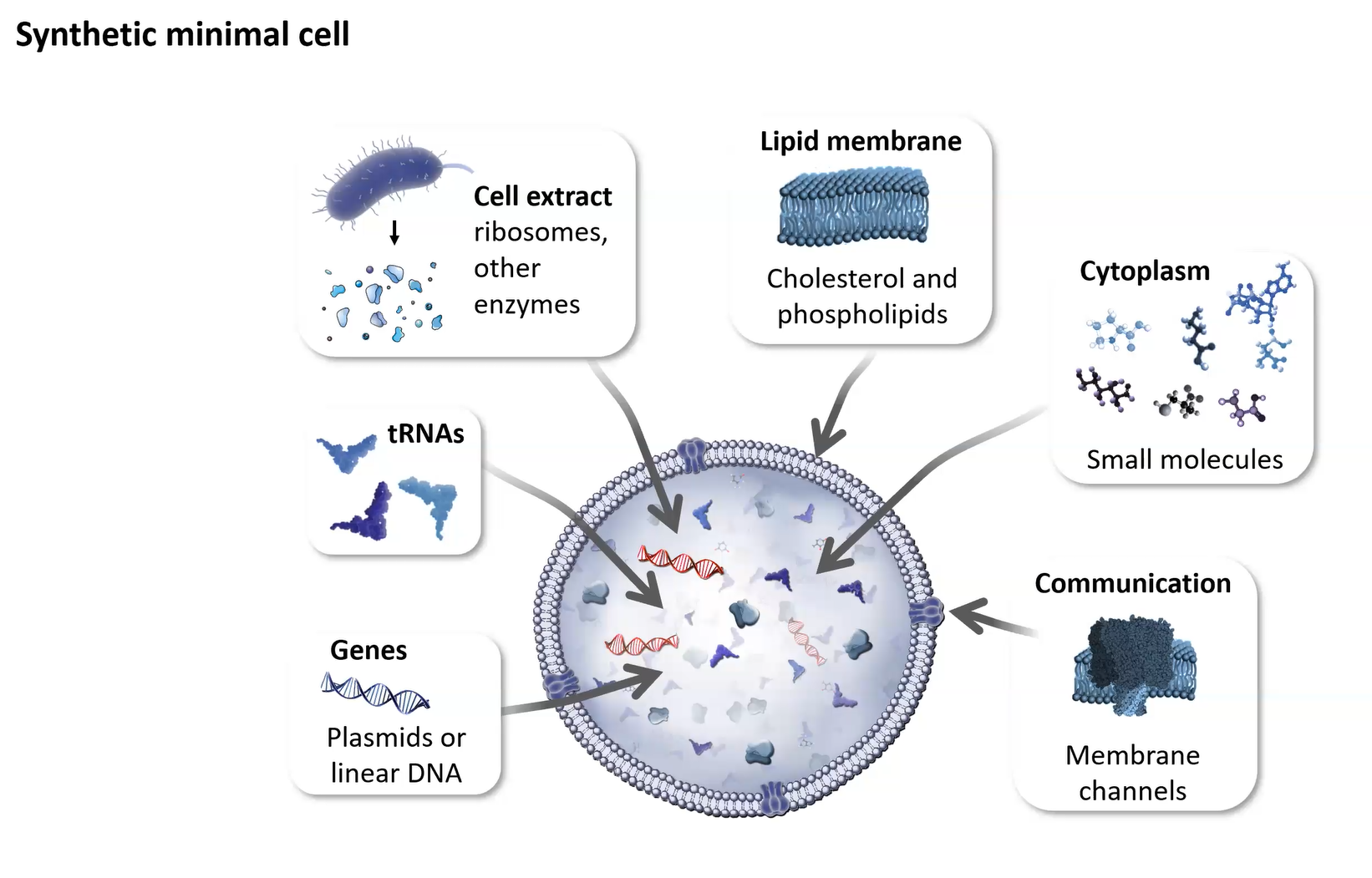
Image credit: Kate Adamala’s lab
2a. Membrane Composition
According to Kate Adamala’s lecture, the membrane of the synthetic cell should be made of phospholipids and cholesterol.
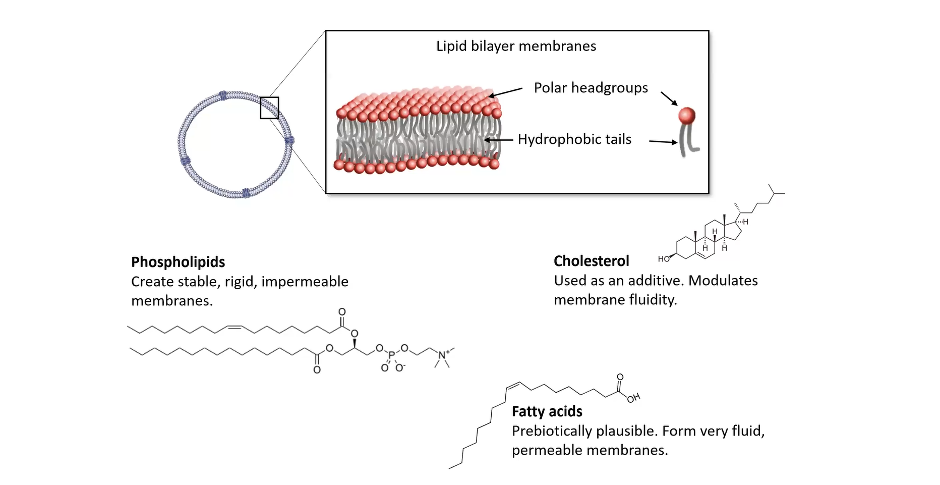 Image credit: Kate Adamala’s lab
2b. Encapsulated Contents
- Cell-free Tx/Tl system (e.g. E. coli PURE system) incl. RNA polymerase, co-factors, ribosomes, tRNAs, amino acids, ATP/GTP regeneration system
- Plasmid DNA encoding the genetic circuit (e.g. E2 sensing > expression of NLuc, see above)
- Pre-synthesised transcription factor protein (e.g. GEV) to accelerate the sensing response
- Possibly NLuc’s substrate (e.g. furimazine) acting as small reservoir before the intra- and extra-cellular concentrations equilibrate naturally
2c. System Type
Normally, human hormone receptors need many helper proteins and complicated processes inside human cells to work properly. But when integrating the GEV system, the protein can directly recognize and bind to the hormone, so there is no need for complicated cell machinery.
2d. Environmental Communication
- Extracellular oestradiol (input molecule) passively diffuses across the lipid bilayer due to its high lipophilicity, so there is no active transporter required here.
- The membrane is also permeable to furizamine (NLuc substrate).
- While the membrane is not an obstacle to the bioluminescent emission (photons), one needs to consider how the composition of the extracellular environment may affect the intensity of the output signal.
- One may consider adding nanopores to the membrane to enhance signal if oestradiol/furizamine might become limiting factors, but this would have to be finely tuned as nanopores could also cause components of the cell-free system to leak out.
3. Experimental Details
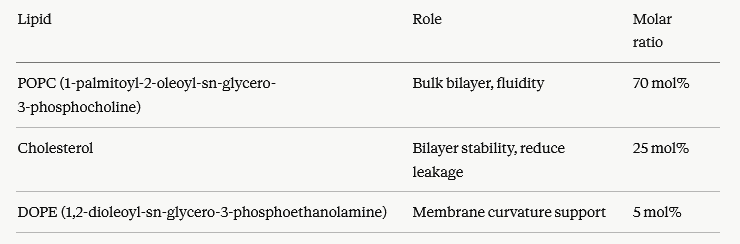
3a. Lipids and Genes
Giant Unilamellar Vesicles (GUVs) will be produced by emulsion phase transfer or microfluidic double-emulsion encapsulating the E. coli PURE system + DNA + pre-made GEV protein + furimazine.
Lipids:
According to Claude:
Genes:
- Gene 1 : Chimeric transcription factor (sensor module): GEV, Z₃EV or LexA-HBD(hERα)-B42). This protein is pre-made and encapsulated (part of GUV formation) to bypass the lag of de novo expression.
- Gene 2 : Reporter (output module): NanoLuc under the control of a synthetic promoter containing LexA operator arrays.
- Gene 3 : Repressor module: LexA-HBD(hERα)-KRAB (to implement a BAND-PASS circuit: at high oestradiol concentrations, the activator becomes out-competed by the repressor, suppressing NLuc expression above a saturation threshold).
- Gene 4 (optional) : Nanopores: hla (low expression, for furimazine equilibration without lysis).
3b. Output Measurements
The bioluminescent signal is detected by a luminometer, bioluminescence imaging system, or photonic sensor.
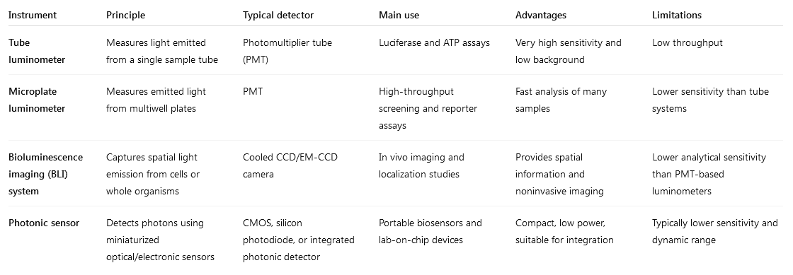
Chart created with ChatGPT. Reference: Instrumentation for Chemiluminescence and Bioluminescence
Measurement protocol suggested by Claude:
- Instrument calibration
- Single-vesicle imaging: Use bioluminescence microscopy to visualise individual GUVs responding to E2 gradients. This validates encapsulation efficiency and cell-to-cell signal heterogeneity.
- Selectivity control: Test against structurally related steroids (e.g. testosterone, progesterone, cortisol, oestrone E1, oestriol E3) at equimolar concentrations to confirm specificity of the binding for E2.
- Band-pass validation: Confirm that signal returns to baseline above a certain threshold.
- Negative controls: for instance, absence of plasmid, denatured GUV.