Measurements Final project
1. Types of measurements
Biosensor types To be confirmed after for class on biosensors
- G protein–coupled receptors (GPCRs)
- Bacterial two-component system histidine kinase receptors (e.g., EnvZ, NarX, PhoQ-type sensors)
- Ligand-gated ion channels (LGICs) or transporters (e.g., GLUT transporters, engineered channels)
GLYCOGEN measurements Membrane proteins that can detect glycogen levels in the environment or on the cell surface are generally designed as engineered biosensors, as natural extracellular glycogen sensors are rare. Key proteins and strategies identified in research include:
- Stbd1 (Starch-binding domain-containing protein 1): Stbd1 is a human protein featuring a Carbohydrate-Binding Module (CBM20) that binds specifically to glycogen and is targeted to membranes via an N-terminal hydrophobic sequence. It has been utilized to create a fusion protein, GYSC, which serves as a probe to detect glycogen in mammalian cells and muscle fibers.
- Engineered Glycogen-Binding Probes (e.g., Patent Blue V): While not a traditional membrane protein, Patent Blue V (PBV) is a fluorescent probe that binds specifically to glycogen and can be used in complex environments, making it a powerful tool for monitoring extracellular glycogen.
- AMPK β-Subunit: The carbohydrate-binding domain of the AMP-activated protein kinase (AMPK) β-subunit acts as a sensor for intracellular energy reserves by binding to glycogen. Although typically intracellular, this mechanism highlights the protein’s ability to sense glycogen levels.
- Lectin-Based Biosensors: Lectins are a family of proteins with strong binding affinity to specific carbohydrates, including glycogen and other glycans, making them useful in designing biosensors to detect carbohydrate levels in various environments. These sensors are often targeted to the cell surface, allowing researchers to monitor extracellular glycogen levels or cell-surface glycogen-binding activities.
Membrane protein-based biosensors https://pmc.ncbi.nlm.nih.gov/articles/PMC5938585/
A generic method for fluorescence monitoring glycogen through patent blue V triggered supramolecular switching https://www.sciencedirect.com/science/article/abs/pii/S0925400522002726
A red fluorescent genetically encoded biosensor for in vivo imaging of extracellular l-lactate dynamics https://www.nature.com/articles/s41467-025-64484-x
Protein Targeting to Glycogen (PTG): A Promising Player in Glucose and Lipid Metabolism https://www.mdpi.com/2218-273X/12/12/1755
A yeast FRET biosensor enlightens cAMP signaling https://www.molbiolcell.org/doi/10.1091/mbc.E20-05-0319
Novel method for detection of glycogen in cells https://pmc.ncbi.nlm.nih.gov/articles/PMC5444244/
In vivo biochemistry: Applications for small molecule biosensors in plant biology https://pmc.ncbi.nlm.nih.gov/articles/PMC3679211/
ACID LACTIC measurements (Next step)
BIOLUMINESCENCE
Membrane proteins play a crucial role in bioluminescence, acting as transporters for substrates, anchoring luciferases for localized signaling, or as part of energy transfer complexe.
Key membrane proteins and related mechanisms include:
- Oatp1 (Organic Anion Transporting Polypeptide 1): Identified as a plasma membrane transporter for D-luciferin. Expressing Oatp1 alongside luciferase significantly increases light output in vivo by facilitating substrate entry into cells.
- PDGFR Transmembrane Domain: Used to anchor luciferase enzymes, such as dinoflagellate luciferase, to the plasma membrane to monitor cell surface expression kinetics.
- HaloTag/NanoLuc Fusion Proteins: GPCRs (G protein-coupled receptors) are often fused with NanoLuc (a bright luciferase) and HaloTag (a self-labeling protein) to measure cell surface expression, trafficking, and interactions.
- Antenna Proteins (e.g., Lumazine protein): While sometimes soluble, some antenna proteins are membrane-associated. They receive energy from the excited state of the luciferase-luciferin complex and shift the emission color.
- Q-BOLT (Quenching Bioluminescent Voltage Indicator): A hybrid system using a HaloTag-NanoLuc fusion localized to the plasma membrane via a pDisplay sequence, which acts as a membrane potential reporter.
- Luminopsins: Fusion proteins combining a luciferase with light-sensitive ion channels (like channelrhodopsin) on the membrane, enabling artificial light generation to control membrane voltage.
In marine organisms, these membrane proteins are essential for the high-intensity, controlled flashes of light often seen in deep-sea creatures, where localization of the reaction to the membrane maximizes light output efficiency.
Bioluminescent and Fluorescent Proteins: Molecular Mechanisms and Modern Applications https://pmc.ncbi.nlm.nih.gov/articles/PMC9820413/
Bioluminescence Assay for Detecting Cell Surface Membrane Protein Expression https://pmc.ncbi.nlm.nih.gov/articles/PMC3064531/
2. Measurement methods
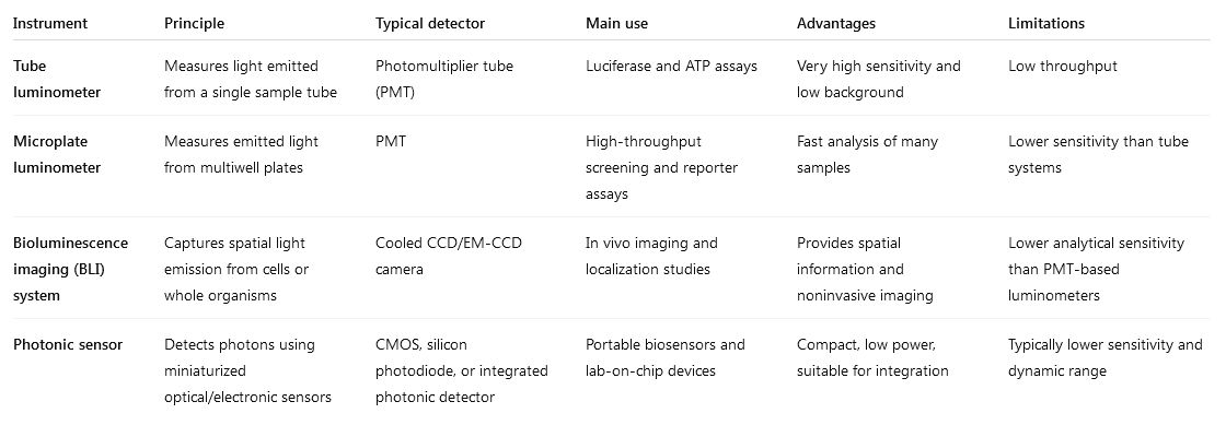
Quantifying bioluminescence involves measuring the photons emitted from the luciferase-substrate reaction. The two primary methods are luminometry (for bulk samples in vitro) and bioluminescence imaging (for spatial distribution in cells or whole organisms).
Reference: Quantitative Analysis of Bioluminescence Optical Signal
3. Measurement technologies