James Utley, PhD — HTGAA Spring 2026

⠀⠀⠀⠀⠀⠀⠀⠀⠀⠀⠀⠀⠀⠀⠀⠀⠀⠀⠀⠀⠀⠀⠀⠀⠀⠀⠀⠀⠀⠀⠀⠀⠀⠀⠀⠀⠀⠀⠀⠀⠀⠀⠀⠀⠀⠀⠀⠀⠀⠀⠀⠀⢀⠀⠀
⠀⠀⠀⠀⠀⠀⠀⠀⠀⠀⠀⠀⠀⠀⠀⠀⠀⠀⠀⠀⠀⠀⠀⠀⠀⠀⠀⠀⠀⠀⠀⠀⠀⠀⠀⠀⠀⠀⠀⠀⠀⠀⠀⢀⠀⠀⢠⣄⠀⠀⠀⠀⣿⣷⣦
⠀⠀⠀⠀⠀⠀⠀⠀⠀⠀⠀⠀⠀⠀⠀⠀⠀⠀⠀⠀⠀⠀⠀⠀⠀⠀⠀⠀⠀⠀⠀⠀⠀⠀⠀⠀⠀⠀⠀⠀⠀⠀⣦⠘⢶⣄⠀⠙⠳⣤⣀⠀⣿⣿⡇
⠀⠀⠀⠀⠀⠀⠀⠀⠀⠀⠀⠀⠀⠀⠀⠀⠀⠀⠀⠀⠀⠀⠀⠀⠀⠀⠀⠀⠀⠀⠀⠀⠀⠀⠀⠀⠀⠀⠀⠀⠀⠀⣿⣄⠀⠉⠛⠦⣄⡀⠉⢱⡿⣹⠁
⠀⠀⠀⠀⠀⠀⠀⠀⠀⠀⠀⠀⠀⠀⠀⠀⠀⠀⠀⠀⠀⠀⠀⠀⠀⠀⠀⠀⠀⠀⠀⠀⠀⠀⠀⠀⠀⠀⠀⢀⢀⠀⣿⣿⡗⠦⣄⠀⠀⢉⣴⣟⡴⠃⠀
⠀⠀⠀⠀⠀⠀⠀⠀⠀⠀⠀⠀⠀⠀⠀⠀⠀⠀⠀⠀⠀⠀⠀⠀⠀⠀⠀⠀⠀⠀⠀⠀⣠⣤⢶⡚⣿⣯⣭⣽⣯⣽⣿⡇⣷⠲⣾⣒⣿⡯⠟⠋⠀⠀⠀
⠀⠀⠀⠀⠀⠀⠀⠀⠀⠀⠀⠀⠀⠀⠀⠀⠀⠀⠀⠀⠀⠀⠀⠀⠀⠀⠀⠀⠀⢀⡴⠻⣍⡼⠿⣯⠉⠈⠛⢦⡈⠛⢦⣿⣸⡄⠀⠀⠀⠀⠀⠀⠀⠀⠀
⠀⠀⠀⠀⠀⠀⠀⠀⠀⠀⠀⠀⠀⠀⠀⠀⠀⠀⠀⠀⠀⠀⠀⠀⠀⠀⠀⠀⢠⡏⣰⠞⢿⣄⠀⠈⠳⣄⠀⠀⠙⠶⡄⢸⡇⣇⠀⠀⠀⠀⠀⠀⠀⠀⠀
⠀⠀⠀⠀⠀⠀⠀⠀⠀⠀⠀⠀⠀⠀⠀⠀⠀⠀⠀⠀⠀⠀⠀⠀⠀⠀⠀⠀⡿⢠⡏⠀⠀⠙⢷⣄⠀⠈⠳⣄⠀⠀⠘⢺⡇⣿⠀⠀⠀⠀⠀⠀⠀⠀⠀
⠀⠀⠀⠀⠀⠀⠀⠀⠀⠀⠀⠀⠀⠀⠀⠀⠀⠀⠀⠀⠀⠀⠀⠀⠀⠀⠀⠐⣇⢸⠻⣦⡀⠀⠀⠈⠳⣄⠀⠈⠳⣄⠀⣼⠁⡟⠀⠀⠀⠀⠀⠀⠀⠀⠀
⠀⠀⠀⠀⠀⠀⠀⠀⠀⠀⠀⠀⠀⠀⠀⠀⠀⠀⠀⠀⠀⠀⠀⠀⠀⠀⠀⠀⣿⣼⡆⠈⠛⣦⡀⠀⠀⠈⠑⢤⡀⠈⢳⠟⣸⠃⠀⠀⠀⠀⠀⠀⠀⠀⠀
⠀⠀⠀⠀⠀⠀⠀⠀⠀⠀⠀⠀⠀⠀⠀⠀⠀⠀⠀⠀⠀⠀⠀⠀⠀⠀⠀⠀⢹⠹⡷⣄⡀⠀⠙⠶⣄⡀⠀⠀⢙⠶⠋⣠⡟⠀⠀⠀⠀⠀⠀⠀⠀⠀⠀
⠀⠀⠀⠀⠀⠀⠀⠀⠀⠀⠀⠀⠀⠀⠀⠀⠀⠀⠀⠀⠀⠀⠀⠀⠀⠀⠀⠀⢸⡄⢧⠈⠛⢦⣀⠀⣀⣩⡷⠞⣁⣤⠾⠋⠀⠀⠀⠀⠀⠀⠀⠀⠀⠀⠀
⠀⠀⠀⠀⠀⠀⠀⠀⠀⠀⠀⠀⠀⠀⠀⠀⠀⠀⠀⢀⣠⡤⠶⢶⣚⣉⣹⣿⣿⡇⢸⣖⣲⡶⣟⣯⣯⣶⠷⠛⠉⠀⠀⠀⠀⠀⠀⠀⠀⠀⠀⠀⠀⠀⠀
⠀⠀⠀⠀⠀⠀⠀⠀⠀⠀⠀⠀⠀⠀⠀⠀⣠⢾⣿⣁⣠⠴⠞⠛⢻⣍⠀⠀⠀⣧⣸⠉⠉⠉⠉⠁⠀⠀⠀⠀⠀⠀⠀⠀⠀⠀⠀⠀⠀⠀⠀⠀⠀⠀⠀
⠀⠀⠀⠀⠀⠀⠀⠀⠀⠀⠀⠀⠀⠀⢰⡾⠁⣿⠞⠹⣦⡀⠀⠀⠀⠈⠳⣄⠀⣿⣸⡆⠀⠀⠀⠀⠀⠀⠀⠀⠀⠀⠀⠀⠀⠀⠀⠀⠀⠀⠀⠀⠀⠀⠀
⠀⠀⠀⠀⠀⠀⠀⠀⠀⠀⠀⠀⠀⢠⡏⢹⣾⡁⠀⠀⠈⠻⣦⡀⠀⠀⠀⠈⠳⡿⢹⠀⠀⠀⠀⠀⠀⠀⠀⠀⠀⠀⠀⠀⠀⠀⠀⠀⠀⠀⠀⠀⠀⠀⠀
⠀⠀⠀⠀⠀⠀⠀⠀⠀⠀⠀⠀⢀⡿⢀⡟⠀⠹⢦⡀⠀⠀⠀⠙⢦⡀⠀⠀⠀⣿⢸⠀⠀⠀⠀⠀⠀⠀⠀⠀⠀⠀⠀⠀⠀⠀⠀⠀⠀⠀⠀⠀⠀⠀⠀
⠀⠀⠀⠀⠀⠀⠀⠀⠀⠀⠀⠀⢺⢧⡼⠀⠀⠀⠀⠙⠷⣄⠀⠀⠀⠙⢦⡀⢀⡿⣸⠀⠀⠀⠀⠀⠀⠀⠀⠀⠀⠀⠀⠀⠀⠀⠀⠀⠀⠀⠀⠀⠀⠀⠀
⠀⠀⠀⠀⠀⠀⠀⠀⠀⠀⠀⠀⣾⠈⡟⠷⣤⡀⠀⠀⠀⠈⠙⢦⣀⠀⠀⠙⡾⢠⡇⠀⠀⠀⠀⠀⠀⠀⠀⠀⠀⠀⠀⠀⠀⠀⠀⠀⠀⠀⠀⠀⠀⠀⠀
⠀⠀⠀⠀⠀⠀⠀⠀⠀⠀⠀⠀⢸⠀⣇⠀⠀⠙⠢⣄⠀⠀⠀⠀⠈⣳⣤⠞⣡⠟⠀⠀⠀⠀⠀⠀⠀⠀⠀⠀⠀⠀⠀⠀⠀⠀⠀⠀⠀⠀⠀⠀⠀⠀⠀
⠀⠀⠀⠀⠀⠀⠀⠀⠀⠀⣀⣠⣼⣷⣿⣿⣿⣿⣿⣛⣛⣛⣻⣻⣋⣡⠾⠛⠁⠀⠀⠀⠀⠀⠀⠀⠀⠀⠀⠀⠀⠀⠀⠀⠀⠀⠀⠀⠀⠀⠀⠀⠀⠀⠀
⠀⠀⠀⠀⠀⢀⣠⡤⠞⣿⣿⣷⡟⢻⣿⠁⠀⠀⠀⠀⠀⠀⠀⠀⠀⠀⠀⠀⠀⠀⠀⠀⠀⠀⠀⠀⠀⠀⠀⠀⠀⠀⠀⠀⠀⠀⠀⠀⠀⠀⠀⠀⠀⠀⠀
⠀⠀⠀⣠⣶⣟⣡⠶⠋⠁⠀⠈⠳⣼⣿⡇⠀⠀⠀⠀⠀⠀⠀⠀⠀⠀⠀⠀⠀⠀⠀⠀⠀⠀⠀⠀⠀⠀⠀⠀⠀⠀⠀⠀⠀⠀⠀⠀⠀⠀⠀⠀⠀⠀⠀
⠀⢀⡼⣻⡿⠋⠻⢦⣀⠀⠀⠀⠀⠈⣿⣧⠀⠀⠀⠀⠀⠀⠀⠀⠀⠀⠀⠀⠀⠀⠀⠀⠀⠀⠀⠀⠀⠀⠀⠀⠀⠀⠀⠀⠀⠀⠀⠀⠀⠀⠀⠀⠀⠀⠀
⠀⣾⣻⠋⠀⠀⠀⠀⠈⠛⠦⣄⡀⣰⣿⡟⠀⠀⠀⠀⠀⠀⠀⠀⠀⠀⠀⠀⠀⠀⠀⠀⠀⠀⠀⠀⠀⠀⠀⠀⠀⠀⠀⠀⠀⠀⠀⠀⠀⠀⠀⠀⠀⠀⠀
⢸⣿⡿⠦⢤⣘⣢⠄⠀⠀⠀⠈⠙⣿⣳⠇⠀⠀⠀⠀⠀⠀⠀⠀⠀⠀⠀⠀⠀⠀⠀⠀⠀⠀⠀⠀⠀⠀⠀⠀⠀⠀⠀⠀⠀⠀⠀⠀⠀⠀⠀⠀⠀⠀⠀
⢸⣿⠂⠀⠀⠈⠉⠓⠒⠂⠀⠀⠀⠈⠉⠀⠀⠀⠀⠀⠀⠀⠀⠀⠀⠀⠀⠀⠀⠀⠀⠀⠀⠀⠀⠀⠀⠀⠀⠀⠀⠀⠀⠀⠀⠀⠀⠀⠀⠀⠀⠀⠀⠀⠀
⠈⠁⠀⠀⠀⠀⠀⠀⠀⠀⠀⠀⠀⠀⠀⠀⠀⠀⠀⠀⠀⠀⠀⠀⠀⠀⠀⠀⠀⠀⠀⠀⠀⠀⠀⠀⠀⠀⠀⠀⠀⠀⠀⠀⠀⠀⠀⠀⠀⠀⠀⠀⠀⠀⠀

🧬 HATERS GONNA HATE, CRISPR GONNA CRISPATE 🧬

About me

Greetings! I’m James Utley, PhD.

I wear many unusually shaped hats — find the full picture at drutley.com.

I hold a PhD in Health Science with a multidisciplinary background rooted in the Clinical Laboratory. I am a board-certified Longevity Scientist (ABAAHP) and a Certified Advanced Biotherapies Professional (CABP) Currently, I’m based in Panama City, Panama, serving as CSO for MSC (Stem Cell) Research Center.

I also founded Syndicate Laboratories about 5 years ago now — technically a research company, but we do far more. Basically robust future tech for survival. Check us out at syndicate-labs.io.

Other skills: Longevity Scientist \ [Bio]hacker \ AI Engineer \ Cool Human.

Contact info

I look forward to learning with everyone, and getting a global node started here in Panama.

Cheers to the good life!

Homework

Labs

Projects

Subsections of James Utley, PhD — HTGAA Spring 2026

Homework

Weekly homework submissions:

  • Week 1 HW: Principles and Practices

    🧬 Week 1 Homework Components 📋 Professor Questions & Answers Detailed scientific answers to questions from: Professor Jacobson — DNA polymerase error rates, genetic code degeneracy Dr. LeProust — Oligonucleotide synthesis methods and limitations Professor Church — Essential amino acids and the “Lysine Contingency” 🔬 BioVolt Project - DIY Electroporation Device Complete end-to-end project documentation including governance assessment and interactive Python application.

  • Week 2 HW: DNA Read, Write, & Edit

    🧬 Week 2 Homework Components DNA Read, Write, & Edit — sequencing and synthesis workflows, restriction digests and gel electrophoresis, genome-editing frameworks. 📋 Overview This week covers: Part 0: Basics of Gel Electrophoresis Part 1: Benchling & In-silico Gel Art ✓ Part 2: Gel Art — Restriction Digests and Gel Electrophoresis (wet lab, optional with lab access) Part 3: DNA Design Challenge ✓ Part 4: Prepare a Twist DNA Synthesis Order ✓ Part 5: DNA Read/Write/Edit ✓ Content to be added as you complete each part.

  • Week 3 HW: Lab Automation

    Published paper on automation for novel biological applications; automation project description for gumol MD simulations + ECSOD/MSC + new-Clara microfluidic validation.

Subsections of Homework

Week 1 HW: Principles and Practices

cover image cover image

🧬 Week 1 Homework Components

📋 Professor Questions & Answers

Detailed scientific answers to questions from:

  • Professor Jacobson — DNA polymerase error rates, genetic code degeneracy
  • Dr. LeProust — Oligonucleotide synthesis methods and limitations
  • Professor Church — Essential amino acids and the “Lysine Contingency”

🔬 BioVolt Project - DIY Electroporation Device

Complete end-to-end project documentation including governance assessment and interactive Python application.

     /\_/\  
    ( o.o ) 
     > ^ <
    /|   |\
   (_|   |_)
   
   "Meow! Check out both sections above!"

BioVolt Governance Assessment — Policy Options Comparison - I filled this out anyways but the project is located in the Sub as the Cat suggested- Meow

The table below compares three governance approaches for the BioVolt DIY electroporation device across multiple criteria. Scoring: 3 = Best, 2 = Moderate, 1 = Worst.

CriteriaOption 1:
Community
Self-Governance
Option 2:
Safety Warnings
& Labels
Option 3:
Regulatory
Licensing
ENHANCE BIOSECURITY
• By preventing incidents223
• By helping respond312
FOSTER LAB SAFETY
• By preventing incident223
• By helping respond313
PROTECT THE ENVIRONMENT
• By preventing incidents223
• By helping respond312
OTHER CONSIDERATIONS
• Minimizing costs and burdens321
• Feasibility?331
• Not impede research321
• Promote constructive applications321

Option Summaries

Option 1 — Community-Led Self-Governance:
✓ Best for: response capacity, feasibility, minimizing burdens, not impeding research
✗ Weaker on: prevention (relies on voluntary participation; rogue actors may ignore)

Option 2 — Targeted Product Restrictions (Safety Warnings/Labels):
✓ Best for: feasibility, moderate prevention without bans
✗ Weaker on: response capacity (warnings don’t help after incidents), limited impact on determined bad actors

Option 3 — Regulatory Classification (Licensing/HVA Review):
✓ Best for: prevention (permits, training, HVA peer review blocks worst misuse)
✗ Weaker on: costs, feasibility, impedes DIY research, harms global equity

Recommendation: Prioritize Option 1 (community self-governance) as primary, combine with Option 2 (warnings) as secondary safeguard. Avoid Option 3 unless clear evidence of high-risk proliferation emerges.

Subsections of Week 1 HW: Principles and Practices

DIY Electroporation Project: BioVolt - First rolled out at DEFCON 32- Now revisted from END to END

Project Overview: BioVolt - DIY Electroporation Device & Full Transformation Pipeline

Biological engineering application/tool to develop:
BioVolt is a portable, ultra-low-cost DIY electroporation device (~$10-20 in parts) that uses a piezoelectric crystal from a barbecue lighter to generate ~2,000 V pulses for temporary cell membrane permeabilization. This enables DNA/RNA uptake in bacteria (e.g., E. coli), yeast, plant protoplasts, or even stem cells for genetic transformation. Inspired by the DEFCON 32 talk “You got a lighter I need to do some Electroporation” (presented by Dr. James Utley (Me), Phil Rhodes, and Josh Hill from Viva Securus/Syndicate Laboratories), it builds on frugal biohacking principles: piezoelectric trigger pulsing, custom microfluidic cuvettes from aluminum tape/magnets/glass slides, and simple high-voltage testing.

DEFCON 32 Presentation — Where It Started for me

At DEFCON 32 the talk I presented focused on the device itself — proving that a barbecue lighter’s piezoelectric crystal could generate sufficient voltage to temporarily permeabilize cell membranes for DNA uptake. The talk covered design details, demos, troubleshooting (e.g., arc gap tuning with Post-it notes), and the biohacking ethos behind building a ~$10 electroporator.

Key highlights from the talk: ~2,000 V pulses via lighter clicks, high cell mortality (50-70%) but viable transformants, GFP reporter demos, open protocols encouraged.

Next Phase: End-to-End Pipeline with Efficiency Focus

The next phase of BioVolt moves beyond the device and brings the entire workflow end to end, with a focus on efficiency and frugal validation. The goal: take a piezoelectric electroporator built from a barbecue lighter and prove — through a full pipeline — that it actually works. The pipeline includes:

  1. Plasmid amplification via thermal cycling — Before electroporation, the initial plasmid source will be amplified using the MJ Research PTC-100 thermal cycler (Peltier-effect programmable controller) available in the lab. This ensures sufficient plasmid DNA concentration for transformation.

  2. DNA concentration measurement — Using the Rodeo open colorimeter (visible light version for OD600 cell density measurements) and, if possible, the UV version for DNA concentration quantification. This provides pre- and post-transformation metrics.

  3. Electroporation — Transformation of cells with the amplified plasmid DNA using the BioVolt piezoelectric device, followed by recovery and plating.

  4. Post-transformation PCR verification — For good measure, PCR will be run after transformation using the same thermal cycler to check whether the insert is present in the recovered cells. This triangulates and correlates with plating results to provide a hasty “close enough” frugal validation.

  5. Gel electrophoresis confirmation — Agarose gel electrophoresis to visualise PCR products and verify successful transformation (e.g., presence of reporter genes like GFP via band patterns under UV).

The aim is to triangulate multiple data points — plasmid amplification, colorimetric/UV measurement, transformation plating, and post-transformation PCR — to build confidence that the piezo electroporator from a lighter actually delivers. Fingers crossed, this provides a credible, frugal, end-to-end validation of a DIY electroporation workflow.

This democratizes synthetic biology for education, citizen science, and personal biohacking in resource-limited settings.

Lab Setup & Tools in Action - You can see I got some goods to work with!

My biohacker lab integrates the device with the full verification pipeline.

Working in the lab — handling samples and preparing equipment for the electroporation pipeline Working in the lab — handling samples and preparing equipment for the electroporation pipelineIO Rodeo open colorimeter — visible light version for OD600 cell density and downstream assays; UV version targeted for DNA concentration measurement IO Rodeo open colorimeter — visible light version for OD600 cell density and downstream assays; UV version targeted for DNA concentration measurement

On to the assignement - Interactive Governance Assessment Form

An interactive Python application (app.py) is provided to assess governance and risk mitigation strategies for the BioVolt project. The form uses a block-based rating scale where more filled blocks mean more effective:

BlocksRatingMeaning
●○○Minimally EffectiveLow impact — unlikely to achieve the goal
●●○Moderately EffectiveModerate impact — partial success likely
●●●Most EffectiveHigh impact — highly likely to achieve goal

Project File Structure

BioVolt_week_01_hw_principles_and_practices/
├── _index.md                      # This file — project documentation (Hugo page)
├── app.py                         # Interactive governance assessment application
├── requirements.txt               # Python dependencies
├── Biohacker_Lab.jpeg             # Lab overview photo
├── in_da_lab.jpeg                 # Working in the lab photo
├── Volt_Test.jpeg                 # High-voltage testing with insulation tester
├── rodeo-colorimeter.png          # IO Rodeo open colorimeter
├── BioVolt_govern_UI.png          # Screenshot of the application UI
└── Biovolt_Govern_Report.png      # Screenshot of the PDF report output

Prerequisites

  • Python 3.x installed on your system
  • tkinter (usually included with Python; on Linux you may need python3-tk)

Installation

  1. Navigate to the project directory:

    cd BioVolt_week_01_hw_principles_and_practices
  2. Install required dependencies:

    pip install -r requirements.txt

Running the Application

python app.py

How to Use the Form

  1. Launch — The application opens a dark-themed window with the assessment matrix.

  2. Read the instructions — System instructions are displayed at the top of the form explaining the block-based rating system.

  3. Review each concern category — Three categories are presented, each with context questions:

    • Biosecurity Concerns — preventing GMO release, high-voltage mishandling, pathogen engineering
    • Equity Concerns — access, regulation, educational barriers, global equity
    • Environmental Concerns — microbial activity, non-human organisms, public concerns
  4. Rate each action — For every action under each stakeholder (Researchers, Manufacturers, Industry, Organizations), click one of three block-rating buttons:

    • ●○○ — Minimally Effective (button highlights red)
    • ●●○ — Moderately Effective (button highlights amber)
    • ●●● — Most Effective (button highlights green)
  5. Visual feedback — When you click a rating:

    • The selected button stays highlighted with its rating colour
    • A status indicator appears to the right showing your selection
    • Other buttons in the same row reset to their default state
  6. Export to PDF — Click the “EXPORT TO PDF” button to generate a report containing:

    • Cover page with assessment date and completion count
    • Rating scale legend with colour-coded descriptions
    • Full assessment tables for each concern category
    • Colour-coded rows: green tint for Most Effective, amber for Moderate, red for Minimal
    • Block indicators (●●● / ●●○ / ●○○) printed in every row
    • Summary page with counts and percentages for each rating level
  7. Reset — Click “RESET MATRIX” to clear all selections and start over.

Application Features

  • Block-based rating scale — intuitive system where more blocks = more effective (no ambiguity)
  • Dark theme UI — dark background with neon accent colours for readability
  • Persistent button state — selected buttons remain highlighted with their rating colour
  • Status indicators — each row shows the current selection in text beside the buttons
  • Scrollable interface — mouse wheel support for navigating the full assessment matrix
  • Neon accent bars — left-side accent bars on each concern card for visual hierarchy
  • Colour-coded PDF output — rating cells are tinted to match their effectiveness level
  • Summary statistics — PDF includes a final page with counts and percentages
  • Empty export protection — warns you if no ratings are selected before exporting
  • Form reset — one-click reset with confirmation dialog

Screenshots

Application UI — Dark-themed interface with block-based rating buttons and colour-coded status indicators:

BioVolt Governance Assessment Matrix UI BioVolt Governance Assessment Matrix UI

PDF Report Output — Exported assessment with colour-coded rows, block indicators, and stakeholder ratings:

BioVolt Governance Assessment PDF Report BioVolt Governance Assessment PDF Report

Governance / Policy Goals (Preventing Harm)

Focus on non-tool-function risks: Prevent environmental release of unintended GMOs, biosafety incidents from mishandling high-voltage + microbes, escalation to unsafe self-experimentation/human applications, or biosecurity concerns (e.g., pathogen engineering).
Core aims: Minimize biosafety/biosecurity harms, promote responsible use, avoid stifling innovation with heavy regulation, encourage informed DIYbio practices, and address public/environmental concerns.

Three Potential Governance/Policy Actions

Action 1: Community-Led Self-Governance with Voluntary Guidelines and Reporting

Goal: Foster peer accountability and safe practices through DIYbio networks, reducing risks via shared norms without external mandates.

Design:

  • Opt-in: DIYbio communities, forums (e.g., Discord, Reddit, The ODIN users), and makerspaces.
  • Fund: Crowdfunding, donations, or volunteer time.
  • Approve: Community-elected moderators or biosafety working groups.
  • Implement: Publish voluntary guidelines (e.g., “BioVolt Safety Protocol” on protocols.io or GitHub), require protocol sharing for builds, anonymous incident reporting (expand “Ask a Biosafety Expert” services).

Risks / What could go wrong (incorrect assumptions, uncertainties):
Assumes broad ethical participation - rogue actors may ignore; self-reporting misses hidden issues; low adoption if seen as “extra work.”

Assumptions, “Success” and “Failure” rubric:

  • Success (best - 1): High adoption -> fewer accidents, strong norms against risky uses (e.g., no human trials), community self-corrects.
  • Mid (2): Partial uptake -> safety improvements in visible projects, but gaps remain.
  • Failure (worst - 3): Guidelines ignored -> no risk reduction, or “forbidden fruit” effect increases experimentation.
  • Unintended consequences: Overly cautious norms suppress legitimate educational uses.

Action 2: Targeted Product Restrictions (e.g., Safety Warnings / Age Limits on Kits & Components)

Goal: Reduce impulsive or uninformed misuse by requiring clear hazard labels on high-voltage components (e.g., piezoelectric lighters, capacitors) or full kits, without banning access.

Design:

  • Opt-in/compliance: Online sellers (Amazon, AliExpress), hardware stores, kit makers.
  • Fund: Seller-borne costs.
  • Approve: Consumer safety agencies or state-level consumer protection (e.g., modeled on CRISPR kit labeling laws).
  • Implement: Mandatory labels (“Not for human use; biological hazard when combined with genetic material; 18+ recommended”).

Risks / What could go wrong:
Warnings may not deter determined users (parts sourced separately); patchy enforcement online/global; could increase black-market activity.

Assumptions, “Success” and “Failure” rubric:

  • Success (best - 1): Warnings raise awareness, reduce naive accidents while preserving access.
  • Mid (2): Labels added but often ignored by experienced users.
  • Failure (worst - 3): Little impact on bad actors; adds cost/delays for legitimate builders.
  • Unintended consequences: Drives activity underground, reducing community visibility/oversight.

Action 3: Treat as if it has a Regulatory Classification as Restricted Biotech Equipment (e.g., Licensing for High-Voltage Builds) Pledge reporting and Safe use.

Goal: Treat advanced DIY electroporators like controlled lab tools - require permits/training for >1,000 V devices to prevent proliferation to high-risk genetic work.

Design:

  • Opt-in: Individual builders/users via registration.
  • Fund: User fees.
  • Approve: Government agencies (e.g., expanding CDC/NIH biosafety rules or local health depts).
  • Implement: Permits, training requirements, inspections for community labs/shared spaces.
  • Hazard Vulnerability Assessment (HVA) and Peer Review: Conduct a comprehensive HVA and require peer review through a pseudo-IRB-like entity - a multidisciplinary and independent review board focusing on environmental and human safety. This entity would evaluate proposed uses, assess risks, and provide guidance on safe protocols before high-voltage builds are deployed.

Risks / What could go wrong:
Hard to define safe thresholds; bureaucracy kills accessibility; overreach chills innovation globally.

Assumptions, “Success” and “Failure” rubric:

  • Success (best - 1): Blocks worst misuse (e.g., pathogen work), funnels activity to supervised settings.
  • Mid (2): Some compliance, but many unlicensed builds continue.
  • Failure (worst - 3): Broad restrictions eliminate DIY benefits, push activity to unregulated regions.
  • Unintended consequences: Harms global equity/education; favors institutional labs only.

Overall Tradeoffs & Prioritization

Prioritize Action 1 (community self-governance) as primary: Lowest overregulation risk, aligns with DIY ethos, adaptable to low current misuse evidence, leverages community goodwill.

Combine with Action 2 (targeted warnings) as secondary: Adds minimal external safeguard for public health, deters casual risks without bans.

Avoid/minimize Action 3 unless clear evidence of high-risk proliferation: Highest chance of killing accessibility and innovation, poor fit for low-harm tool like BioVolt.

Key uncertainties (misuse rates, community response, enforcement feasibility) favor lighter interventions. Monitor via voluntary reporting; escalate only if serious incidents arise. This balances empowerment with responsible governance for biosafety and preventing broader DIY genetic risks.

Made with love and the AI Slop is from Cursor-GLM 4.7

Week 1: Professor Questions

Answers organized by instructor, please click the question to reveal the answer!

Instructions: Click the triangle (▶) or question text to expand and view the full answer.


[SECTION 1] Questions from Professor Jacobson

Source: Lecture 2 slides


▶ Question 1: Nature's machinery for copying DNA is called polymerase. What is the error rate of polymerase? How does this compare to the length of the human genome? How does biology deal with that discrepancy?

Answer

Executive Summary:
DNA polymerase intrinsic error rate (~10⁻⁷) would cause ~320 errors per human genome replication (3.2 × 10⁹ bp). Biology employs multilayer error correction (proofreading, mismatch repair, excision repair) to achieve final fidelity of ~10⁻⁹ to 10⁻¹⁰ errors per base per division, yielding 0.3-3 errors per replication in normal somatic cells.


Error Rate of DNA Polymerase

DNA polymerase has an intrinsic error rate of approximately 1 error per 10⁷ nucleotides during DNA synthesis. With integrated 3’ to 5’ exonuclease proofreading activity, this improves to approximately 1 error per 10⁸-10⁹ nucleotides.

Comparison to Human Genome Length

The human genome contains approximately 3.2 × 10⁹ base pairs.

Without proofreading:

  • Error rate: ~10⁻⁷ per nucleotide
  • Expected errors per replication: ~320 errors per genome copy

With proofreading:

  • Error rate: ~10⁻⁸ to 10⁻⁹ per nucleotide
  • Expected errors per replication: ~3-32 errors per genome copy

How Biology Deals with This Discrepancy

Biology employs multiple layers of error correction that act sequentially:

  1. Proofreading (3’ → 5’ exonuclease activity)

    • DNA polymerase detects incorrect base pairing via geometric distortion
    • Removes mismatched nucleotide immediately
    • Reduces error rate by approximately 100-1000-fold
  2. Mismatch Repair (MMR) System

    • Post-replication surveillance mechanism
    • In bacteria (E. coli): MutS, MutL, and MutH proteins
    • In eukaryotes: MSH (MutS homolog), MLH (MutL homolog), and PMS protein families
    • System identifies mismatched base pairs, excises incorrect strand segment, and resynthesizes
    • Further reduces error rate by approximately 100-1000-fold
  3. Nucleotide Excision Repair (NER)

    • Repairs bulky DNA lesions (UV-induced thymine dimers, chemical adducts)
    • Removes damaged nucleotide segments (20-30 nt patches)
  4. Base Excision Repair (BER)

    • Corrects small base modifications (deamination, oxidation, alkylation)
    • DNA glycosylases remove damaged bases; AP endonucleases process abasic sites

Result:
The combined fidelity of replication in eukaryotic somatic cells typically achieves ~10⁻⁹ to 10⁻¹⁰ errors per base per cell division, depending on organism, cell type, and proliferation status. This ensures 0.3-3 errors per genome replication under normal physiological conditions.

Note: Fidelity varies by context. Cancer cells with MMR defects exhibit 100-1000× higher mutation rates. Germline cells employ additional proofreading mechanisms. Some DNA polymerases (e.g., Pol η, translesion synthesis polymerases) have lower fidelity by design for specialized repair functions.


▶ Question 2: How many different ways are there to code (DNA nucleotide code) for an average human protein? In practice what are some of the reasons that all of these different codes don't work to code for the protein of interest?

Answer

Executive Summary:
For a typical 400-residue protein, the number of synonymous DNA sequences (due to codon degeneracy) is astronomically large—on the order of 10¹⁰⁰ or more, calculated as the product of synonymous codon counts across all positions. In practice, most sequences fail due to codon usage bias, mRNA secondary structure, RNA instability, splicing interference, cryptic regulatory elements, and synthesis/cloning constraints.


Number of Different Ways to Code for a Protein

The genetic code is degenerate—61 sense codons encode 20 standard amino acids plus start/stop signals. Each amino acid (except Met and Trp) has multiple synonymous codons:

  • Leucine, Serine, Arginine: 6 codons each
  • Isoleucine: 3 codons
  • Methionine, Tryptophan: 1 codon each

For an average human protein (~400 amino acids):

The total number of synonymous DNA sequences is the product of synonymous codon counts across all positions:

N = ∏(i=1 to 400) n_i

where n_i = number of synonymous codons for amino acid i.

Rough estimate:

  • Average degeneracy per amino acid ≈ 3 codons (weighted by frequency)
  • Total combinations ≈ 3⁴⁰⁰ ≈ 10¹⁹⁰ possible DNA sequences

Even conservative estimates (e.g., leucine-rich proteins) yield 10¹⁰⁰+ combinations.

Why All These Different Codes Don’t Work in Practice

Even though multiple sequences encode the same amino acid sequence, the vast majority fail to express functional protein due to:

1. Codon Usage Bias
  • Each organism has preferred codons reflecting tRNA abundance (Plotkin & Kudla 2011)
  • E. coli prefers different codons than humans (e.g., AGG/AGA rare in bacteria, common in mammals)
  • Rare codons → ribosome stalling → may alter co-translational folding kinetics
  • Using non-optimal codons can reduce expression 10-1000-fold (Gustafsson et al. 2004)
2. mRNA Secondary Structure
  • Certain nucleotide sequences form stem-loops or hairpins
  • Strong secondary structures can:
    • Block ribosome binding
    • Stall translation
    • Trigger mRNA degradation
3. RNA Stability
  • AU-rich sequences → rapid mRNA degradation
  • GC-rich sequences → more stable mRNA
  • Wrong codon choice can drastically reduce mRNA half-life
4. Splicing Interference
  • Certain sequences create cryptic splice sites
  • Can cause exon skipping or intron retention
  • Results in truncated or non-functional protein
5. Ribosome Binding Sites (RBS) Interference
  • Shine-Dalgarno sequences (prokaryotes) or Kozak sequences (eukaryotes)
  • Internal RBS-like sequences can cause premature translation initiation
  • Results in truncated proteins
6. Restriction Enzyme Sites
  • Cloning often requires avoiding certain restriction sites
  • Limits sequence choices for practical molecular biology
7. Repetitive Sequences
  • Long homopolymer runs (e.g., AAAAAA) cause synthesis/sequencing errors
  • Can trigger recombination or replication errors

Quantitative Example: For a 10-amino acid peptide (assuming average 3-fold degeneracy), there are theoretically 3¹⁰ ≈ 59,000 synonymous sequences. However, accounting for all the constraints listed above, only an estimated 10²-10³ sequences (~1-2%) would be practically functional.


[SECTION 2] Questions from Dr. LeProust

Source: Lecture 2 slides


▶ Question 3: What's the most commonly used method for oligo synthesis currently?

Answer

Executive Summary:
Phosphoramidite chemistry on solid-phase support (Caruthers method, 1981) is the current industry standard, with typical coupling efficiency of 98.5-99.5% per cycle and practical length ceiling of 150-200 nucleotides.


Phosphoramidite Chemistry (Solid-Phase Synthesis)

The phosphoramidite method on solid support is the dominant technology for oligonucleotide synthesis worldwide.

Key Features:

  • Invented: Marvin Caruthers and colleagues (1981)
  • Platform: Solid-phase synthesis on controlled-pore glass (CPG) or polystyrene beads
  • Direction: 3’ → 5’ synthesis (chain grows from 3’-OH to 5’ end)
  • Cycle efficiency: Typically 98.5-99.5% per nucleotide addition
  • Practical length limit: 150-200 nucleotides for routine synthesis

Four-Step Cycle:

  1. Detritylation (acid treatment)

    • Removes DMT (dimethoxytrityl) protecting group from 5’-OH
    • Exposes reactive hydroxyl for next nucleotide
  2. Coupling (phosphoramidite addition)

    • Protected phosphoramidite monomer + tetrazole activator
    • Forms phosphite triester linkage
    • ~98-99.5% coupling efficiency
  3. Capping (acetic anhydride)

    • Blocks unreacted 5’-OH groups
    • Prevents deletion sequences
  4. Oxidation (iodine/water)

    • Converts unstable phosphite (P³⁺) to stable phosphate (P⁵⁺)
    • Forms phosphate backbone

Advantages:

  • High throughput (96-384 well formats)
  • Automated
  • Scalable (nmol to µmol scale)
  • Well-established chemistry

Current Platforms: Commercial platforms include BioAutomation and ABI/Applied Biosystems synthesizers for traditional column-based synthesis. Newer high-throughput approaches include Twist Bioscience (silicon-based microarray synthesis) and Custom Array (electrochemical synthesis on chips).


▶ Question 4: Why is it difficult to make oligos longer than 200nt via direct synthesis?

Answer

Executive Summary:
Cumulative coupling inefficiency (even at 99% per cycle) yields only ~13% full-length product at 200 nt. Dominant failure modes are deletion sequences from incomplete coupling, depurination during detritylation, and increasing purification difficulty as n-1, n-2… products accumulate.


Cumulative Coupling Errors and Deletion Sequences

The primary challenge is imperfect coupling efficiency in each phosphoramidite addition cycle.

The Mathematics of Error Accumulation:

  • Coupling efficiency per cycle: typically 98.5-99.5%
  • Stepwise failure rate: 0.5-1.5% per cycle
  • Yield of full-length product = (coupling efficiency)^n where n = oligo length

Yield Calculation:

LengthCoupling EfficiencyFull-Length Yield
50 nt99%60%
100 nt99%37%
150 nt99%22%
200 nt99%13%
300 nt99%5%

At 200 nucleotides with 99% efficiency:

  • Only 13% of molecules are full-length correct sequence
  • 87% are deletion products (n-1, n-2, n-3… truncations)

Specific Problems Beyond 200nt (in order of impact):

  1. Deletion Sequences from Incomplete Coupling

    • Failed coupling at position i → all subsequent additions build on truncated chain
    • Creates heterogeneous mixture of n-1, n-2, n-3… products
    • Capping step blocks these from extending, but they remain in final pool
  2. Depurination During Acid Treatment

    • Detritylation uses trichloroacetic acid or dichloroacetic acid
    • Causes glycosidic bond cleavage at purines (A, G)
    • Cumulative damage over 200+ cycles
    • Results in abasic sites and chain breaks
  3. Purification Difficulty

    • Full-length (200 nt) vs. n-1 (199 nt) differ by <0.5% in mass
    • HPLC and PAGE separation becomes marginal
    • Impure product affects downstream applications
  4. Secondary Structure Formation

    • Long single-stranded oligos form intramolecular hairpins during synthesis
    • Blocks reagent access to growing 3’-OH end (on solid support, growing from 3’ end)
    • Reduces effective coupling efficiency in later cycles
  5. Synthesis Time and Cost

    • 200 cycles × 10-15 min/cycle = 33-50 hours continuous synthesis
    • Reagent consumption scales linearly
    • Low yields require larger scale synthesis → higher cost

Practical Solutions: Modern approaches avoid direct synthesis beyond 200 nt by using gene assembly from overlapping 60-80 nt oligos (polymerase cycling assembly, Gibson assembly), column-based assembly methods (e.g., Twist Bioscience chip synthesis followed by assembly), or emerging enzymatic synthesis using terminal deoxynucleotidyl transferase-based methods.


▶ Question 5: Why can't you make a 2000bp gene via direct oligo synthesis?

Answer

Executive Summary:
Direct phosphoramidite synthesis of 2000 nt is practically infeasible due to vanishingly low yields (0.99^2000 ≈ 10⁻⁹), prohibitive synthesis time (~2-3 weeks continuous), cumulative depurination, and insurmountable purification challenges. Modern gene synthesis uses hierarchical assembly of 60-80 nt oligos into fragments, then full-length genes.


Practical Infeasibility with Current Phosphoramidite Chemistry

Making a 2000 bp gene via direct oligonucleotide synthesis is practically infeasible with standard phosphoramidite chemistry due to insurmountable yield, time, and purification barriers.

Yield Barriers:

At 99% coupling efficiency (best-case scenario):

  • Yield = 0.99^2000 ≈ 2 × 10⁻⁹ (0.0000002%)
  • To obtain 1 picomole of full-length product requires ~0.5 moles of starting material
  • Equivalent to ~660 grams of protected nucleotide phosphoramidites
  • Material cost alone: ~$500,000 - $1,000,000

Even at 99.5% efficiency (exceptional, rarely achieved):

  • Yield = 0.995^2000 ≈ 5 × 10⁻⁵ (0.005%)
  • Still economically and practically prohibitive

Physical/Chemical Barriers:

  1. Synthesis Time

    • Typical cycle time: 10-15 minutes per nucleotide addition
    • 2000 cycles = 20,000-30,000 minutes = 14-21 days continuous synthesis
    • Reagent degradation over extended periods
    • Instrument reliability over multi-week runs
  2. Cumulative Depurination

    • 2000 acid detritylation steps
    • Each cycle causes low-frequency glycosidic bond cleavage at purines
    • Accumulates to extensive abasic sites and strand breaks
  3. Secondary Structure Collapse

    • Long single-stranded DNA forms extensive intramolecular structure
    • Hairpins and G-quadruplexes block reagent access
    • Synthesis typically stalls beyond 300-400 nt even with optimized conditions
  4. Solubility and Handling

    • Very long oligos can precipitate on solid support
    • Reduced accessibility to coupling reagents
    • Cleavage and deprotection become inefficient

Practical Solution: Hierarchical Gene Assembly

Modern commercial gene synthesis uses multi-step assembly:

Step 1: Oligo Synthesis

  • Synthesize 30-50 oligonucleotides (60-80 nt each, with 20-40 nt overlaps)
  • Yield per oligo: 60-95% (high quality)

Step 2: Fragment Assembly

  • Assemble oligos into 4-6 intermediate fragments (400-600 bp each)
  • Methods: Polymerase cycling assembly (PCA), Gibson assembly, Golden Gate
  • Yield per fragment: 70-90%

Step 3: Final Assembly

  • Combine fragments into full 2000 bp gene
  • Gibson assembly or restriction enzyme-based methods
  • Final yield: 60-85% overall

Example for 2000 bp gene:

  • 40 oligos × 70 nt average = 2800 nt synthesized capacity
  • Assemble into 5 fragments (~400 bp each)
  • Final Gibson assembly into 2000 bp construct
  • Overall yield: ~70% (vs. 10⁻⁹% for direct synthesis)

Commercial Gene Synthesis: Major vendors (Twist Bioscience, IDT, GenScript, Thermo Fisher) offer typical academic pricing of $0.07-0.20/bp, though this is highly variable depending on sequence complexity (GC content, repeats, secondary structure), turnaround time (5-10 days standard, 2-3 days expedited), and order volume. Standard turnaround is 5-10 days with rush options of 2-3 days.


[SECTION 3] Question from Professor George Church

Source: Lecture 2 slides


▶ Question 6: [Using Google & Prof. Church's slide #4] What are the 10 essential amino acids in all animals and how does this affect your view of the "Lysine Contingency"?

(I chose this question from the three options)

Answer

Executive Summary:
The commonly listed essential amino acids in vertebrates include His, Ile, Leu, Lys, Met, Phe, Thr, Trp, Val, and conditionally Arg. The “Lysine Contingency” from Jurassic Park is scientifically flawed because lysine is already naturally essential in all vertebrates—the genetic modification provides zero additional biocontainment. Moreover, lysine is abundant in all natural food sources, and deficiency takes months to years to be lethal.


The Commonly Listed Essential Amino Acids in Vertebrates

Essential amino acids cannot be synthesized de novo by vertebrate metabolism and must be obtained from diet. The standard list for humans and most vertebrates includes: Histidine (His, H), Isoleucine (Ile, I), Leucine (Leu, L), Lysine (Lys, K) [focus of Jurassic Park scenario], Methionine (Met, M), Phenylalanine (Phe, F), Threonine (Thr, T), Tryptophan (Trp, W), Valine (Val, V), and Arginine (Arg, R), which is conditionally essential—essential in juveniles, young/growing animals, and during illness, though adults can synthesize limited amounts via the urea cycle.

Mnemonic: “PVT TIM HALL” (Phe, Val, Thr, Trp, Ile, Met, His, Arg, Leu, Lys)

Note: The classification varies slightly by species and life stage. Arginine is typically considered semi-essential or conditionally essential in adult mammals.


The “Lysine Contingency” from Jurassic Park

In Jurassic Park (Michael Crichton, 1990), InGen implemented a “Lysine Contingency” as a biocontainment measure. The plan involved genetically engineering dinosaurs unable to synthesize lysine, making them dependent on lysine supplements in their food. The theory was that if they escaped, they would die from lysine deficiency. As Dr. Wu stated: “The lysine contingency is intended to prevent the spread of the animals is case they ever got off the island.”


Why the Lysine Contingency is Scientifically Flawed

Critical Problem: ALL ANIMALS ALREADY REQUIRE DIETARY LYSINE

1. Lysine is Naturally Essential in All Vertebrates

Humans, dinosaurs, birds, and mammals cannot synthesize lysine de novo. Animals lost the lysine biosynthesis pathway approximately 500 million years ago during early vertebrate evolution. The dinosaurs would have required dietary lysine regardless of any genetic modification. Therefore, the “contingency” provides zero additional biocontainment—it is entirely redundant.


2. Lysine is Abundant in Natural Food Sources

Based on USDA nutritional databases, lysine is widespread in both plant and animal food sources. Plant sources include legumes (soybeans, lentils, beans) containing 1-2% lysine by dry weight, seeds and grains with 0.2-0.8% lysine, and grasses and leafy vegetation with 0.3-0.6% lysine. Animal sources are even richer: insects contain approximately 2-3% lysine by dry weight, while vertebrate muscle tissue, fish, and eggs contain 1.5-2.5% lysine by weight.

Estimated lysine intake for large theropods (carnivorous dinosaurs):

Note: The following are rough extrapolations from modern vertebrate nutritional requirements and are not based on direct measurements of dinosaur metabolism. Assuming an estimated daily food intake of 50-100 kg meat (scaled from modern large carnivores) and lysine content of meat at approximately 1.5-2.0 g/100g, the estimated daily lysine intake would be 750-2000 g. Compared to an estimated lysine requirement of approximately 10-50 g/day (scaled from mammals, though highly uncertain), even conservative estimates suggest 10-100× excess lysine intake.

Estimated lysine intake for herbivorous dinosaurs:

Assuming estimated daily vegetation consumption of hundreds of kg for sauropods and lysine content in plant matter of 0.3-1.0% dry weight, the estimated daily lysine intake would be hundreds of grams. This substantially exceeds the likely requirement of 50-200 g/day when scaled from large herbivorous mammals.

Key Point: Even consuming exclusively grass, leaves, or insects would likely provide sufficient lysine to meet metabolic needs, assuming dinosaur requirements scaled similarly to modern vertebrates.


3. Timescale of Lysine Deficiency is Impractical

Lysine deficiency symptoms develop slowly: immune system impairment occurs over weeks to months, growth retardation takes months, and muscle wasting progresses over months to years. Lethality from severe deficiency requires months to years. A dinosaur escaping into the wild would eat naturally available food and immediately obtain sufficient lysine, never developing deficiency symptoms. The timescale mismatch is fatal to the strategy: containment must occur in minutes to hours (the escape window), while lysine deficiency lethality takes months to years. The result is a completely ineffective biocontainment strategy.


4. Better Biocontainment Strategies

If the goal is preventing escaped dinosaurs from surviving or reproducing, several approaches would be more effective than the lysine contingency.

Metabolic Dependencies: Creating auxotrophy for synthetic amino acids not found in nature (such as D-amino acids or unnatural amino acids requiring continuous supplementation), nucleotide auxotrophy (e.g., thymine requirement), or vitamin/cofactor dependencies (e.g., engineered B12 requirement) would provide genuine containment.

Genetic Kill Switches: Conditional lethality genes requiring antidote molecules, thermosensitive essential genes that allow survival only at controlled temperatures, or light-dependent survival mechanisms requiring specific UV or wavelength exposure offer programmed containment.

Reproductive Control: All-female populations (as attempted in Jurassic Park), meiotic drive systems ensuring sterility, or genetic incompatibility with wild relatives would prevent population establishment.

Environmental Dependencies: Temperature-sensitive phenotypes surviving only in controlled climates or organisms requiring specific atmospheric pressure or composition would restrict habitat range.


Conclusion: How This Affects My View of the Lysine Contingency

The Lysine Contingency is scientifically flawed as a biocontainment strategy and represents a misunderstanding of vertebrate nutritional biochemistry. The strategy fails on four fundamental levels: (1) it is not a contingency since lysine is already naturally essential in all vertebrates, making the modification redundant; (2) it is not limiting since lysine is abundant in nearly all natural food sources; (3) it is not fast-acting since lysine deficiency takes months to years to be lethal in large vertebrates; and (4) it provides no additional biocontainment barrier beyond natural biology.

From a biosafety perspective, the lysine contingency demonstrates the risk of “security theater” in synthetic biology—creating the appearance of control without meaningful containment. Real biocontainment requires dependencies on synthetic or artificial inputs not present in natural ecosystems. Modern synthetic biology approaches include unnatural amino acid dependencies (e.g., amber suppressor systems with synthetic tRNAs), genetic kill switches (toxin-antitoxin modules, essential gene knockout with complementation), orthogonal genetic systems (expanded genetic code, xenobiology with XNA), and metabolic dependencies on synthetic nutrients or specific light wavelengths.

Narrative function in Jurassic Park: The flawed lysine contingency serves as a plot device illustrating InGen’s overconfidence and foreshadows that all their control measures will fail (“Life finds a way”). It highlights the dangers of inadequate risk assessment and overconfidence in genetic engineering safeguards.

Lessons for modern synthetic biology: Biological containment is extremely difficult and requires multiple redundant safeguards. Single-point dependencies, especially on naturally occurring molecules, are inadequate. Rigorous testing and evolutionary escape rate measurements are essential for any containment strategy.


[REFERENCES]

Primary Literature and Resources

DNA Replication Fidelity (Q1):

  • Alberts B, Johnson A, Lewis J, et al. Molecular Biology of the Cell. 6th edition. Garland Science, 2014. Chapter 5: DNA Replication, Repair, and Recombination.
  • Kunkel TA, Bebenek K. DNA replication fidelity. Annu Rev Biochem. 2000;69:497-529. doi:10.1146/annurev.biochem.69.1.497
  • Iyer RR, Pluciennik A, Burdett V, Modrich PL. DNA mismatch repair: functions and mechanisms. Chem Rev. 2006;106(2):302-323. doi:10.1021/cr0404794

Genetic Code and Translation (Q2):

  • Plotkin JB, Kudla G. Synonymous but not the same: the causes and consequences of codon bias. Nat Rev Genet. 2011;12(1):32-42. doi:10.1038/nrg2899
  • Gustafsson C, Govindarajan S, Minshull J. Codon bias and heterologous protein expression. Trends Biotechnol. 2004;22(7):346-353. doi:10.1016/j.tibtech.2004.04.006
  • Tuller T, Carmi A, Vestsigian K, et al. An evolutionarily conserved mechanism for controlling the efficiency of protein translation. Cell. 2010;141(2):344-354. doi:10.1016/j.cell.2010.03.031

Oligonucleotide Synthesis (Q3-Q5):

  • Caruthers MH. Gene synthesis machines: DNA chemistry and its uses. Science. 1985;230(4723):281-285. doi:10.1126/science.3863253
  • Kosuri S, Church GM. Large-scale de novo DNA synthesis: technologies and applications. Nat Methods. 2014;11(5):499-507. doi:10.1038/nmeth.2918
  • Hughes RA, Ellington AD. Synthetic DNA synthesis and assembly: putting the synthetic in synthetic biology. Cold Spring Harb Perspect Biol. 2017;9(1):a023812. doi:10.1101/cshperspect.a023812

Amino Acid Nutrition and Biosafety (Q6):

  • Reeds PJ. Dispensable and indispensable amino acids for humans. J Nutr. 2000;130(7):1835S-1840S. doi:10.1093/jn/130.7.1835S
  • WHO/FAO/UNU Expert Consultation. Protein and amino acid requirements in human nutrition. WHO Technical Report Series 935. Geneva: World Health Organization; 2007.
  • USDA National Nutrient Database for Standard Reference (Release 28). Agricultural Research Service, U.S. Department of Agriculture. 2015.
  • Crichton M. Jurassic Park. New York: Alfred A. Knopf; 1990.
  • Mandell DJ, Lajoie MJ, Mee MT, et al. Biocontainment of genetically modified organisms by synthetic protein design. Nature. 2015;518(7537):55-60. doi:10.1038/nature14121 [Modern unnatural amino acid containment systems]

Document created: February 10, 2026
Author: James Utley, PhD
Affiliation: Syndicate Laboratories, Panama City, Panama
Course: HTGAA 2026 Spring — Week 1 Homework

Week 2 HW: DNA Read, Write, & Edit

cover image cover image

🧬 Week 2 Homework Components

DNA Read, Write, & Edit — sequencing and synthesis workflows, restriction digests and gel electrophoresis, genome-editing frameworks.

📋 Overview

This week covers:

Content to be added as you complete each part.

Subsections of Week 2 HW: DNA Read, Write, & Edit

Part 1: Benchling & In-silico Gel Art

Part 1: Benchling & In-silico Gel Art

Simulated restriction enzyme digestion with the seven enzymes specified in this week’s lab protocol: SalI, SacI, EcoRV, KpnI, BamHI, HindIII, and EcoRI. Used both the DNA Gel Art Interface (λ DNA) and Benchling (lambda phage genome NC_001416) to visualize digest patterns and verify cut-site predictions.

Lab protocol: Gel Art: Restriction Digests and Gel Electrophoresis


Benchling Digest — NC_001416 (Lambda Phage Genome)

Sequence: NC_001416 — Escherichia phage lambda, 48,502 bp (linear).

Benchling digest link: NC_001416 Digest — Benchling


Proof of Work — Screenshots

1. DNA Gel Art Interface — λ DNA Restriction Digests

Simulated gel electrophoresis using the DNA Gel Art tool. λ DNA was digested with various enzyme combinations (EcoRV + SacI, HindIII + PvuII, NdeI + SalI, etc.) across lanes 2–10. The table documents water, CutSmart buffer, λ DNA, and enzyme volumes per lane.

DNA Gel Art Interface — simulated restriction digests of λ DNA with multiple enzyme combinations; lanes 2–10 show fragment patterns; restriction digest table documents reagents per lane DNA Gel Art Interface — simulated restriction digests of λ DNA with multiple enzyme combinations; lanes 2–10 show fragment patterns; restriction digest table documents reagents per lane

2. Benchling — NC_001416 Sequence Map with Restriction Sites

Linear map of NC_001416 in Benchling showing the raw sequence, annotated genetic features (e.g., xis, nul, lambdap genes), and restriction enzyme cut sites (PciI, AscI, PmeI, BsaI, KpnI, SacI, SalI, and others) along the 48.5 kb genome.

Benchling NC_001416 — sequence map and linear map with restriction enzyme cut sites and genetic features Benchling NC_001416 — sequence map and linear map with restriction enzyme cut sites and genetic features

3. Virtual Digest Gel — NC_001416 with All Seven Required Enzymes

Simulated gel (Life 1 kb Plus ladder) showing digest results for NC_001416 with each of the seven required enzymes:

LaneEnzymeFragment pattern
1HindIII3 bands (~11 kb, ~6.5 kb, ~2.1 kb)
2BamHI3 bands (~11.5 kb, ~7 kb, ~5.8 kb)
3KpnI2 bands (~12 kb, ~1.7 kb)
4EcoRVMultiple bands (many cut sites)
5SacI2 bands (~11.5 kb, ~1 kb)
6SalI2 bands (~11.5 kb, ~550 bp)
7EcoRIMultiple bands (~12 kb, ~9.5 kb, ~8.5 kb, ~7.5 kb, ~6 kb, ~3.5 kb)
Virtual digest gel — NC_001416 digested with HindIII, BamHI, KpnI, EcoRV, SacI, SalI, EcoRI; Life 1 kb Plus ladder Virtual digest gel — NC_001416 digested with HindIII, BamHI, KpnI, EcoRV, SacI, SalI, EcoRI; Life 1 kb Plus ladder

Enzymes Simulated

EnzymeRecognition siteNotes
SalIG^TCGAC6-cutter
SacIGAGCT^C6-cutter
EcoRVGAT^ATC6-cutter, blunt
KpnIGGTAC^C6-cutter
BamHIG^GATCC6-cutter
HindIIIA^AGCTT6-cutter
EcoRIG^AATTC6-cutter

Part 3: DNA Design Challenge

Part 3: DNA Design Challenge

3.1 Choose Your Protein

Protein chosen: Superfolder Green Fluorescent Protein (sfGFP)

Why: sfGFP is a robust, rapidly maturing fluorescent protein derived from Aequorea victoria (Pédelacq et al., 2005). It is widely used in synthetic biology as a reporter—when expressed in cells, it fluoresces bright green under blue/UV light, enabling real-time visualization of gene expression, protein localization, and cell tracking. Its “superfolder” mutations improve folding efficiency in diverse hosts (including E. coli), making it ideal for expression experiments. It also connects directly to Part 4, where we build an expression cassette to make E. coli glow green.

Source: FPbase — Superfolder GFP | UniProt | GenBank: ASL68970

Protein sequence (amino acids):

MSKGEELFTGVVPILVELDGDVNGHKFSVRGEGEGDATNGKLTLKFICTTGKLPVPWPTLVTTLTYGVQCFSRYPDHMKRHDFFKSAMPEGYVQERTISFKDDGTYKTRAEVKFEGDTLVNRIELKGIDFKEDGNILGHKLEYNFNSHNVYITADKQKNGIKANFKIRHNVEDGSVQLADHYQQNTPIGDGPVLLPDNHYLSTQSVLSKDPNEKRDHMVLLEFVTAAGITHGMDELYK

(238 amino acids, ~26.8 kDa)


3.2 Reverse Translate: Protein → DNA

Using the Central Dogma in reverse: given a protein sequence, we infer a possible DNA sequence that could encode it. Because the genetic code is degenerate (multiple codons encode the same amino acid), many DNA sequences can produce the same protein. A simple reverse translation uses one valid codon per amino acid—here, E. coli preferred codons (most frequently used in highly expressed genes).

Tool used: Reverse translation with E. coli codon preferences (e.g., ExPASy Translate or similar tools; can also be done manually with a codon usage table).

Reverse-translated DNA sequence (one possible encoding):

ATGTCAAAAGGTGAAGAACTGTTTACCGGTGTGGTGCCGATTCTGGTGGAACTGGATGGTGATGTGAACGGTCACAAATTTTCAGTGCGTGGTGAAGGTGAAGGTGATGCTACCAACGGTAAACTGACCCTGAAATTTATTTGCACCACCGGTAAACTGCCGGTGCCGTGGCCGACCCTGGTGACCACCCTGACCTACGGTGTGCAGTGCTTTTCACGTTACCCGGATCACATGAAACGTCACGATTTTTTTAAATCAGCTATGCCGGAAGGTTACGTGCAGGAACGTACCATTTCATTTAAAGATGATGGTACCTACAAAACCCGTGCTGAAGTGAAATTTGAAGGTGATACCCTGGTGAACCGTATTGAACTGAAAGGTATTGATTTTAAAGAAGATGGTAACATTCTGGGTCACAAACTGGAATACAACTTTAACTCACACAACGTGTACATTACCGCTGATAAACAGAAAAACGGTATTAAAGCTAACTTTAAAATTCGTCACAACGTGGAAGATGGTTCAGTGCAGCTGGCTGATCACTACCAGCAGAACACCCCGATTGGTGATGGTCCGGTGCTGCTGCCGGATAACCACTACCTGTCAACCCAGTCAGTGCTGTCAAAAGATCCGAACGAAAAACGTGATCACATGGTGCTGCTGGAATTTGTGACCGCTGCTGGTATTACCCACGGTATGGATGAACTGTACAAA

(714 bp)


3.3 Codon Optimization

Why optimize codon usage? Different organisms prefer different codons for the same amino acid, based on tRNA abundance and other factors. Using rare codons can slow translation, cause ribosome stalling, and reduce protein yield. Codon optimization replaces codons with those most frequently used in the target organism, improving expression levels and folding. It also allows us to avoid restriction enzyme recognition sites (e.g., BsaI, BsmBI, BbsI) that would interfere with Golden Gate or other assembly methods.

Organism chosen: Escherichia coli (K-12)

Why E. coli? It is the standard workhorse for recombinant protein expression: well-characterized genetics, fast growth, simple culture, and widely available vectors and protocols. The HTGAA Part 4 exercise uses E. coli for the sfGFP expression cassette, so optimizing for E. coli keeps the workflow consistent.

Tool used: Twist Bioscience Codon Optimization Tool (avoiding Type IIs sites BsaI, BsmBI, BbsI as recommended).

Codon-optimized DNA sequence (for E. coli):

Using Twist Codon Optimization Tool, avoiding Type IIs sites BsaI, BsmBI, BbsI:

ATGAGCAAAGGAGAAGAACTTTTCACTGGAGTTGTCCCAATTCTTGTTGAATTAGATGGTGATGTTAATGGGCACAAATTTTCTGTCCGTGGAGAGGGTGAAGGTGATGCTACAAACGGAAAACTCACCCTTAAATTTATTTGCACTACTGGAAAACTACCTGTTCCGTGGCCAACACTTGTCACTACTCTGACCTATGGTGTTCAATGCTTTTCCCGTTATCCGGATCACATGAAACGGCATGACTTTTTCAAGAGTGCCATGCCCGAAGGTTATGTACAGGAACGCACTATATCTTTCAAAGATGACGGGACCTACAAGACGCGTGCTGAAGTCAAGTTTGAAGGTGATACCCTTGTTAATCGTATCGAGTTAAAGGGTATTGATTTTAAAGAAGATGGAAACATTCTTGGACACAAACTCGAGTACAACTTTAACTCACACAATGTATACATCACGGCAGACAAACAAAAGAATGGAATCAAAGCTAACTTCAAAATTCGCCACAACGTTGAAGATGGTTCCGTTCAACTAGCAGACCATTATCAACAAAATACTCCAATTGGCGATGGCCCTGTCCTTTTACCAGACAACCATTACCTGTCGACACAATCTGTCCTTTCGAAAGATCCCAACGAAAAGCGTGACCACATGGTCCTTCTTGAGTTTGTAACTGCTGCTGGGATTACACATGGCATGGATGAGCTCTACAAA

(717 bp; optimized for E. coli expression, restriction-site free — same sequence used in Part 4 expression cassette)


3.4 You Have a Sequence! Now What?

Technologies to produce sfGFP from this DNA:

  1. Cell-dependent (recombinant expression in E. coli):

    • Clone the codon-optimized gene into an expression vector (e.g., pTwist Amp High Copy) with a constitutive or inducible promoter (e.g., BBa_J23106), RBS (e.g., BBa_B0034), and terminator (e.g., BBa_B0015).
    • Transform the plasmid into E. coli (e.g., DH5α, BL21).
    • Grow cells; the host RNA polymerase transcribes the DNA into mRNA, and ribosomes translate the mRNA into sfGFP.
    • The protein folds and forms its chromophore; cells fluoresce green under blue light (~488 nm excitation, ~510 nm emission).
  2. Cell-free (in vitro transcription–translation):

    • Use a cell-free system (e.g., E. coli lysate, PURE system) with the DNA template.
    • Add NTPs, amino acids, and energy sources; the system transcribes and translates the gene without living cells.
    • Useful for rapid prototyping, toxic proteins, or when cell growth is impractical.
  3. DNA synthesis (Twist, IDT, etc.):

    • Order the gene as a clonal or linear fragment from a synthesis provider.
    • Use it directly for cloning or cell-free expression, avoiding PCR or cloning from natural sources.

Flow: DNA → (RNA polymerase) → mRNA → (ribosomes + tRNAs + amino acids) → polypeptide → (folding + chromophore formation) → fluorescent sfGFP.


3.5 [Optional] How Does It Work in Nature?

Alignment of DNA, RNA, and protein: In the Central Dogma, DNA is transcribed to RNA (T→U), and RNA is translated to protein (3 nt → 1 aa). Tools like Benchling or Ronan’s gel art site can visualize this alignment.

Single gene → multiple proteins: Alternative splicing (eukaryotes) or alternative start codons/ribosomal frameshifting can produce multiple proteins from one gene. sfGFP is a single open reading frame, but in general, one gene can yield multiple isoforms through these mechanisms.

Part 4: Prepare a Twist DNA Synthesis Order

Part 4: Prepare a Twist DNA Synthesis Order

Practice exercise — building an sfGFP expression cassette in Benchling, preparing a mock Twist order, and annotating the plasmid.


4.1–4.2 Accounts & Build Your DNA Insert Sequence

Created Twist and Benchling accounts. Built the sfGFP expression cassette in Benchling with annotated parts:

  • Promoter (BBa_J23106)
  • RBS (BBa_B0034)
  • Start codon (ATG)
  • Coding sequence (codon-optimized sfGFP from Part 3)
  • 7× His tag
  • Stop codon (TAA)
  • Terminator (BBa_B0015)

Proof of Annotation in Benchling

Benchling sequence link: sfGFP_expression_cassette · Benchling

Screenshot: Annotated Sequence Map in Benchling

The sequence map shows the sfGFP expression cassette (924 bp) with promoter, RBS, and sfGFP CDS annotated, plus restriction enzyme cut sites.

Benchling sfGFP expression cassette — sequence map and linear map with annotated promoter (BBa_J23106), RBS (BBa_B0034), sfGFP CDS, and restriction enzyme sites Benchling sfGFP expression cassette — sequence map and linear map with annotated promoter (BBa_J23106), RBS (BBa_B0034), sfGFP CDS, and restriction enzyme sites

Screenshot: Circular Plasmid Map (sfGFP in pTwist Amp High Copy)

The full construct (3145 bp) in pTwist Amp High Copy, with insert, source, AmpR promoter, and vector backbone annotated.

Note: The color choices for the plasmid annotations are a reflection of my cringe-worthy color skills — consider yourself warned.

Circular plasmid map — sfGFP_expression_cassette in pTwist Amp High Copy with annotated regions and restriction enzyme sites Circular plasmid map — sfGFP_expression_cassette in pTwist Amp High Copy with annotated regions and restriction enzyme sites

4.3–4.6 Twist Order Flow

  • Selected GenesClonal Genes on Twist
  • Uploaded FASTA (sfGFP expression cassette)
  • Chose vector: pTwist Amp High Copy from Twist Vector Catalog
  • Downloaded GenBank construct and imported into Benchling

Screenshot: Sequence Upload to Twist

Twist Genes — HTGAA-Wk-2 upload interface showing sfgfp_expression_cassette successfully uploaded Twist Genes — HTGAA-Wk-2 upload interface showing sfgfp_expression_cassette successfully uploaded

Design Notes: Manual vs. Programmatic

Efficiency: Designing expression cassettes and plasmids can be far more efficient with Python and/or R — tools like DNA Chisel, PyDNA, or SynBioHub enable scripted design, validation, and export. Batch operations, automated codon optimization, and constraint checking become straightforward.

Learning value: Building the construct manually in Benchling — clicking through each part, copying sequences, and annotating by hand — offers a different kind of learning. You develop intuition for how promoters, RBSs, and CDSs fit together, where restriction sites fall, and what the plasmid “looks like” at each step. That tactile understanding is harder to get from a script. For a first expression cassette, the manual approach is worth the extra time.

    MANUAL (Benchling)              PROGRAMMATIC (Python/R)
    ─────────────────               ─────────────────────
    Click, paste, annotate           Script → design → export
    Slow, one construct at a time    Fast, many constructs
    Deep, tactile understanding     Scalable, reproducible
    "I built this"                   "I designed 50 of these"
    
    Both have their place. Start manual; scale with code.

Documented Deliverables

ItemStatus
Desired Twist cloning vectorpTwist Amp High Copy
Fully annotated Benchling insert fragmentsfGFP_expression_cassette
GenBank construct imported

Part 5: DNA Read, Write, & Edit

Part 5: DNA Read, Write, & Edit

Answers framed around the BioVolt DIY electroporation pipeline: plasmid amplification → transformation → PCR verification → gel electrophoresis. What DNA would we read, write, and edit to make this frugal pipeline sing?

     ╔═══════════════════════════════════════════════════════════════╗
     ║  🧬 THE CENTRAL DOGMA MEETS BIOVOLT 🧬                         ║
     ║                                                               ║
     ║     READ          WRITE         EDIT                          ║
     ║       │              │             │                          ║
     ║       ▼              ▼             ▼                          ║
     ║   [Sequence]   [Synthesize]   [CRISPR]                        ║
     ║       │              │             │                          ║
     ║       └──────────────┼─────────────┘                          ║
     ║                      │                                        ║
     ║                      ▼                                        ║
     ║            ⚡ BIOVOLT ZAPS IT IN ⚡                             ║
     ║                 (E. coli glows green)                         ║
     ╚═══════════════════════════════════════════════════════════════╝

5.1 DNA Read

(i) What DNA would you want to sequence and why?

In the BioVolt pipeline: After electroporation, we transform E. coli with plasmids (e.g., sfGFP expression cassette). We run post-transformation PCR and gel electrophoresis to infer success—but we don’t know the exact sequence. Sequencing the plasmid (or PCR amplicon) confirms that:

  • The insert is correct (no truncations, no wrong gene)
  • Electroporation didn’t introduce mutations (high voltage can stress DNA)
  • The expression cassette is intact for downstream experiments

Broader applications (aligned with BioVolt’s democratization goals):

  • Environmental monitoring — e.g., sewage/wastewater DNA for microbiome analysis in Panama; biodiversity surveys
  • Human health — disease-associated genes, pharmacogenomics
  • DNA data storage — archival sequences in synthetic DNA
  • Biobank validation — verifying stored samples
    ┌─────────────────────────────────────────────────────────────┐
    │  BIOVOLT PIPELINE: WHERE SEQUENCING FITS                    │
    │                                                             │
    │   Plasmid ──► PCR amp ──► BioVolt zap ──► Plate ──► Colonies│
    │      │                         │                    │       │
    │      │                         │                    │       │
    │      └─────────────┬────────────┴────────────────────┘      │
    │                    │                                        │
    │                    ▼                                        │
    │              "Did it work?"  ──►  SEQUENCE IT! 🔬           │
    │              (gel = maybe)       (sequence = certainty)     │
    └─────────────────────────────────────────────────────────────┘

(ii) What technology would you use and why?

Technology chosen: Oxford Nanopore (MinION) — third-generation sequencing

Why Nanopore for BioVolt / frugal labs:

  • Portable — USB-sized device; runs on laptop; fits in a backpack. Ideal for Panama, field sites, or home labs.
  • Real-time — base calling as reads stream; no batch wait.
  • Long reads — can span full plasmids; fewer assembly gaps.
  • Low capital — compared to Illumina, much cheaper to get started.
  • No PCR required for some workflows — direct DNA sequencing possible (native DNA).
QuestionAnswer
Output?FASTQ files (reads + quality scores); can be base-called in real time to BAM/FASTA.
Essential steps & base calling?(1) DNA passes through a nanopore; (2) each base disrupts ionic current differently; (3) base caller (e.g., Guppy) converts current traces → A/T/G/C; (4) reads assembled/compared to reference.
Input & preparation?Option A (PCR amplicon): PCR product → end-prep → adapter ligation → load onto flow cell. Option B (native): Fragment DNA (e.g., g-TUBE or sonication) → repair ends → adapter ligation → load. Key: adapters enable motor protein to thread DNA through pore.
First-, second-, or third-generation?Third-generation. Single-molecule, real-time; no amplification required for some lib preps; long reads; portable form factor.
         NANOPORE SEQUENCING (simplified)
         
              ╭───-╮
    DNA ────► │ ▓▓ │  ← pore in membrane
              │ ▓▓ │     (ionic current changes per base)
              ╰───-╯
                 │
                 ▼
           ╔═══════════╗
           ║  A T G C  ║  ← base caller (Guppy, etc.)
           ║  ▓ ▓ ▓ ▓  ║     converts squiggle → sequence
           ╚═══════════╝

5.2 DNA Write

(i) What DNA would you want to synthesize and why?

For BioVolt: The expression cassettes we electroporate! Specifically:

  • sfGFP plasmid — promoter + RBS + sfGFP CDS + terminator (e.g., BBa_J23106, BBa_B0034, sfGFP, BBa_B0015). This is the “make E. coli glow green” construct we build in Part 4.
  • Custom reporters — e.g., biosensors that fluoresce in response to environmental cues (pH, metals, toxins) for citizen-science monitoring.
  • Validation controls — known sequences for PCR/gel positive controls in the frugal pipeline.

Broader: Therapeutics (mRNA vaccines), genetic circuits, DNA origami, gene clusters for metabolic engineering.

    WHAT WE SYNTHESIZE FOR BIOVOLT:
    
    ┌────────────────────────────────────────────────────────────┐
    │  [Promoter]─[RBS]─[ATG]─[sfGFP]─[His]─[TAA]─[Terminator]   │
    │       │                    │                               │
    │       └── always on        └── glows green under UV        │
    │                                                            │
    │  Twist / IDT makes this. BioVolt zaps it in. Done. 🟢      │
    └────────────────────────────────────────────────────────────┘

(ii) What technology would you use and why?

Technology: Column-based phosphoramidite synthesis (e.g., Twist Bioscience, IDT) — the industry standard for gene synthesis.

Why: High fidelity, scalable, cost-effective for genes and gene fragments. Twist can deliver clonal genes (circular) ready for transformation—perfect for BioVolt.

QuestionAnswer
Limitations?Speed: days to weeks. Accuracy: ~1 error per 1–3 kb; may need sequencing to confirm. Scalability: great for genes; whole genomes get expensive. Length: very long constructs may need assembly.
Essential steps?(1) Design sequence (e.g., codon-optimized); (2) split into overlapping oligos; (3) synthesize oligos (phosphoramidite chemistry, base-by-base); (4) assemble oligos (PCR, Gibson, or enzymatic); (5) clone into vector; (6) sequence to verify.
    PHOSPHORAMIDITE SYNTHESIS (cartoon)
    
    Base + Base + Base + ...  →  oligo  →  assemble  →  gene
    
        A   T   G   C   A   T   ...
        │   │   │   │   │   │
        ▼   ▼   ▼   ▼   ▼   ▼
    ┌───┴───┴───┴───┴───┴───┴───----┐
    │  ████ ████ ████ ████ ████     │  ← solid support (column)
    │  add → couple → oxidize → cap │  (repeat ~hundreds of times)
    └─────────────────────────────- ┘

5.3 DNA Edit

(i) What DNA would you want to edit and why?

For BioVolt:

  • Improve electroporation efficiency — edit E. coli to knock out or modify genes that affect membrane composition, cell wall, or DNA repair (e.g., recA, mutS) to get more transformants per zap.
  • Biosensor chassis — edit strains to express reporter circuits (e.g., GFP under metal-responsive promoter) for environmental sensing in the DIY pipeline.
  • Safety — auxotrophic markers, kill switches, or containment edits for responsible DIYbio.

Broader: Human therapeutics (e.g., sickle cell), agriculture (nitrogen fixation, disease resistance), conservation (genetic rescue), longevity research.

    EDIT E. coli FOR BETTER BIOVOLT TRANSFORMATION?
    
         Wild-type E. coli              Edited E. coli
              │                              │
              │  "Membrane too tough"        │  "Softer membrane?"
              │  "DNA repair too good?"      │  "Fewer repair enzymes?"
              │                              │
              ▼                              ▼
         ⚡ BioVolt ⚡                  ⚡ BioVolt ⚡
              │                              │
              ▼                              ▼
         10³ CFU/µg                    10⁵ CFU/µg?  🎯
              │                              │
            "Meh"                      "Now we're talking!"

(ii) What technology would you use and why?

Technology: CRISPR/Cas9 (with HDR for precise edits) — or base editors for single-nucleotide changes without double-strand breaks.

Why: Programmable, precise, widely adopted. gRNA design is straightforward; many tools (Benchling, etc.) support it.

QuestionAnswer
Limitations?Efficiency: not 100%; mixed populations. Precision: off-target cuts possible; PAM requirement constrains target sites. Delivery: need to get Cas9 + gRNA into cells (electroporation works!).
Preparation & input?Design: gRNA(s) targeting locus; donor template (ssODN or plasmid) for HDR. Input: DNA template, Cas9 nuclease, gRNA (or plasmid expressing both), cells. Optional: base editor (e.g., ABE, CBE) for point mutations.
Essential steps?(1) Design gRNA (avoid off-targets; check PAM, e.g., NGG for SpCas9); (2) deliver Cas9 + gRNA + donor (electroporation, conjugation, etc.); (3) Cas9 cuts DNA; (4) cell repairs via NHEJ or HDR; (5) screen for edits (PCR, sequencing).
    CRISPR/Cas9 IN ACTION (simplified)
    
    gRNA:  "Find this sequence"  ── ┐
                                    ├──►  Cas9  ──►  CUT! ✂️
    DNA:   ...TARGET...PAM...     ──┘
    
    Before:  ────[TARGET]────
    After:   ────╲     ╱────   (cell repairs: NHEJ or HDR)
                  ╲   ╱
                   gap
    
    BioVolt could deliver Cas9 RNP + donor via electroporation! ⚡

Summary: Read, Write, Edit → BioVolt

    ╔════════════════════════════════════════════════════════════════╗
    ║                     BIOVOLT + DNA TOOLKIT                      ║
    ║                                                                ║
    ║   WRITE (Twist)     ──►  plasmid with sfGFP                    ║
    ║         │                                                      ║
    ║         ▼                                                      ║
    ║   EDIT (optional)   ──►  tune E. coli for better zapping       ║
    ║         │                                                      ║
    ║         ▼                                                      ║
    ║   ⚡ BIOVOLT ⚡     ──►  transform cells                         ║
    ║         │                                                      ║
    ║         ▼                                                      ║
    ║   READ (Nanopore)   ──►  confirm plasmid sequence              ║
    ║                                                                ║
    ║   Result: Frugal, validated, democratized synthetic biology.   ║
    ╚════════════════════════════════════════════════════════════════╝

Week 3 HW: Lab Automation

cover image cover image

🤖 Week 3 Homework: Lab Automation

Find and describe a published paper utilizing automation for novel biological applications; describe automation tools for your final project.

📋 Overview

     ╔═══════════════════════════════════════════════════════════════╗
     ║  🤖 LAB AUTOMATION: PAPER + PROJECT 🤖                         ║
     ║                                                               ║
     ║   Part 1                    Part 2                            ║
     ║      │                         │                              ║
     ║      ▼                         ▼                              ║
     ║   [Microfluidics]          [gumol + new-Clara]                 ║
     ║   Synthetic cells          MD → oxidative surrogate            ║
     ║   (automation tool)        (validation pipeline)               ║
     ╚═══════════════════════════════════════════════════════════════╝

This week covers:

  • Part 1: Published paper — synthetic cells via droplet-based microfluidics
  • Part 2: Automation project description — gumol + ECSOD/MSC + new-Clara validation pipeline

Part 1: Published Paper — Automation for Novel Biological Applications

Paper Citation

Title: Synthetic cells and droplet-based microfluidics (review)
Journal: Small
DOI: 10.1002/smll.202400086
Year: 2024

Abstract Summary

Synthetic cells function as biological mimics of natural cells by mimicking salient features such as metabolism, response to stimuli, gene expression, direct metabolism, and high stability. Droplet-based microfluidic technology presents the opportunity for encapsulating biological functional components in uni-lamellar liposome or polymer droplets. Verified by its success in the fabrication of synthetic cells, microfluidic technology is widely replacing conventional labor-intensive, expensive, and sophisticated techniques justified by its ability to miniaturize and perform batch production operations.

Automation Tool

Droplet-based microfluidics — lab-on-chip systems that automate encapsulation, mixing, and batch production of synthetic cell constructs. Microfluidics serves as the automation platform: it replaces manual, labor-intensive methods with reproducible, tunable, high-throughput workflows.

    DROPLET MICROFLUIDICS: MANUAL → AUTOMATED
    
    Before (manual):              After (microfluidic):
    
      🧪 Hand pipetting             ╭─────╮  ╭─────╮  ╭─────╮
      tedious, variable             │ ○ ○ │  │ ○ ○ │  │ ○ ○ │  ← droplets
      batch-to-batch                ╰──┬──╯  ╰──┬──╯  ╰──┬──╯
                                       │        │        │
      "Labor-intensive"                └────────┼────────┘
                                               │
                                               ▼
                                        ┌─────────────┐
                                        │  CHIP       │  ← reproducible
                                        │  (automated)│     tunable
                                        └─────────────┘     batch production

Biological Applications

Synthetic Cell TypeDescription
Lipid vesicles (liposomes)Uni-lamellar lipid bilayers encapsulating biological components
Polymer vesicles (polymersomes)Polymer-based membranes for encapsulation
Coacervate microdropletsLiquid-liquid phase separation compartments
ColloidosomesColloidal particle-stabilized droplets

The review discusses microfluidic chip design for synthetic cell preparation, the combination of microfluidics with bottom-up synthetic biology for reproductive and tunable construction, and advances in biosensors and biomedical applications.

Novel Aspects

  • Reproducible, tunable construction — Batch production from simple structures to higher hierarchical structures
  • Miniaturization — Replaces conventional expensive techniques
  • Integration — Design, assembly, manipulation, and analysis within lab-on-chip devices
  • Biomedical relevance — Biosensors, drug delivery, therapeutic applications

Why This Paper Fits the Assignment

Microfluidics is an automation tool that achieves novel biological applications: it automates the fabrication of synthetic cells at scale, enabling research that would otherwise be labor-intensive and costly. The paper provides an overview of how this automation enables bottom-up synthetic biology and biomedical innovation.

    ╔═══════════════════════════════════════════════════════════════╗
    ║  SYNTHETIC CELLS: MICROFLUIDICS AS AUTOMATION                 ║
    ║                                                               ║
    ║   [Droplet microfluidics]  ──►  Liposome | Polymersome |      ║
    ║   (automation tool)              Coacervate | Colloidosome     ║
    ║                    │                      │                    ║
    ║                    └──────────────────────┘                    ║
    ║                               │                               ║
    ║                               ▼                               ║
    ║              Biosensors & biomedical applications             ║
    ╚═══════════════════════════════════════════════════════════════╝

Part 2: Automation Tools for Final Project — gumol + ECSOD + new-Clara

Project Overview

Project in development: A combined computational–experimental pipeline to study ECSOD (extracellular superoxide dismutase) overexpression from mesenchymal stem cells (MSCs) in acute radiation environments, with microfluidic validation serving as a surrogate for radiation exposure.

     ╔═══════════════════════════════════════════════════════════════╗
     ║  🔬 GUMOL + ECSOD + new-Clara PIPELINE 🔬                     ║
     ║                                                               ║
     ║   Rust MD engine          Microfluidic validation             ║
     ║   (radiation sim)         (oxidative surrogate)               ║
     ║        │                           │                          ║
     ║        └───────────┬───────────────┘                          ║
     ║                    ▼                                          ║
     ║            ECSOD from MSC  ──►  Correlation & validation      ║
     ╚═══════════════════════════════════════════════════════════════╝

Pipeline Components

ComponentRole
gumolCustom MD simulation engine in Rust for molecular dynamics in acute radiation environments
ECSOD / MSCSimulated overexpression of extracellular superoxide dismutase from MSC cells (mechanism still being refined)
new-ClaraMicrofluidic system for controlled validation runs
Surrogate modelMicrofluidic oxidative stress used as a surrogate for radioactive conditions

Workflow: Simulation → Validation → Correlation

┌─────────────────────────────────────────────────────────────────────────────┐
│  COMPUTATIONAL ARM                    │  EXPERIMENTAL ARM (AUTOMATION)       │
│                                       │                                       │
│  gumol (Rust MD engine)               │  new-Clara microfluidic system        │
│       │                                │       │                               │
│       ▼                                │       ▼                               │
│  Acute radiation environment          │  Simulated oxidative environment      │
│  simulations                          │  (surrogate for radiation)            │
│       │                                │       │                               │
│       ▼                                │       ▼                               │
│  ECSOD overexpression from MSC      │  Validation runs: controlled           │
│  (mechanism in refinement)            │  oxidative stress delivery            │
│       │                                │       │                               │
│       └────────────────────────────────┼───────┘                               │
│                                        ▼                                       │
│                              CORRELATION & VALIDATION                          │
│                              (MD predictions ↔ microfluidic data)              │
└─────────────────────────────────────────────────────────────────────────────┘

Automation Tool: new-Clara Microfluidic System

new-Clara is the primary automation tool in this project. It provides:

  • Controlled oxidative stress — Reproducible delivery of oxidative conditions as a surrogate for radiation
  • Precision and throughput — Automated, repeatable runs instead of manual handling
  • Data alignment — Outputs that can be directly compared with gumol MD results

Because radiation experiments are costly and regulated, the microfluidic oxidative environment acts as a surrogate for acute radiation, enabling validation of computational predictions under safer, more accessible conditions.

    SURROGATE VALIDATION: Radiation ↔ Oxidative stress
    
    Radiation (expensive, regulated)     Oxidative stress (accessible)
              │                                    │
              │    "Same downstream damage         │
              │     pathways (ROS, etc.)"          │
              │                                    │
              └──────────────┬────────────────────┘
                             │
                             ▼
                    ┌─────────────────┐
                    │  new-Clara      │  ← controlled, reproducible
                    │  microfluidic   │     surrogate runs
                    └─────────────────┘

What Will Be Automated

  1. Microfluidic runs — new-Clara controls flow, dosing, and timing of oxidative stress
  2. Data collection — Automated or semi-automated readouts (e.g., fluorescence, viability) for correlation with MD
  3. Parameter sweeps — Systematic variation of oxidative stress levels to map dose–response and compare with simulation

Connection to Part 1 (Synthetic Cells Paper)

The synthetic cells / droplet microfluidics review supports this project by demonstrating how microfluidics enables:

  • Reproducible, tunable conditions — Aligned with the need for controlled oxidative stress
  • Lab-on-chip workflows — Similar to new-Clara’s role in validation
  • Biosensor and biomedical applications — Relevant to ECSOD and MSC-based therapies for radiation injury

Current Status & Next Steps

  • gumol — MD engine in Rust, in development
  • ECSOD/MSC mechanism — Still being refined
  • new-Clara — Microfluidic system for validation runs
  • Surrogate design — Oxidative stress protocol as radiation surrogate

Example Pseudocode (Conceptual)

# Pseudocode: new-Clara validation run aligned with gumol MD output
# Input: MD simulation predicts ECSOD protection at oxidative stress level X
# Output: Microfluidic validation at equivalent oxidative dose

def run_validation(md_stress_level, n_replicates=3):
    """
    Map MD-predicted stress to microfluidic oxidative surrogate.
    Run n_replicates for statistical correlation.
    """
    oxidative_dose = map_md_to_oxidative_surrogate(md_stress_level)
    
    for rep in range(n_replicates):
        new_clara.set_oxidative_conditions(oxidative_dose)
        new_clara.run_flow_protocol()
        data = new_clara.collect_readouts()  # e.g., viability, ROS markers
        log_for_correlation(md_stress_level, oxidative_dose, data)
    
    return correlate_with_md_predictions()

Summary

    ╔═══════════════════════════════════════════════════════════════╗
    ║  WEEK 3 HOMEWORK SUMMARY                                      ║
    ║                                                               ║
    ║   Part 1: Paper                                               ║
    ║   ┌─────────────────────────────────────────────────────┐    ║
    ║   │ Microfluidics → synthetic cells (liposomes, etc.)     │    ║
    ║   │ Automation for reproducible, tunable fabrication    │    ║
    ║   └─────────────────────────────────────────────────────┘    ║
    ║                                                               ║
    ║   Part 2: Project                                             ║
    ║   ┌─────────────────────────────────────────────────────┐    ║
    ║   │ gumol (MD) ──► new-Clara (microfluidic) ──► validate │    ║
    ║   │ Oxidative surrogate for radiation; ECSOD/MSC focus    │    ║
    ║   └─────────────────────────────────────────────────────┘    ║
    ╚═══════════════════════════════════════════════════════════════╝
PartContent
Part 1Synthetic cells via droplet microfluidics — microfluidics as automation for reproducible, tunable biological fabrication
Part 2gumol (Rust MD) + ECSOD/MSC + new-Clara microfluidic validation — oxidative surrogate for radiation, MD–experiment correlation

This homework does not need to be tested on the Opentrons yet; it describes the intended automation workflow for the final project.

Labs

Lab writeups:

  • Week 1 Lab: Pipetting

  • Week 3 Lab: Opentrons Fluorescent Bacteria Pixel Artwork

    Opentrons Bio-Art Lab Program the Opentrons OT-2 pipetting robot to create glowing designs by depositing genetically engineered E. coli onto black (charcoal) agar plates. Fluorescent proteins form bio-art that comes alive under UV light. Lab Overview This two-day lab combines synthetic biology, automation, and art:

Subsections of Labs

Week 1 Lab: Pipetting

cover image cover image

Week 3 Lab: Opentrons Fluorescent Bacteria Pixel Artwork

cover image cover image

Opentrons Bio-Art Lab

Program the Opentrons OT-2 pipetting robot to create glowing designs by depositing genetically engineered E. coli onto black (charcoal) agar plates. Fluorescent proteins form bio-art that comes alive under UV light.


Lab Overview

This two-day lab combines synthetic biology, automation, and art:

  • Opentrons OT-2 — liquid handling robot for precise pipetting
  • Fluorescent E. coli — R/G/B/Orange/YFP bacteria on black agar
  • Your design — custom Python protocol to deposit any pattern

Workflow: Paper Protocol → Opentrons Protocol → Compiled Protocol

  1. Paper Protocol — plain-language steps (e.g., “pipette 100 µL Green into well A1”)
  2. Opentrons Protocol — Python code the robot understands
  3. Compiled Protocol — validated and run on the OT-2

Key Actions

ActionFunction
Pick up tippick_up_tip()
Aspirateaspirate(volume, location_of_color('Green'))
Dispensedispense_and_detach(pipette, volume, location)
Drop tipdrop_tip()

Design: T-Rex QR Code

This lab implements a QR code with an embedded T-Rex — a 33×33 pixel grid where black modules are deposited as fluorescent bacteria. Under UV light, the QR code glows and remains scannable.

T-Rex QR code design — monochrome pixel art combining QR code structure with a central dinosaur silhouette T-Rex QR code design — monochrome pixel art combining QR code structure with a central dinosaur silhouette

Lab Components

Part 1: Protocol Script

The Python protocol that maps the QR code pixel grid to agar plate coordinates and deposits fluorescent bacteria at each black pixel. Uses a single color (Green) for the monochrome design.


Submission & Running

  • Submit your protocol to your TA or publish to GinkgoArtworks
  • Sign up for a robot time slot (MIT/Harvard: during Lab hours)
  • Submit at least one day before your robot slot via the course Form

Post-Lab (All Students)

  1. Automation for final project — Describe what you intend to automate (procedures, 3D-printed holders, pseudocode)
  2. Published paper — Find and describe a paper using Opentrons or similar automation for novel biological applications (e.g., automated PACE)

Subsections of Week 3 Lab: Opentrons Fluorescent Bacteria Pixel Artwork

Part 1: Protocol Script

Part 1: Opentrons Protocol — T-Rex QR Code

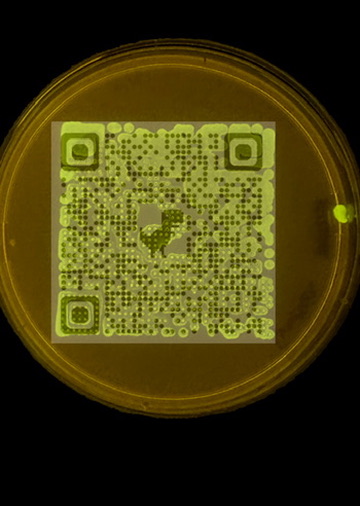
Python protocol for the Opentrons OT-2 that deposits fluorescent bacteria to reproduce the T-Rex QR code on a black agar plate. Black pixels in the 33×33 grid are mapped to plate coordinates; one color (Green) is used for the monochrome design.


Protocol Code Block 1 — Main Protocol

Copy this into the first code block of the HTGAA26 Opentrons Colab notebook (after the prerequisite setup):

from opentrons import types

metadata = {
    'author': 'James Utley',
    'protocolName': 'T-Rex QR Code Bio-Art',
    'description': 'Deposits fluorescent E. coli to create a scannable QR code with T-Rex on black agar',
    'source': 'HTGAA 2026 Opentrons Lab',
    'apiLevel': '2.20'
}

##############################################################################
###   Robot deck setup constants - don't change these
##############################################################################

TIP_RACK_DECK_SLOT = 9
COLORS_DECK_SLOT = 6
AGAR_DECK_SLOT = 5
PIPETTE_STARTING_TIP_WELL = 'A1'

well_colors = {
    'A1': 'Red',
    'B1': 'Green',
    'C1': 'Orange'
}

# 33x33 QR code pixel grid (1 = black/deposit, 0 = white/skip)
# Extracted from T-Rex QR code image
QR_PIXELS = [
    [0, 0, 0, 0, 0, 0, 0, 0, 0, 0, 0, 0, 0, 0, 0, 0, 0, 0, 0, 0, 0, 0, 0, 0, 0, 0, 0, 0, 0, 0, 0, 0, 0],
    [0, 0, 0, 0, 0, 0, 0, 0, 0, 0, 0, 0, 0, 0, 0, 0, 0, 0, 0, 0, 0, 0, 0, 0, 0, 0, 0, 0, 0, 0, 0, 0, 0],
    [0, 0, 0, 0, 0, 0, 0, 0, 0, 0, 0, 0, 0, 0, 0, 0, 0, 0, 0, 0, 0, 0, 0, 0, 0, 0, 0, 0, 0, 0, 0, 0, 0],
    [0, 0, 0, 1, 1, 1, 1, 1, 0, 0, 0, 0, 0, 0, 0, 0, 0, 0, 0, 0, 0, 0, 0, 0, 0, 1, 1, 1, 1, 1, 0, 0, 0],
    [0, 0, 0, 1, 0, 1, 0, 1, 0, 0, 0, 1, 0, 1, 0, 0, 0, 0, 1, 1, 0, 1, 0, 0, 0, 1, 0, 1, 0, 1, 0, 0, 0],
    [0, 0, 0, 1, 1, 1, 1, 1, 0, 0, 1, 1, 0, 0, 1, 1, 0, 0, 1, 1, 0, 1, 0, 0, 0, 1, 1, 1, 1, 1, 0, 0, 0],
    [0, 0, 0, 1, 0, 1, 0, 1, 0, 0, 0, 0, 0, 1, 0, 0, 0, 0, 0, 1, 0, 0, 0, 0, 0, 1, 0, 1, 0, 1, 0, 0, 0],
    [0, 0, 0, 1, 1, 1, 1, 1, 0, 0, 1, 0, 0, 1, 0, 0, 1, 0, 0, 1, 0, 0, 1, 0, 0, 1, 1, 1, 1, 1, 0, 0, 0],
    [0, 0, 0, 0, 0, 0, 0, 0, 0, 0, 1, 0, 0, 1, 1, 1, 1, 1, 0, 1, 0, 0, 1, 0, 0, 0, 0, 0, 0, 0, 0, 0, 0],
    [0, 0, 0, 0, 0, 1, 0, 0, 1, 0, 0, 0, 0, 0, 0, 0, 0, 0, 0, 1, 0, 0, 0, 0, 1, 0, 0, 0, 0, 0, 0, 0, 0],
    [0, 0, 0, 0, 0, 0, 0, 1, 0, 0, 1, 0, 0, 1, 1, 0, 1, 0, 1, 0, 0, 1, 0, 0, 1, 1, 0, 1, 0, 1, 0, 0, 0],
    [0, 0, 0, 1, 1, 0, 1, 0, 0, 0, 0, 0, 0, 0, 0, 1, 0, 1, 0, 1, 0, 0, 1, 0, 1, 1, 1, 1, 0, 1, 0, 0, 0],
    [0, 0, 0, 0, 0, 0, 0, 0, 0, 0, 0, 0, 0, 0, 0, 0, 0, 0, 0, 0, 0, 0, 0, 0, 0, 0, 0, 0, 0, 0, 0, 0, 0],
    [0, 0, 0, 0, 1, 1, 1, 1, 1, 0, 0, 0, 0, 0, 0, 0, 0, 1, 1, 0, 0, 1, 0, 0, 0, 1, 1, 1, 1, 1, 0, 0, 0],
    [0, 0, 0, 0, 1, 1, 1, 0, 1, 0, 1, 0, 0, 0, 0, 0, 0, 1, 1, 0, 0, 0, 0, 0, 1, 1, 0, 1, 1, 1, 0, 0, 0],
    [0, 0, 0, 0, 0, 0, 0, 0, 0, 0, 0, 0, 0, 0, 0, 0, 0, 1, 1, 0, 0, 0, 0, 0, 0, 0, 0, 0, 0, 0, 0, 0, 0],
    [0, 0, 0, 0, 1, 1, 1, 1, 0, 0, 1, 1, 0, 0, 0, 1, 1, 1, 0, 0, 0, 1, 1, 0, 1, 1, 1, 0, 1, 1, 0, 0, 0],
    [0, 0, 0, 0, 1, 0, 1, 0, 0, 0, 0, 1, 0, 0, 1, 1, 1, 1, 0, 0, 0, 1, 0, 0, 1, 1, 1, 0, 0, 1, 0, 0, 0],
    [0, 0, 0, 0, 0, 0, 0, 0, 0, 0, 0, 0, 0, 0, 0, 1, 1, 0, 0, 0, 0, 0, 0, 0, 0, 0, 0, 0, 0, 0, 0, 0, 0],
    [0, 0, 0, 1, 0, 0, 0, 1, 0, 0, 1, 1, 0, 0, 0, 0, 0, 0, 0, 0, 0, 1, 0, 0, 0, 0, 0, 1, 0, 0, 0, 0, 0],
    [0, 0, 0, 0, 0, 0, 0, 0, 0, 0, 1, 1, 0, 0, 0, 0, 0, 0, 0, 0, 0, 1, 0, 0, 1, 0, 0, 1, 0, 0, 0, 0, 0],
    [0, 0, 0, 0, 0, 0, 0, 0, 0, 0, 0, 0, 0, 0, 0, 0, 0, 0, 0, 0, 0, 0, 0, 0, 0, 0, 0, 0, 0, 0, 0, 0, 0],
    [0, 0, 0, 1, 0, 1, 0, 1, 1, 0, 1, 0, 0, 1, 1, 0, 0, 0, 0, 0, 0, 1, 0, 0, 1, 1, 1, 1, 0, 1, 0, 0, 0],
    [0, 0, 0, 1, 0, 1, 0, 0, 0, 0, 0, 1, 0, 1, 0, 0, 0, 0, 0, 1, 0, 0, 0, 0, 1, 0, 0, 1, 0, 0, 0, 0, 0],
    [0, 0, 0, 0, 0, 0, 0, 0, 0, 0, 0, 0, 0, 0, 0, 0, 0, 0, 0, 0, 0, 0, 0, 0, 0, 0, 0, 0, 0, 0, 0, 0, 0],
    [0, 0, 0, 1, 1, 1, 1, 1, 0, 0, 0, 1, 0, 1, 1, 1, 1, 1, 0, 0, 0, 1, 1, 0, 0, 1, 1, 0, 0, 1, 0, 0, 0],
    [0, 0, 0, 1, 0, 0, 0, 1, 0, 0, 0, 1, 0, 1, 0, 0, 0, 0, 1, 0, 0, 1, 0, 0, 0, 0, 0, 0, 0, 0, 0, 0, 0],
    [0, 0, 0, 1, 1, 1, 1, 1, 0, 0, 0, 0, 0, 0, 0, 0, 0, 0, 0, 0, 0, 0, 0, 0, 0, 0, 0, 0, 0, 0, 0, 0, 0],
    [0, 0, 0, 1, 1, 1, 1, 1, 0, 0, 0, 1, 0, 0, 0, 1, 0, 0, 1, 0, 0, 1, 0, 0, 1, 0, 1, 0, 0, 1, 0, 0, 0],
    [0, 0, 0, 1, 0, 0, 0, 1, 0, 0, 1, 1, 0, 1, 1, 0, 1, 0, 0, 0, 0, 0, 0, 0, 1, 1, 0, 0, 0, 1, 0, 0, 0],
    [0, 0, 0, 0, 0, 0, 0, 0, 0, 0, 0, 0, 0, 0, 0, 0, 0, 0, 0, 0, 0, 0, 0, 0, 0, 0, 0, 0, 0, 0, 0, 0, 0],
    [0, 0, 0, 0, 0, 0, 0, 0, 0, 0, 0, 0, 0, 0, 0, 0, 0, 0, 0, 0, 0, 0, 0, 0, 0, 0, 0, 0, 0, 0, 0, 0, 0],
    [0, 0, 0, 0, 0, 0, 0, 0, 0, 0, 0, 0, 0, 0, 0, 0, 0, 0, 0, 0, 0, 0, 0, 0, 0, 0, 0, 0, 0, 0, 0, 0, 0],
]


def run(protocol):
  ##############################################################################
  ###   Load labware, modules and pipettes
  ##############################################################################

  tips_20ul = protocol.load_labware('opentrons_96_tiprack_20ul', TIP_RACK_DECK_SLOT, 'Opentrons 20uL Tips')
  pipette_20ul = protocol.load_instrument("p20_single_gen2", "right", [tips_20ul])
  temperature_module = protocol.load_module('temperature module gen2', COLORS_DECK_SLOT)
  temperature_plate = temperature_module.load_labware('opentrons_96_aluminumblock_generic_pcr_strip_200ul', 'Cold Plate')
  color_plate = temperature_plate
  agar_plate = protocol.load_labware('htgaa_agar_plate', AGAR_DECK_SLOT, 'Agar Plate')
  center_location = agar_plate['A1'].top()
  pipette_20ul.starting_tip = tips_20ul.well(PIPETTE_STARTING_TIP_WELL)

  ##############################################################################
  ###   Helper functions
  ##############################################################################

  def location_of_color(color_string):
    for well, color in well_colors.items():
      if color.lower() == color_string.lower():
        return color_plate[well]
    raise ValueError(f"No well found with color {color_string}")

  def dispense_and_detach(pipette, volume, location):
    above_location = location.move(types.Point(z=location.point.z + 5))
    pipette.move_to(above_location)
    pipette.dispense(volume, location)
    pipette.move_to(above_location)

  ##############################################################################
  ###   T-Rex QR Code patterning
  ##############################################################################

  PIXEL_SIZE_MM = 2.0   # spacing between pixels (mm)
  VOLUME_UL = 0.5       # 0.5 uL per drop (~117 uL total, fits in 200uL PCR strip well)
  COLOR = 'Green'

  # Collect (row, col) for all black pixels
  black_pixels = []
  for row in range(len(QR_PIXELS)):
    for col in range(len(QR_PIXELS[row])):
      if QR_PIXELS[row][col] == 1:
        black_pixels.append((row, col))

  # One tip, one color - aspirate in batches of 20uL (max pipette volume)
  pipette_20ul.pick_up_tip()
  source = location_of_color(COLOR)

  for i in range(0, len(black_pixels), 20):
    batch = black_pixels[i:i+20]
    vol = min(len(batch) * VOLUME_UL, 20)
    pipette_20ul.aspirate(vol, source)

    for row, col in batch:
      # Map pixel (row,col) to plate coords: center at (16,16), 2mm per pixel
      x_mm = (col - 16) * PIXEL_SIZE_MM
      y_mm = (row - 16) * PIXEL_SIZE_MM
      loc = center_location.move(types.Point(x=x_mm, y=y_mm))
      dispense_and_detach(pipette_20ul, VOLUME_UL, loc)

  pipette_20ul.drop_tip()

Protocol Code Block 2 — Simulation / Visualization

Copy this into the second code block (runs after the first):

# Execute Simulation / Visualization -- don't change this code block
protocol = OpentronsMock(well_colors)
run(protocol)
protocol.visualize()

Notes

  • 233 black pixels — uses ~117 µL total (0.5 µL per drop); aspirated in batches of 20 µL; fits in 200 µL PCR strip well.
  • Single color (Green) — monochrome design; change COLOR to 'Red' or 'Orange' if desired
  • 2 mm pixel spacing — fits within ~64 mm on a 90 mm plate
  • One tip per color — avoids cross-contamination; only one color used here

Standalone Protocol File

For Opentrons App validation or direct upload: trex_qr_protocol.py

Google Colab Notebook

HTGAA26 Opentrons Colab — make a copy and paste the protocol code blocks above into the notebook.

Subsections of Projects

Individual Final Project: BioVolt - DIY Electroporation Device

Project Overview: BioVolt - DIY Electroporation Device & Full Transformation Pipeline

Biological engineering application/tool to develop:
BioVolt is a portable, ultra-low-cost DIY electroporation device (~$10-20 in parts) that uses a piezoelectric crystal from a barbecue lighter to generate ~2,000 V pulses for temporary cell membrane permeabilization. This enables DNA/RNA uptake in bacteria (e.g., E. coli), yeast, plant protoplasts, or even stem cells for genetic transformation. Inspired by the DEFCON 32 talk “You got a lighter I need to do some Electroporation” (presented by Dr. James Utley (Me), Phil Rhodes, and Josh Hill from Viva Securus/Syndicate Laboratories), it builds on frugal biohacking principles: piezoelectric trigger pulsing, custom microfluidic cuvettes from aluminum tape/magnets/glass slides, and simple high-voltage testing.

DEFCON 32 Presentation — Where It Started for me

At DEFCON 32 the talk I presented focused on the device itself — proving that a barbecue lighter’s piezoelectric crystal could generate sufficient voltage to temporarily permeabilize cell membranes for DNA uptake. The talk covered design details, demos, troubleshooting (e.g., arc gap tuning with Post-it notes), and the biohacking ethos behind building a ~$10 electroporator.

Key highlights from the talk: ~2,000 V pulses via lighter clicks, high cell mortality (50-70%) but viable transformants, GFP reporter demos, open protocols encouraged.

Next Phase: End-to-End Pipeline with Efficiency Focus

The next phase of BioVolt moves beyond the device and brings the entire workflow end to end, with a focus on efficiency and frugal validation. The goal: take a piezoelectric electroporator built from a barbecue lighter and prove — through a full pipeline — that it actually works. The pipeline includes:

  1. Plasmid amplification via thermal cycling — Before electroporation, the initial plasmid source will be amplified using the MJ Research PTC-100 thermal cycler (Peltier-effect programmable controller) available in the lab. This ensures sufficient plasmid DNA concentration for transformation.

  2. DNA concentration measurement — Using the Rodeo open colorimeter (visible light version for OD600 cell density measurements) and, if possible, the UV version for DNA concentration quantification. This provides pre- and post-transformation metrics.

  3. Electroporation — Transformation of cells with the amplified plasmid DNA using the BioVolt piezoelectric device, followed by recovery and plating.

  4. Post-transformation PCR verification — For good measure, PCR will be run after transformation using the same thermal cycler to check whether the insert is present in the recovered cells. This triangulates and correlates with plating results to provide a hasty “close enough” frugal validation.

  5. Gel electrophoresis confirmation — Agarose gel electrophoresis to visualise PCR products and verify successful transformation (e.g., presence of reporter genes like GFP via band patterns under UV).

The aim is to triangulate multiple data points — plasmid amplification, colorimetric/UV measurement, transformation plating, and post-transformation PCR — to build confidence that the piezo electroporator from a lighter actually delivers. Fingers crossed, this provides a credible, frugal, end-to-end validation of a DIY electroporation workflow.

This democratizes synthetic biology for education, citizen science, and personal biohacking in resource-limited settings.

Lab Setup & Tools in Action

My biohacker lab at Syndicate Laboratories integrates the device with the full verification pipeline.


Project Timeline & Milestones

This section will be updated throughout the semester as the project progresses.

Completed Milestones

  • DEFCON 32 presentation and initial device concept
  • Basic proof-of-concept: piezoelectric voltage generation
  • Initial transformation experiments with GFP reporter

Current Phase (Week 1-4)

  • Acquire and test MJ Research PTC-100 thermal cycler
  • Optimize plasmid amplification protocols
  • Set up Rodeo colorimeter for OD600 measurements
  • Source UV colorimeter module for DNA quantification

Future Milestones

  • Complete end-to-end pipeline testing
  • Document reproducibility across multiple transformation attempts
  • Optimize efficiency and reduce cell mortality
  • Create comprehensive open-source protocol documentation
  • Final presentation and demonstration

Documentation & Resources

Project documentation will be added here as the project develops throughout the semester.

  • Week 1 homework includes initial governance assessment
  • Future updates will include protocols, data, images, and results
  • Final documentation will include complete build instructions and validation data

Project Status: In Progress (Week 1)
Location: Syndicate Laboratories, Panama City, Panama
Researcher: James Utley, PhD

Group Final Project

cover image cover image