Individual Final Project
SynStromatolite
Abstract
Stromatolites are pillars of mineralized biofilm laid down by communities of photosynthetic bacteria. The dominant visible evidence of life on Earth for billions of years, stromatolites now only form in environments like hyper-saline water that exclude multi-cellular animals. Like tree rings, the records left in the layers of ancient stromatolites can be used to infer information about their environment like hypersalinity (Zhu et al., 2021) or fact that the Earth was spinning faster 2.5 billion years ago and an Earth year was 435 days instead of 365 ([(Vanyo & Awramik, 1985)(#vanyo1985)]). A synthetically engineered consortia that mimics the layer formation ability of stromatolites enables the development of low cost self-powered biological biosensors that record a time series of their outputs as mineralized biofilm layers. [Combining with existing water quality biosensors can help address water quality health problems in resource constrained areas] As a feasibility demonstration this projects engineers a two-strain Esceheriichia coli consortium that that use quorum-triggered curli amyloid secretion to lay down biofilm layers with enhanced mineralization capabilities. The projects’s central hypothesis is that separating biofilm generation and reporting responsibilities across multiple strains will be superior to trying to put all the functions in a single organism in terms of growth (as measured by OD900 curves), biofilm production (as measured by a Congo Red depletion assay), sensor signal strength (as measured by 590nm absorption), and mineralization (as measured by a calcium depletion precipitation assay). The project also lays out a road map to further develop this consortia into a deployable biosensor recorder platform.
Project Aims
Aim 1: Consortia With Triggered Biofilm Formation
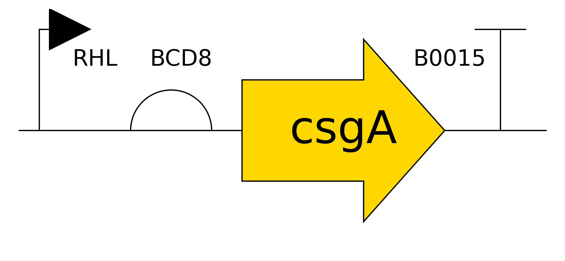
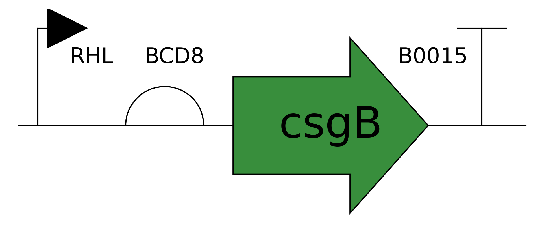
Design and assemble a consortia of two Esceheriichia coli strains where one strain (the trigger strain) specializes in producing sensor output and triggering biofilm layer formation while they other strain(builder strain) specializes in producing the majority of the mineralizable protein mass that makes up the biofilm layers. Create the two E. coli strains by separately introducing engineered trigger and builder plasmids into the BL21 E. Coli chassis using chemical transformation. Produce both plasmids using Golden Gate assembly of parts plasmids compatible with the JUMP (Valenzuela-Ortega & French, 2021) assembly vectors in the igem 2025 distribution kit. Design custom JUMP-compatible DNA for curlin biofilm and quorum-signaling proteins that are missing in the igem 2025 kit by downloading amino acid sequences from Uniprot and using Twist Biosciences to generate DNA vectors that are JUMP compatible and optimized for E. Coli.
Validate that the consortia functions and outperforms alternative implementations in growth, biofilm production, sensor signal strength, and mineralization activity. Evaluate growth of cultures by using spectrophotometer to measure optical density with infrared (900 nm) light to avoid absoprtion by the sensor color signal. Evaluate biofilm production by depletion of Congo Red via absorption of red (490nm) light in supernatant of culture exposed to a known concentration of Congo Red. Evaluate the strength of sensor signals by absorption of purple light (580 nm). Evalute mineralization activity by depletion of calcium ions from supernatant exposed to a known concentration of calcium chloride using an analytical balance to measure the mass of precipitate formed with excess sodium carbonate.
Aim 2: Long-Term Reliable Biosensor Recording in Biofilm Layers
Optimize the function of the consortia and add features required to support field deployment by adding biosensor integration, reliable layer generation, recording durability, and autonomous power generation.
Optimize Core Functions
Leverage lab automation and the modular nature of the Golden Gate assembly to optimize the four basic functions by varying promoter and RBS strength.
Biosensor Integration
Replace the tsPurple biosensor proxy in the prototype with a true biosensor that detects a condition and switches color of biofilm layer being formed depending on the current state it detects. Validate sensor response strength in biofilm layer by measuring relflectance at relevant wavelength. Ensure the consortia can support multiple different types of water quality biosensors by developing multiple variants with biosensors for water-borne pathogens like cholera quorum sensing molecules and toxic pollutants like arsenic. Using a remote cloud lab will again be important for this work as the community lab will not have access to chemicals to run toxin detection experiments.
Reliable Layer Generation
Ensure the consortia lays down distinct layers of biofilm results by engineering a detection cycle in the consortia that has distinct stages: growth -> sensor trigger -> biofilm production -> settling and layer formation -> reset stage -> growth [MISSING PICTURE]. Validate the sensor cycle by introducing trigger chemicals at known times and concentrations and validating the sensor color response by in situ reflectance of the biofilm at the appropriate wavelength. To avoid cross-cycle interference, add additional components to the consortia that clear the medium of the biosensor chemical signal during the reset stage by degradation or sequestration in the biofilm layers.
Layer Durability
Ensure recording layers survive typical shipment handling after separation from growth medium, drying, and placement in a box. Measure samples with thirty or more layers of biofilm records under representative compressive, shearing, and tensile loads. If required add consortia members to increase structural integrity of recording layers. For example add cellulose generating genes to the consortia or add an anaerobic bacteria that live in the lower layers and secrete urease to create a more favorable environment for biomineralziation by raising the pH.
Autonmous Power
Support long term operation by adding a photosynthetic member to consortia that has been engineered to secrete sucrose to provide power to the rest of the consortia. Validate integration by comparing photosynthetic consortia growth, biofilm production, and sensor signal growing on minimal mineral media like BG-11 to the results from non-photosynthetic consortia growing on LB broth over one month with the expectation that the photosynethetic consortia performs at a consistent level for weeks and outperforms the hetrotrophic consortia in biofilm generation and sensor signal beyond the first few days of operation.
Aim 3: Deployment for Water-Quality Monitoring in Resource Constrained Environments
Package the synthetic stromatolite consortia into a hydrogel container (Tang et al., 2021 and field deploy a water quality recording system into resource constrained environments. The containment capsule can keep the consortium contained while allowing small molecules (less than 22 nm) to traverse the permeable membrane and interact with the sensor. The sensors can give immediate warning to people that there water supply is tainted and at the same time provide an inexpensive way for public health officials to gather comprehensive records of water quality across households, water systems, and watersheds.
The two main evaluation metrics for this are ease of use and cost. End user usage should be as simple as:
Float the sensor on top of the water
Leave the sensor in partial sunlight
If the bottom of the sensor is red the water is currently bad
Sensors should operate for at least a month
Sensor turns black when expended
Domestic Water Quality Monitoring 
Time series collection of water quality by public health officials should be as simple as
Collect expended sensors
Extract biofilm record disk
Break disk in half and take picture with digital camera on phone
Upload picture, geo-location, and sensor id to data collection and analysis app
If available random sample of biofilm disks to lab for microscopic and chemical evaluation for more detailed information.
Watershed Water Quality Monitoring 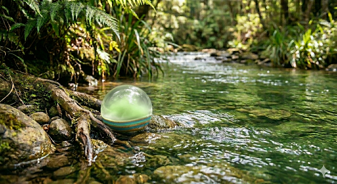
Capturing Water Quality Time Series 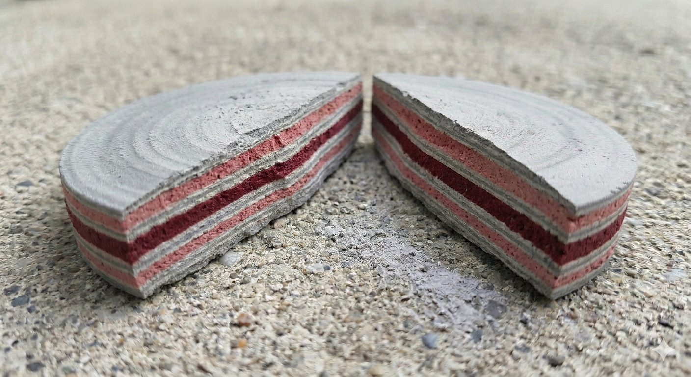
Section 3: Background
The main structural component of E. coli biofilms is curlin protein. The curlin protein system was characterized in (Chapman et al., 2002) and (Robinson et al., 2006) as:
- csgA Main structural amyloid protein that makes up 95% of the E. coli biofilm protein.
- csgB Causes csgA to condense into stacked amyloid sheets.
- csgC A helper protein that prevents the csgA from prematurally folding before it leaves the cell
- csgE A helper protein that helps move csgA and csgB from the cytoplasm to the cell membrane
- csgG A membrane protein that creates pores for the csgA and csgB to exit the cell
- csgF An exterior membrane protein that helps csgA and csgB exit the cell and acts as an anchor point for csgB to bind to the cell membrane.
Modified curlin csgA proteins with histadine tails that can bind to metal have been used to manufacture quantum dots (Chen et al., 2014). This project combines this approach with the observations from mollusks tha amyloid sheets with aspartic acid regions promote biomineralization by attracting calcium ions (Weiner & Hood, 1975).
There are other synthetic biology approaches to recording time series of biosensor readins. Most work in this area builds on George Church’s amazing idea to encode biosensor readings as a time series in the organism’s DNA using Cripsr (Shipman et al., 2017). Using marine biofilms to monitor water quality is a good example of this approach (Nevot et al., 2025). The DNA time series approach gives tight control and digital precision, however it requires extraction of DNA and sequencing which are not generally available in the resource constrained environments that most need ongoing water quality monitoring.
There are also incredible low cost water quality monitoring solutions intended for resource constrained environments. My favorite is the baker’s yeast that turns red when exposed to Cholerla in a water sample (Yudina et al., 2015). In theory this solution should be deployable for pennies per use and solve the problem of cholerla detection in resource constrained environments, however direct addition t well water can trigger the Cartegna Protocol as a release of genetically modified organisms. Avoiding release of genetic modified organsims for water quality biosensors requires cell-free systems or more sophisticated packaging (Siegfried et al., 2012). While the cost of more sophisticated packaging is only a few dollars per use that amount can still be prohibitive for resource constrained environments. Synthetic stromatolites have the same packaging problem but amortize the packaging cost over months of continuous usage reducing the cost of daily usage to a few cents per day. The fact that the same sensor can be used for daily preventive monitoring by distributed users and for time series monitoring by centralized public health officials should also defray costs and boost availability.
The existing cost and labatory requirements of water quality monitoring sensors often limits usage in resource constrained environments to public health officials reactive remediation after a water-borne outbreak or pollution event is severe enough to be noticed by community health outcomes. Cholera alone accounted for more than 600,000 reporteed cases and almost 8000 deaths in 2025 (WHO, 2026). Widespread, low cost, and easy to operate preventive monitoring of water quality pathogens and toxins sensors could repalce reactive remediation to proactive surveillance and improve community health in resource constrained environments.
Given the that the target outcome is improvement in resource constrainted environments a key ethical concern is local empowerment and access. An aproach that is imposed from afar or at substantial financial cost is not a solution. To address this the project should be open and patent free, so that it can be produced and adapted by communities as needed. Furthemore the final sensor package and consortia should be designed to enable production of the sensors is possible as close as possible to where they are deployed without needing sophisticated or expensive equipment.
The other ethical consideration that applies to any genitically modified organsim is containment risk. To avoid risk to the communities using the biosensor there needs to be a multi-level safeguards preventing release. Packaging is the first line of defense. In addition the consortia should be engineered with a built in timed kill switch and mutual dependencies between the the different consortia elements, to make long term survival after an accidentl release unlikely.
Experimental Design
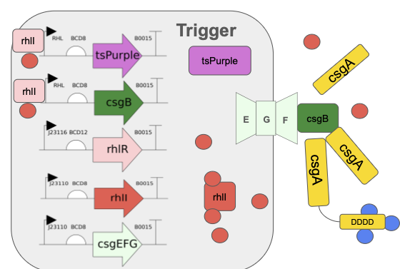
Step 1 — DNA Design: Trigger Strain Plasmid
Purpose: Define the complete genetic architecture of the trigger strain.
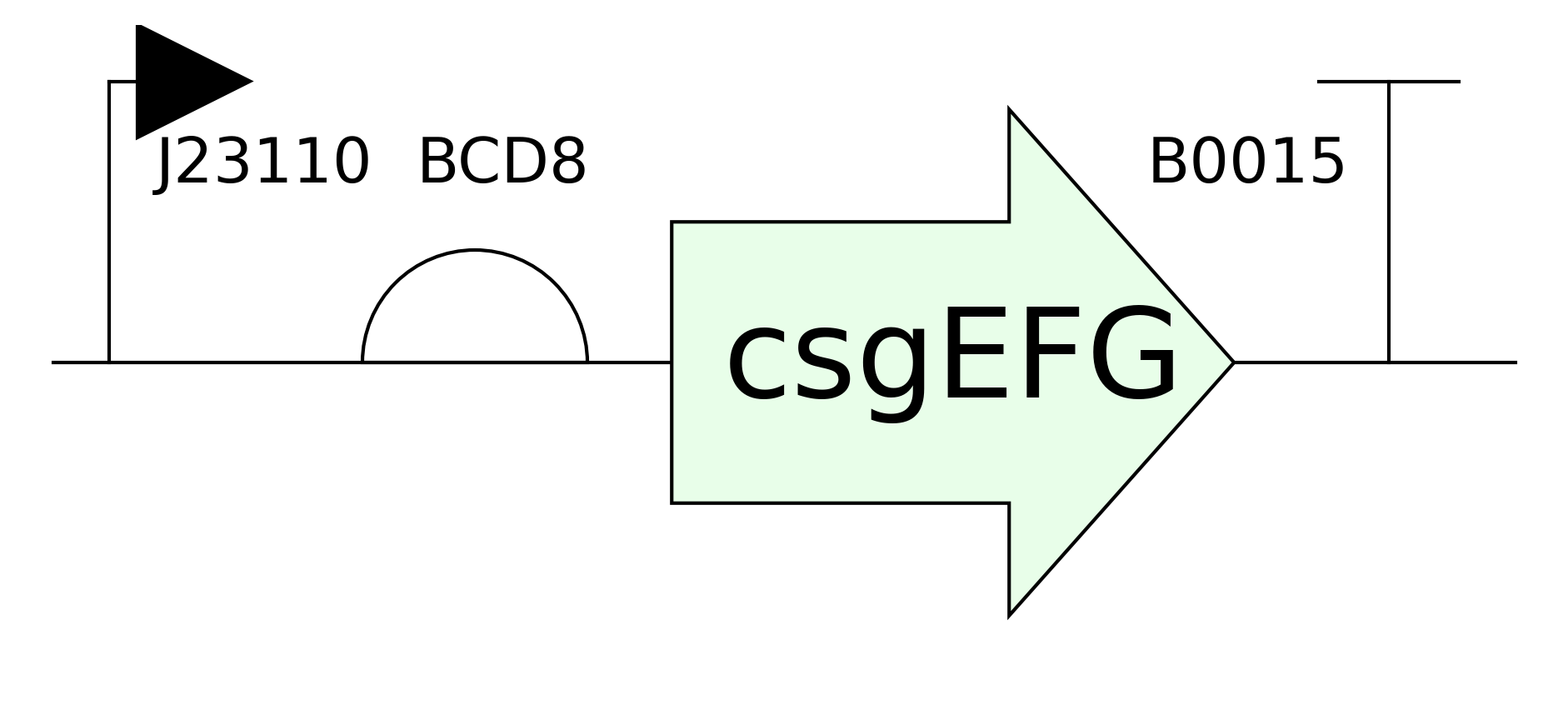
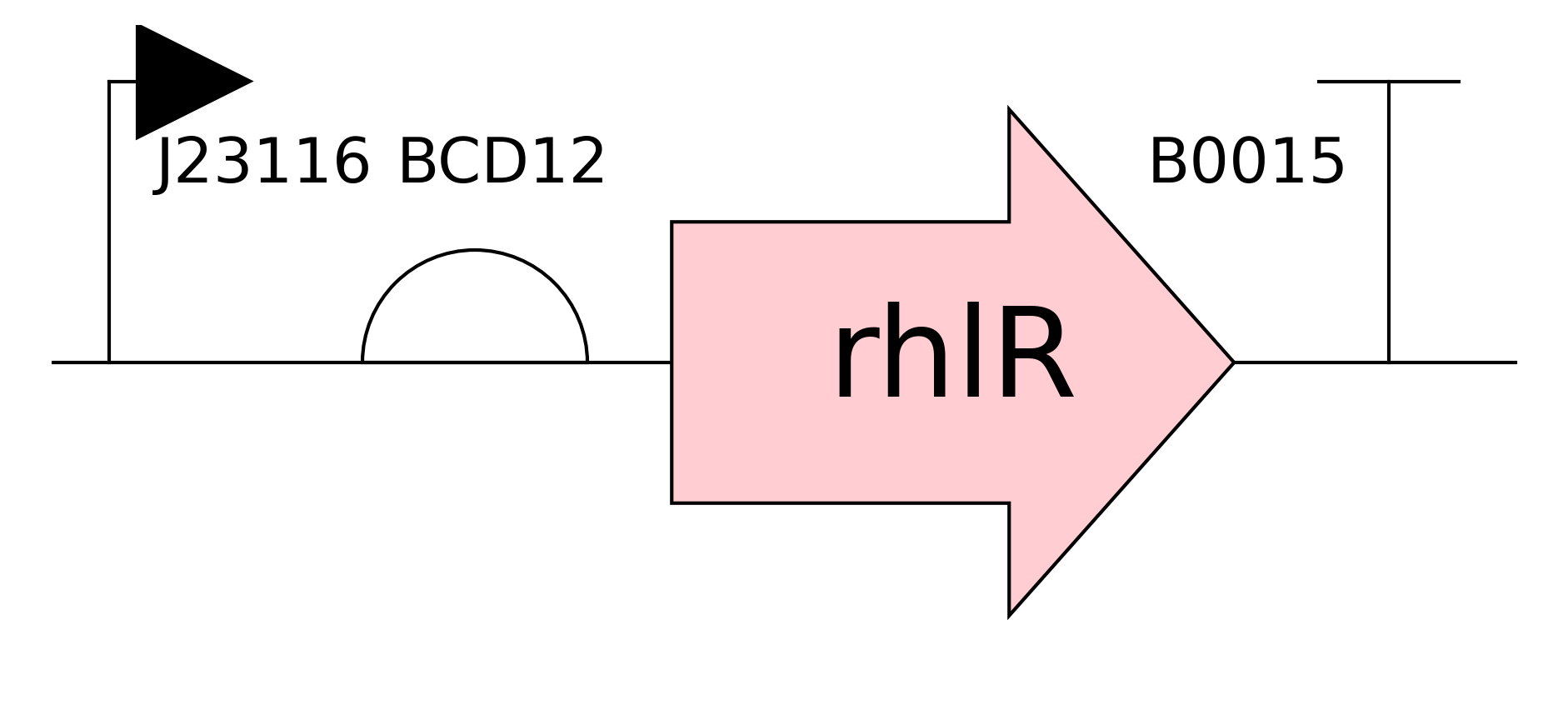
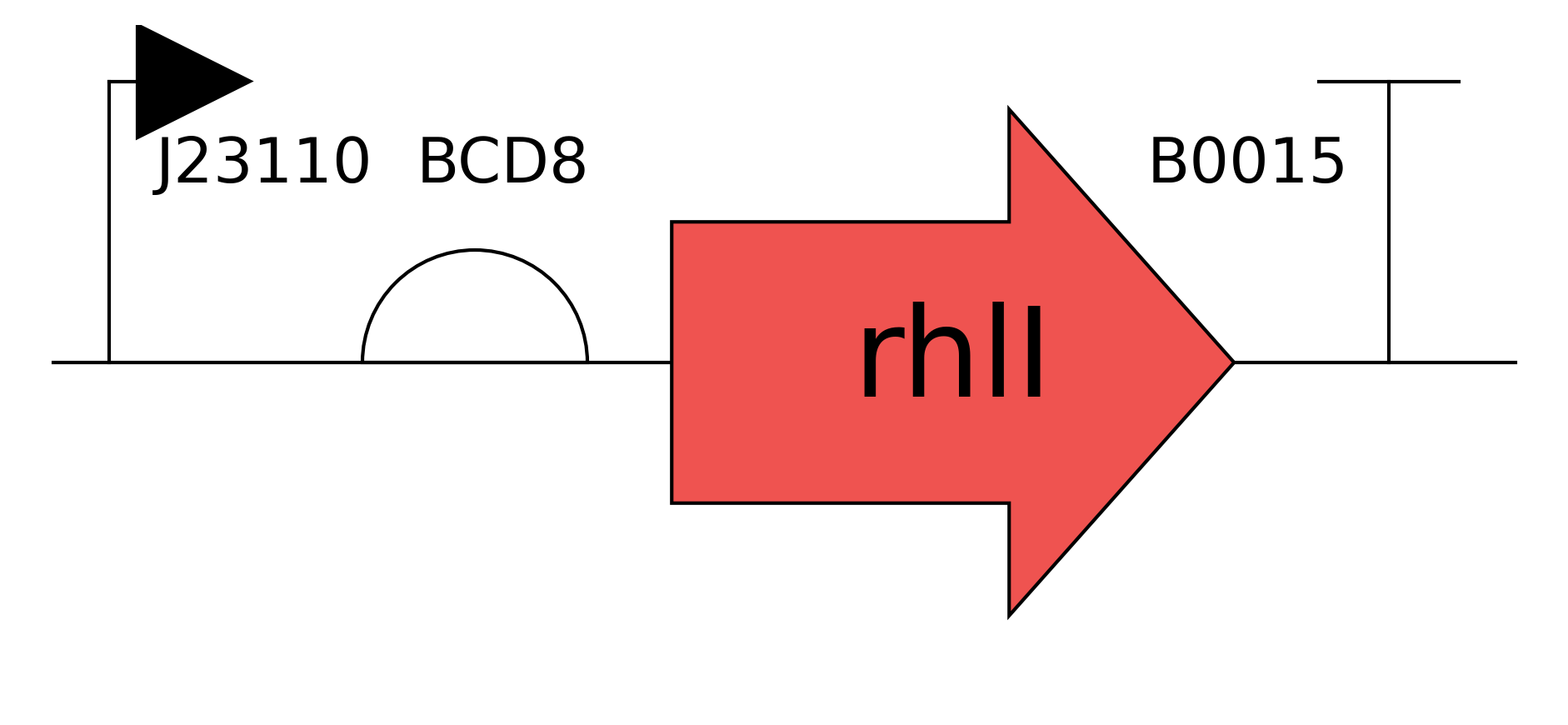
Method: Design a single multi-TU plasmid encoding: (1) rhlI TU [J23110 → BCD8 → rhlI → B0015], (2) rhlR TU [J23116 → BCD12 → BBa_C0070 → B0015], (3) csgB TU [Prhl_NM → BCD8 → csgB → B0015], (4) tsPurple TU [Prhl_NM → BCD1 → tsPurple → B0015], (5) csgEFG TU [J23110 → BCD8 → csgEFG → B0015]. Assembly via MoClo/JUMP Golden Gate using iGEM 2025 distribution kit parts; csgB, rhlI, and csgEFG genes ordered from Twist Bioscience as gene strings.
Automation: Manual assembly at community biolab (Aim 1).
Plate: Standard 1.5 mL microtubes for Golden Gate reactions.
Expected Result: Correct assembly confirmed by colony PCR and Sanger sequencing.
Timeline: Weeks 1–2.
Step 2 — DNA Design: Builder Strain Plasmid
Purpose: Define the complete genetic architecture of the builder strain.
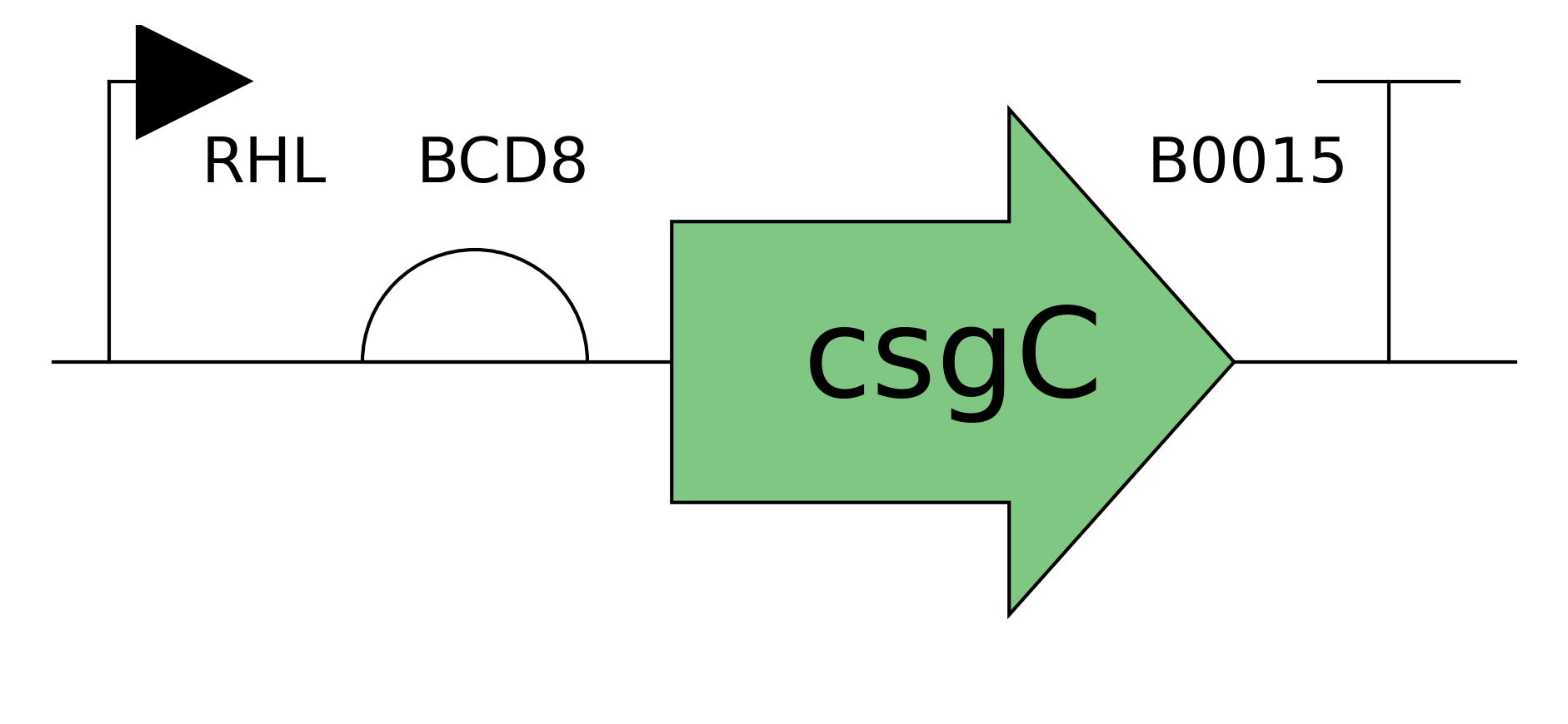
Method: Design a single multi-TU plasmid encoding: (1) rhlR TU [J23116 → BCD12 → BBa_C0070 → B0015], (2) csgC TU [J23116 → BCD12 → csgC → B0015], (3) csgA-Asp TU [Prhl_NM → BCD1 → csgA-Asp-tail → B0015], (4) csgEFG TU [J23110 → BCD8 → csgEFG → B0015]. Two versions of the glue strain plasmid will be made: one with csgA-Asp-tail and one with unmodified csgA, for direct comparison. csgC, csgA variants, and csgEFG ordered from Twist Bioscience.
Automation: Manual assembly at community biolab.
Plate: Standard microtubes.
Expected Result: Two confirmed glue strain plasmids differing only at the csgA CDS.
Timeline: Weeks 1–2.
Twist Bioscience Order Summary:
csgB(E. coli K-12, codon-optimized) — gene stringrhlI(P. aeruginosa PAO1, codon-optimized for E. coli) — gene stringcsgEFGoperon (E. coli K-12) — gene stringcsgC(E. coli K-12) — gene stringcsgAunmodified (E. coli K-12) — gene stringcsgA-Asp-tail: csgA + (Gly₄Asp)₆ C-terminal tail — gene string (custom design)
Translation Units
All synthesized genes will be ordered from Twist Bioscience as gene strings with flanking MoClo BsaI Golden Gate overhangs compatible with the iGEM JUMP assembly standard.
Step 3 — Golden Gate Assembly
Purpose: Assemble multi-TU plasmids from iGEM parts and Twist gene strings.
Method: MoClo/JUMP Golden Gate reactions using BsaI-HFv2 (NEB), T4 DNA ligase, thermocycler cycling protocol (37°C/16°C × 30 cycles). Transform into E. coli DH5α, plate on selective agar.
Automation: Manual at community biolab; ATC Thermal Cycler equivalent used for cycling.
Plate: PCR tubes / 96-well PCR plate.
Expected Result: >10 correct colonies per assembly; correct insert confirmed by colony PCR.
Timeline: Week 2.
 Assembled Trigger Strain In Action
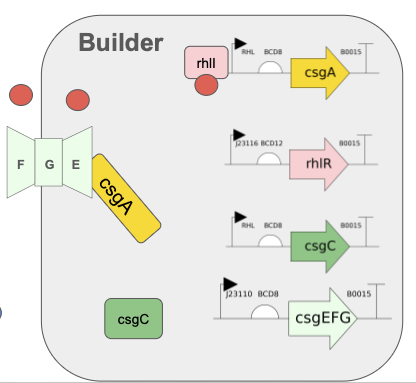 Assembled Builder Strain In Action
Step 4 — Sequence Verification
Purpose: Confirm all TUs are correctly assembled and no mutations introduced.
Method: Sanger sequencing of all five TU junctions per plasmid using standard M13 and custom sequencing primers. Submit to community biolab sequencing service or Plasmidsaurus.
Expected Result: 100% sequence identity to design across all TU junctions.
Timeline: Week 3.
Step 5 — Strain Construction and Glycerol Stocks
Purpose: Generate working bacterial strains carrying each plasmid.
Method: Transform verified plasmids into E. coli BL21(DE3) or MG1655 (curli-competent background). Confirm by PCR. Prepare glycerol stocks at −80°C.
Expected Result: Stable transformants confirmed; glycerol stocks archived.
Timeline: Week 3.
Step 6 — Congo Red Liquid Assay: Strain Preparation
Purpose: Prepare cultures for curli secretion quantification.
Method: Inoculate csgA-Asp-tail glue strain and unmodified csgA glue strain from glycerol stocks into LB + antibiotic. Grow overnight at 30°C (curli-permissive temperature) with shaking. Dilute to OD₆₀₀ = 0.05 in YESCA medium (yeast extract + casamino acids, low salt) to promote curli expression.
Automation: Manual at community biolab.
Expected Result: Cultures at consistent starting OD for assay normalization.
Timeline: Week 4.
Step 7 — Congo Red Liquid Assay: Binding Measurement
Purpose: Quantify curli fiber secretion by Congo red dye binding.
Method: After 48 h growth at 30°C, pellet cells by centrifugation (5,000 × g, 10 min). Resuspend pellet in PBS + 10 µg/mL Congo red. Incubate 30 min at RT with gentle mixing. Centrifuge again; measure absorbance of supernatant at 490 nm. Congo red depletion from supernatant = curli binding. Include: (1) no-cell blank, (2) wild-type MG1655 positive control, (3) ΔcsgA negative control.
Instrument: Spectrophotometer or plate reader at community biolab (96-well format if available).
Plate: 96-well flat-bottom plate.
Expected Result: csgA-Asp-tail strain shows Congo red binding comparable to or greater than unmodified csgA strain, confirming the tail does not disrupt secretion.
Timeline: Week 4–5.
Example Assay Plate Layout (96-well Congo Red Assay):
Step 8 — Calcium Depletion Assay: Setup
Purpose: Quantify calcium-binding enhancement of CsgA-Asp-tail versus unmodified CsgA.
Method: Grow csgA-Asp-tail and unmodified csgA glue strains under identical YESCA conditions for 48 h at 30°C to allow biofilm and curli formation. Add CaCl₂ to a final concentration of 5 mM to each culture and a sterile medium-only control. Incubate 2 h at RT with gentle mixing.
Expected Result: Cultures with established curli biofilm ready for calcium binding measurement.
Timeline: Week 5.
Step 9 — Calcium Depletion Assay: Measurement
Purpose: Measure calcium removed from solution by biofilm binding.
Method: Filter cultures through 0.22 µm syringe filter to remove cells and biofilm. Add Na₂CO₃ (10 mM final) to filtrate to precipitate remaining free calcium as CaCO₃. Collect precipitate by centrifugation; dry at 60°C overnight; weigh on analytical balance. Calculate: calcium bound = (CaCO₃ precipitate from medium control) − (CaCO₃ precipitate from culture filtrate). Include: (1) sterile medium + CaCl₂ + Na₂CO₃ (total calcium control), (2) unmodified csgA culture, (3) csgA-Asp-tail culture, (4) ΔcsgA culture (background binding).
Instrument: Analytical balance; optional ICP-OES for higher precision if available at community biolab partner institution.
Expected Result: csgA-Asp-tail culture shows significantly less free calcium in filtrate (more calcium bound to biofilm) than unmodified csgA or ΔcsgA controls.
Timeline: Week 5–6.
Step 10 — tsPurple Reporter Validation
Purpose: Confirm RHL quorum-sensing trigger is functioning in the curing strain.
Method: Grow curing strain to OD₆₀₀ = 0.1, 0.5, 1.0, 2.0 (representing increasing cell density and RHL accumulation). Measure absorbance at 570 nm (tsPurple) normalized to OD₆₀₀. Include: (1) curing strain without rhlI (no RHL production, negative control), (2) curing strain + exogenous RHL (positive control for Prhl_NM induction).
Expected Result: tsPurple signal increases with cell density, confirming quorum-dependent activation.
Timeline: Week 6.
Step 11 — Two-Strain Co-culture Biofilm Test
Purpose: Verify that the two-strain consortium produces a co-assembled biofilm.
Method: Mix curing and glue strains 1:1 in YESCA medium. Grow in static culture at 30°C for 48–72 h. Observe biofilm formation visually and by crystal violet staining. Confirm tsPurple color in biofilm by visual inspection and absorbance.
Expected Result: Visible purple-tinted biofilm at air-liquid interface; Congo red staining of biofilm confirms curli presence.
Timeline: Week 7.
Aim 2 — Automated CsgA Variant Library Screen (Ginkgo Bioworks Platform)
Step 12 — CsgA Variant Library Design and Twist Order
Purpose: Generate a panel of CsgA-Asp-tail variants to identify optimal tail length and linker for calcium binding.
Method: Design 8 CsgA variants with aspartate tail lengths of 2, 4, 6, 8, 10, 12 repeats of (Gly₄Asp), plus two linker variants (flexible GGGS vs. rigid EAAAK). Order all 8 as gene strings from Twist Bioscience with flanking MoClo overhangs. Assemble into glue strain backbone using Golden Gate. Sequence-verify all constructs.
Automation: Golden Gate assembly in 96-well PCR plate on ATC Thermal Cycler; transformation and plating manual; colony picking manual or with QPix if available.
Expected Result: 8 confirmed glue strain variants ready for screening.
Timeline: Weeks 8–10.
Step 13 — Automated Culture Setup (Ginkgo Platform)
Purpose: Inoculate all 8 variants plus controls in 96-well format for parallel growth.
Method: Use Tempest liquid handler to dispense 150 µL YESCA + antibiotic per well into 96-round-axygen-pdw11cs-halfdeep plates. Use Bravo-96 to inoculate from glycerol stock dilutions. Seal with Plateloc. Incubate in Cytomat at 30°C, 48 h, with orbital shaking via BioshakeD3000.
Automation: Tempest (media dispensing), Bravo-96 (inoculation), Plateloc (sealing), Cytomat (incubation), BioshakeD3000 (mixing).
Plate: 96-round-axygen-pdw11cs-halfdeep.
Expected Result: Uniform growth across all wells; OD₆₀₀ measured at 48 h to confirm comparable growth.
Timeline: Week 10.
Step 14 — Automated Congo Red Secretion Screen
Purpose: Quantify curli secretion for all 8 CsgA variants in parallel.
Method: After 48 h, centrifuge plates on HiG Centrifuge. Use Bravo-96 to transfer supernatant to new 96-well plate. Use Multiflo to add Congo red reagent (10 µg/mL in PBS) to each well. Incubate 30 min at RT on BioshakeD3000. Centrifuge again. Transfer supernatant to 384 Greiner black-well clear-bottom plate using Echo525 for absorbance reading. Read at 490 nm on Spark Plate Reader.
Automation: HiG Centrifuge, Bravo-96, Multiflo, BioshakeD3000, Echo525, Spark Plate Reader.
Plate: 96-round-axygen-pdw11cs-halfdeep (culture); 384 Greiner black-well clear-bottom (absorbance read).
Expected Result: Ranked secretion efficiency across 8 variants; identify top 2–3 variants for calcium assay follow-up.
Timeline: Week 10–11.
Step 15 — Data Analysis and Variant Selection
Purpose: Identify optimal CsgA-Asp-tail design for Aim 3 integration.
Method: Normalize Congo red depletion values to OD₆₀₀ for each variant. Plot secretion efficiency vs. tail length. Select top-performing variant(s) for calcium depletion assay validation (manual, as in Steps 8–9). Export data from Spark Plate Reader to CSV; analyze in Python/R.
Expected Result: Clear optimum tail length identified; data supports rational design principle for Aim 3 consortium.
Timeline: Week 11–12.
Course Technique Checklist
- DNA design and synthesis (Twist Bioscience gene strings)
- Golden Gate / MoClo assembly
- Bacterial transformation
- Colony PCR and Sanger sequencing
- Promoter engineering (constitutive + quorum-sensing inducible)
- Reporter gene assays (tsPurple absorbance)
- Biofilm assays (Congo red)
- Automated liquid handling (Bravo-96, Tempest, Multiflo)
- Plate reader detection (Spark)
- Automated incubation and plate handling (Cytomat, Plateloc, HiG)
- Quorum sensing circuit design
- Protein engineering (CsgA tail fusion)
- Cell-free expression
- CRISPR
- Flow cytometry
Expanded Technique 1: MoClo/JUMP Golden Gate Assembly
MoClo (Modular Cloning) is a hierarchical Golden Gate assembly standard that uses Type IIS restriction enzymes (BsaI, BbsI) to generate defined 4-bp overhangs, enabling scarless, ordered assembly of multiple transcription units into a single plasmid in a single reaction. The JUMP (Joint Universal Modular Parts) extension used in the iGEM 2025 distribution kit standardizes overhang sequences across promoters, RBSs, CDSs, and terminators, making parts from different contributors directly interchangeable. In this project, MoClo/JUMP enables the assembly of 4–5 TUs per plasmid in a single Golden Gate reaction, dramatically reducing cloning time compared to traditional restriction-ligation or Gibson assembly approaches. The fidelity of the assembly is verified by colony PCR across each TU junction and confirmed by Sanger sequencing, ensuring that the final constructs match the computational design exactly before any biological characterization begins.
Expanded Technique 2: Congo Red Biofilm Secretion Assay
Congo red is an azo dye that binds with high affinity to amyloid fibrils, including curli fibers, through intercalation between the β-sheet stacking of the amyloid structure. In the liquid-phase assay format used here, Congo red depletion from the supernatant after incubation with pelleted cells or biofilm material provides a quantitative measure of curli fiber abundance — more curli means more dye bound and less remaining in solution, detected as a decrease in absorbance at 490 nm. This assay is highly amenable to automation: Congo red reagent can be dispensed by Multiflo, incubation handled by BioshakeD3000, and absorbance read in 384-well format on the Spark Plate Reader, enabling screening of dozens of variants in a single run. Critical controls include a ΔcsgA negative control (background dye binding to cell membranes), a wild-type curli-expressing positive control, and a no-cell blank to establish the maximum possible absorbance signal, allowing calculation of percent Congo red depletion as a normalized secretion metric.
Results & Quantitative Expectations
The primary validation experiment for this project is the calcium depletion precipitation assay comparing the CsgA-Asp-tail variant to unmodified CsgA and a ΔcsgA negative control. This experiment directly tests the central engineering hypothesis — that appending an aspartate-rich tail to CsgA will enhance calcium binding and accelerate calcium carbonate mineralization — and provides a quantitative, instrument-independent readout (mass of precipitate on an analytical balance) that is accessible at a community biolab without specialized equipment.
Step-by-Step Validation Protocol
- Grow csgA-Asp-tail glue strain, unmodified csgA glue strain, and ΔcsgA control strain in YESCA medium at 30°C, 48 h, static culture (6 biological replicates each).
- Add CaCl₂ to 5 mM final concentration to all cultures and to a sterile YESCA medium-only control. Incubate 2 h at RT with gentle rocking.
- Filter all cultures through pre-weighed 0.22 µm syringe filters into pre-weighed 1.5 mL microtubes. Record filter + retained biofilm weight.
- Add Na₂CO₃ to filtrate to 10 mM final concentration. Vortex briefly. Incubate 30 min at RT.
- Centrifuge filtrate at 14,000 × g for 10 min. Carefully remove supernatant by pipette.
- Wash pellet twice with deionized water (centrifuge between washes).
- Dry pellet at 60°C overnight in a heat block or oven.
- Weigh dried pellet on analytical balance. Record mass.
- Calculate: free calcium in filtrate = mass of CaCO₃ precipitate (mg) × (40.08/100.09) = mg Ca²⁺ remaining. Calcium bound by biofilm = total calcium added − free calcium in filtrate.
- Perform one-way ANOVA with Tukey post-hoc test across the three conditions (n=6 per group).
Techniques Used
The calcium depletion assay combines microbiology (biofilm culture), analytical chemistry (gravimetric precipitation), and basic statistics (ANOVA) in a workflow that requires no specialized instrumentation beyond an analytical balance and a centrifuge. The use of sodium carbonate precipitation as a proxy for free calcium is a well-established method in water chemistry and has been adapted here for biofilm calcium-binding quantification. Normalization of calcium binding to biofilm dry weight (obtained from the filter mass difference) controls for variation in biofilm abundance between replicates, ensuring that differences in calcium binding reflect the biochemical properties of the CsgA variant rather than differences in biofilm quantity. The inclusion of a ΔcsgA negative control is essential to distinguish specific curli-mediated calcium binding from non-specific binding to cell membranes, extracellular polysaccharides, or other biofilm matrix components.
Troubleshooting
If no difference is observed between csgA-Asp-tail and unmodified csgA, the most likely explanation is that the aspartate tail is being cleaved post-translationally or is misfolded and inaccessible — this can be investigated by running biofilm lysates on SDS-PAGE with anti-His or anti-CsgA antibody (if the tail includes a His-tag for detection) to confirm full-length protein is present. If the ΔcsgA control shows unexpectedly high calcium depletion (>15%), this suggests significant non-specific binding by other biofilm components (e.g., extracellular polysaccharides), and the assay should be repeated with a biofilm-free cell suspension to separate curli-specific from matrix-specific effects. If precipitation is inconsistent between replicates, the Na₂CO₃ addition step should be standardized more carefully — small variations in pH can dramatically affect CaCO₃ precipitation kinetics, so buffering the reaction to pH 9.0 before adding carbonate may improve reproducibility. If the csgA-Asp-tail strain shows reduced Congo red binding relative to unmodified csgA (Step 7), this indicates the tail is interfering with curli polymerization, and a shorter tail or different linker design should be tested — this is precisely the rationale for the Aim 2 variant library screen.
Additional Validation
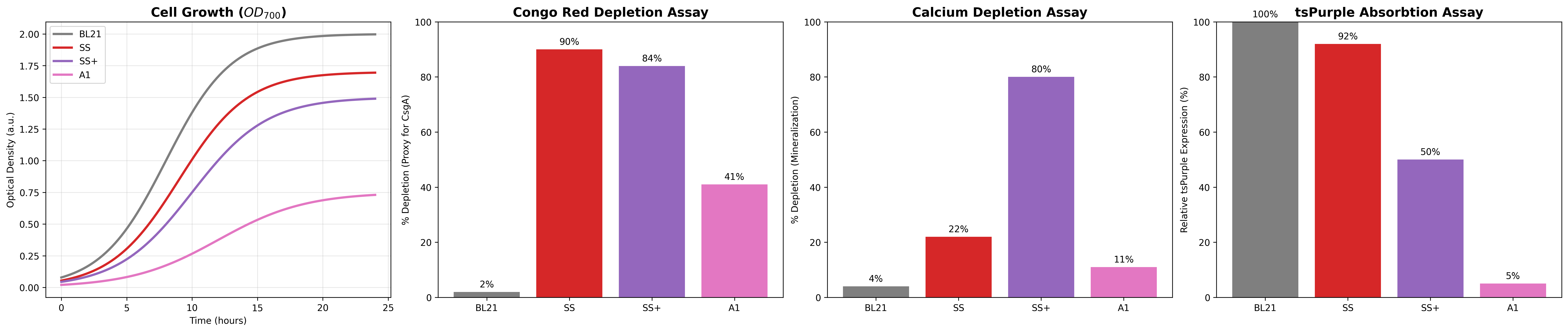
An additional validation for this project is comparing the performance of the consortia to an single E. coli variant that combines all the different genes in one organism. The expected outcome is that the consortia should outperform the A1 strain in all of the core functional aspects.
Expected Results
While the experiements have not been performed yet these graphs reflect the expected outcomes of the Aim 1 synthetic stromatolite consortia versus the control strains:
- SS: Base SynStromatlite
- SS+:SS with D Taili csgA
- A1: All Genes in Single E Coli Strain
- BL21: Unmodified E Coli chassis
Additional Information
References
- Vanyo, J. P., & Awramik, S. M. (1985). Stromatolites and earth—Sun—Moon dynamics. Precambrian Research, 29(1–3), 121–142. https://doi.org/10.1016/0301-9268(85)90064-6
- Zhu, D., Liu, Q., Wang, J., Ding, Q., & He, Z. (2021). Stable carbon and oxygen isotope data of Late Ediacaran stromatolites from a hypersaline environment in the Tarim Basin (NW China) and their reservoir potential. Facies, 67(3), 25. https://doi.org/10.1007/s10347-021-00633-0
- Suarez-Gonzalez, P., Quijada, I. E., Benito, M. I., Mas, R., Merinero, R., & Riding, R. (2014). Origin and significance of lamination in Lower Cretaceous stromatolites and proposal for a quantitative approach. Sedimentary Geology, 300, 11–27. https://doi.org/10.1016/j.sedgeo.2013.11.003
- Daegelen, P., Studier, F. W., Lenski, R. E., Cure, S., & Kim, J. F. (2009). Tracing Ancestors and Relatives of Escherichia coli B, and the Derivation of B Strains REL606 and BL21(DE3). Journal of Molecular Biology, 394(4), 634–643. https://doi.org/10.1016/j.jmb.2009.09.022
- Valenzuela-Ortega, M., & French, C. (2021). Joint universal modular plasmids (JUMP): A flexible vector platform for synthetic biology. Synthetic Biology, 6(1), ysab003. https://doi.org/10.1093/synbio/ysab003
- Tang, T.-C., Tham, E., Liu, X., Yehl, K., Rovner, A. J., Yuk, H., De La Fuente-Nunez, C., Isaacs, F. J., Zhao, X., & Lu, T. K. (2021). Hydrogel-based biocontainment of bacteria for continuous sensing and computation. Nature Chemical Biology, 17(6), 724–731. https://doi.org/10.1038/s41589-021-00779-6
- Zhang, L., Chen, L., Diao, J., Song, X., Shi, M., & Zhang, W. (2020). Construction and analysis of an artificial consortium based on the fast-growing cyanobacterium Synechococcus elongatus UTEX 2973 to produce the platform chemical 3-hydroxypropionic acid from CO2. Biotechnology for Biofuels, 13(1), 82. https://doi.org/10.1186/s13068-020-01720-0
- Zhou, Y., Sun, T., Chen, Z., Song, X., Chen, L., & Zhang, W. (2019). Development of a New Biocontainment Strategy in Model Cyanobacterium Synechococcus Strains. ACS Synthetic Biology, 8(11), 2576–2584. https://doi.org/10.1021/acssynbio.9b00282
- Nevot, G., Pol Cros, M., Toloza, L., Campamà-Sanz, N., Artigues-Lleixà, M., Aguilera, L., & Güell, M. (2025). Engineered Marine Biofilms for Ocean Environment Monitoring. ACS Synthetic Biology, 14(7), 2797–2809. https://doi.org/10.1021/acssynbio.5c00192
- Ostrov, N., Jimenez, M., Billerbeck, S., Brisbois, J., Matragrano, J., Ager, A., & Cornish, V. W. (2017). A modular yeast biosensor for low-cost point-of-care pathogen detection. Science Advances, 3(6), e1603221. https://doi.org/10.1126/sciadv.1603221 ]
- Yudina, N. Yu., Arlyapov, V. A., Chepurnova, M. A., Alferov, S. V., & Reshetilov, A. N. (2015). A yeast co-culture-based biosensor for determination of waste water contamination levels. Enzyme and Microbial Technology, 78, 46–53. https://doi.org/10.1016/j.enzmictec.2015.06.008 * Chapman, M. R., Robinson, L. S., Pinkner, J. S., Roth, R., Heuser, J., Hammar, M., Normark, S., & Hultgren, S. J. (2002). Role of Escherichia coli Curli Operons in Directing Amyloid Fiber Formation. Science, 295(5556), 851–855. https://doi.org/10.1126/science.1067484 * Robinson, L. S., Ashman, E. M., Hultgren, S. J., & Chapman, M. R. (2006). Secretion of curli fibre subunits is mediated by the outer membrane‐localized CsgG protein. Molecular Microbiology, 59(3), 870–881. https://doi.org/10.1111/j.1365-2958.2005.04997.x
- Chen, A. Y., Deng, Z., Billings, A. N., Seker, U. O. S., Lu, M. Y., Citorik, R. J., Zakeri, B., & Lu, T. K. (2014). Synthesis and patterning of tunable multiscale materials with engineered cells. Nature Materials, 13(5), 515–523. https://doi.org/10.1038/nmat3912
- Siegfried, K., Endes, C., Bhuiyan, A. F. Md. K., Kuppardt, A., Mattusch, J., Van Der Meer, J. R., Chatzinotas, A., & Harms, H. (2012). Field Testing of Arsenic in Groundwater Samples of Bangladesh Using a Test Kit Based on Lyophilized Bioreporter Bacteria. Environmental Science & Technology, 46(6), 3281–3287. https://doi.org/10.1021/es203511k
- Guo, Y., Liu, M., Yang, X., Guo, Y., & Hui, C. (2025). Optimized genetic circuitry and reporters for sensitive whole-cell arsenic biosensors: Advancing environmental monitoring. Applied and Environmental Microbiology, 91(8), e00601-25. https://doi.org/10.1128/aem.00601-25
- Weiner, S., & Hood, L. (1975). Soluble Protein of the Organic Matrix of Mollusk Shells: A Potential Template for Shell Formation. Science, 190(4218), 987–989. https://doi.org/10.1126/science.1188379
- Shipman, S. L., Nivala, J., Macklis, J. D., & Church, G. M. (2017). CRISPR–Cas encoding of a digital movie into the genomes of a population of living bacteria. Nature, 547(7663), 345–349. https://doi.org/10.1038/nature23017
- World Health Organization. (2026). Multi-country outbreak of cholera, epidemiological update #33 - 27 January 2026 (Emergency Situation Update Report No. 33). https://www.who.int/publications/m/item/multi-country-outbreak-of-cholera--epidemiological-update--33--27-january-2026
Supplies
Budget
| Item | Quantity | Estimated Cost | Supplier & Link |
|---|---|---|---|
| Twist Bioscience gene strings (csgB, rhlI, csgEFG, csgC, csgA WT, csgA-Asp, 8 variants) | ~15 × 1–3 kb | ~$750 | Twist Bioscience |
| BsaI-HFv2 (Golden Gate) | 1 × 500 U | $65 | NEB R3733S |
| T4 DNA Ligase | 1 × 20,000 U | $55 | NEB M0202S |
| Congo red dye | 5 g | $45 | Millipore Sigma C6767 |
| CaCl₂ (anhydrous) | 100 g | $35 | Millipore Sigma C1016 |
| Na₂CO₃ (anhydrous) | 500 g | $30 | Millipore Sigma S7795 |
| YESCA medium components (yeast extract, casamino acids, agar) | — | $80 | Thermo Fisher Scientific |
| 0.22 µm syringe filters (pack of 50) | 1 pack | $40 | Millipore Sigma SLGP033RS |
| 96-well round-bottom plates (Axygen) | 5 × 50-pack | $75 | Thermo Fisher Scientific |
| 384-well Greiner black clear-bottom plates | 1 × 10-pack | $90 | Millipore Sigma M4187 |
| Sanger sequencing (10 constructs × 3 primers) | 30 reactions | $90 | Azenta/Genewiz |
| Competent cells (E. coli DH5α, NEB 5-alpha) | 6 × 50 µL tubes | $55 | NEB C2987I |
| Analytical balance consumables (weigh boats, etc.) | — | $15 | Thermo Fisher Scientific |
| Total | ~$1,325 |