Week 6 HW: Genetic Circuits Part I
PART 1: Protocol questions
- What are some components in the Phusion High-Fidelity PCR Master Mix and what is their purpose?
The mastermix contains:
- Phusion Hi-Fi DNA Polymerase: It is crucial for completing the amplicons generated during PCR.
- Deoxynucleotides: The building blocks necessary for replicating DNA fragments.
- Buffer including MgCl2: Prevents enzyme denaturation by maintaining pH at a fixed level.
- What are some factors that determine primer annealing temperature during PCR?
The annealing temperature depends on the length of the primers and their GC content. Primers with higher GC content have higher melting temperatures. The sequence of the primer and the presence of mismatches also affect binding. In addition, salt concentration can influence primer stability.
- There are two methods from this class that create linear fragments of DNA: PCR, and restriction enzyme digests. Compare and contrast these two methods, both in terms of protocol as well as when one may be preferable to use over the other
PCR is a method that amplifies a specific DNA fragment using primers and a DNA polymerase. It is very flexible because primers can be designed to target almost any sequence. However, it requires thermal cycling and can introduce small errors.
Restriction enzyme digestion, on the other hand, cuts DNA at specific recognition sites. It is very precise but less flexible because it depends on the presence of those sites in the DNA. PCR is preferred when you need to create or modify fragments, while restriction enzymes are useful when the correct sites already exist.
- How can you ensure that the DNA sequences that you have digested and PCR-ed will be appropriate for Gibson cloning?
To use Gibson cloning, DNA fragments must have overlapping regions of about 20–40 base pairs. These overlaps must be complementary to each other. The overlaps can be added during PCR by designing primers with extra sequences. It is also important to check that the sequences are in the correct orientation and reading frame.
- How does the plasmid DNA enter the E. coli cells during transformation?
The plasmid DNA enters the cells through a process called electroporation where, by means of an externally applied voltage, the membrane permeability increases, allowing the plasmid to enter the bacterial cytosol.
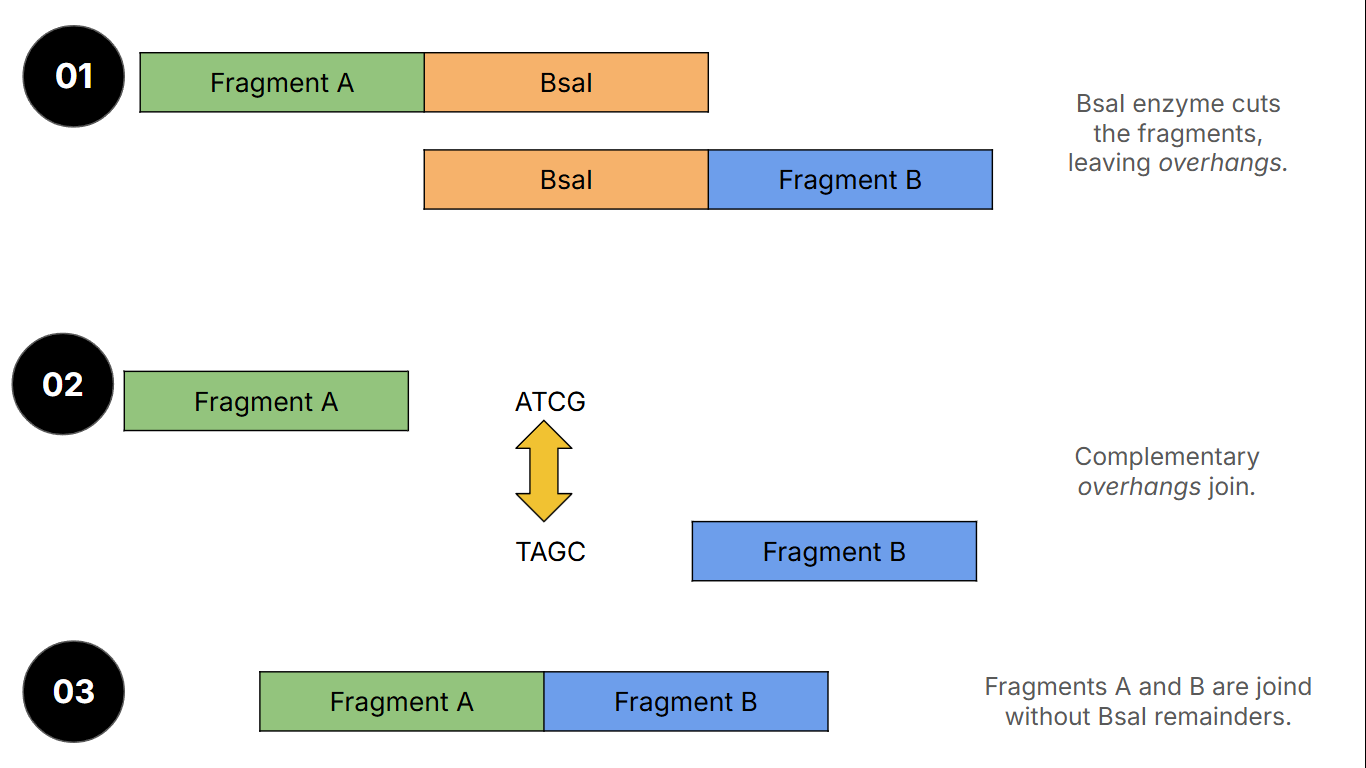
- Describe another assembly method in detail (such as Golden Gate Assembly) 6.1 Explain the other method in 5 - 7 sentences plus diagrams (either handmade or online)
Golden Gate Assembly uses special restriction enzymes that cut outside their recognition sites. This creates custom overhangs that control how DNA fragments join together. The reaction includes both digestion and ligation at the same time. The fragments are designed to have matching overhangs, so they assemble in the correct order. After assembly, the restriction sites are removed, leaving a clean sequence. This method is efficient and allows multiple fragments to be assembled in one step. A diagram is shown below:
6.2. Model this assembly method with Benchling or Asimov Kernel!
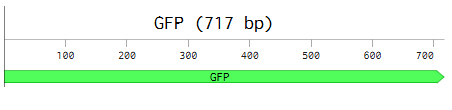
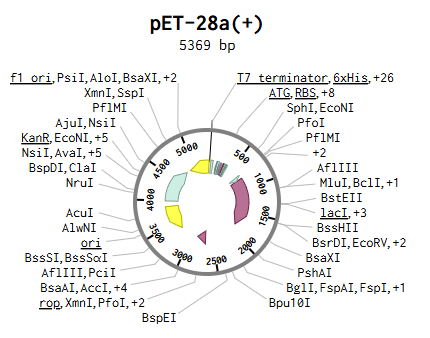
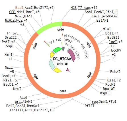
In this case, the GFP protein was selected as the insert and the cloning plasmid pET-28(+). The assembly was done in Benchling.
We click on the assembly wizard option on the lower-right corner of the page and select ‘Golden Gate Assembly’ as shown below:

Then, we select the section outside of the MCS as the backbone, and the full GFP sequence as the fragment to be inserted.
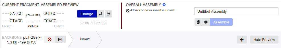
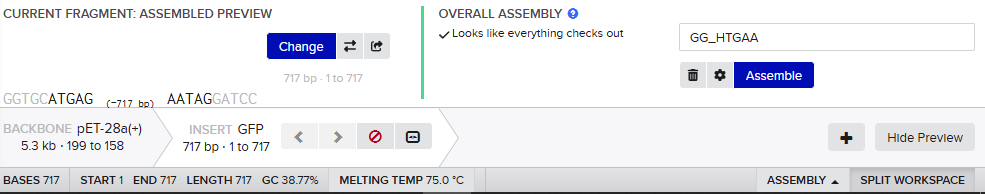
By clicking on the ‘Assemble’, the construct is shown below:
PART 2: Asimov Kernel
- Repressilator
The model was copied form the template and the parts were searched, obtaining, and the simulation with the parameters Simple transfection, E. Coli and 24 h with 10 min timestamps was run, obtaining the figures below:

- Constructs
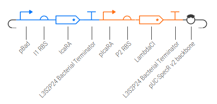
2.1 CONSTRUCT 1
This is a basic construct that expresses LambdaCI. I tried to look fo IPTG sint pTac is an inducible promoter but did not find the protein.
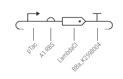
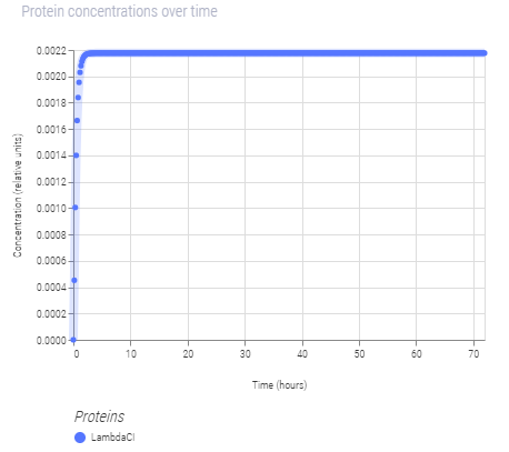
Despite its constituve expression, pTac works when IPTG is induced. Several reasons on why this occurs might be linked to the promoter proerties. pTac might be a leaky promoter because the expression, although low, is constant as the graph shows. This is interesting from an efficiency standpoint. Perpahs when building more sophisticated constructs, pTac should be replaced by a more robust promoter.
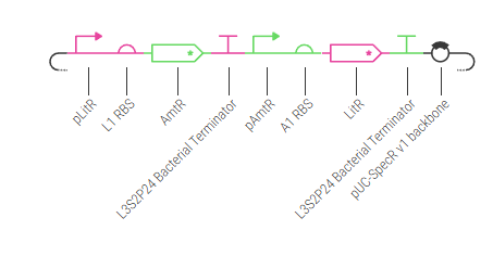
2.2 CONSTRUCT 2
This construct is basically two repressing proteins against each other. The colors show which protein represses which promoter.
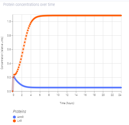
Although both proteins start to express simultaneously, there is a decrease in AmtR and a notable increase in LitR production. This may be due to promoter strength. Since both proteins repress their promoters, small concentrations would be expected. However, the behavior shown in the graph might imply that pAmtR is a stronger promoter than pLitR.
2.3 CONSTRUCT 3
This construct expresses constitutively the protein LambdaCI since pBad is an inducible promoter for L-arabinose.
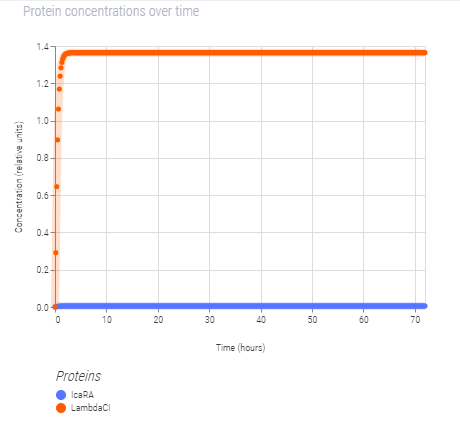
However, when Arabinose for E. Coli is placed in the construct next to LambdaCI, the expression remains unchanged. This may be due to the fact that L-arabinose is the right protein and it is not found in the bacterial parts repository.