Week 3 HW: Lab Automation
Post Lab Questions
Part 1 — Final Project Description: Fluorescent Bio-Art with Opentrons
Project Overview
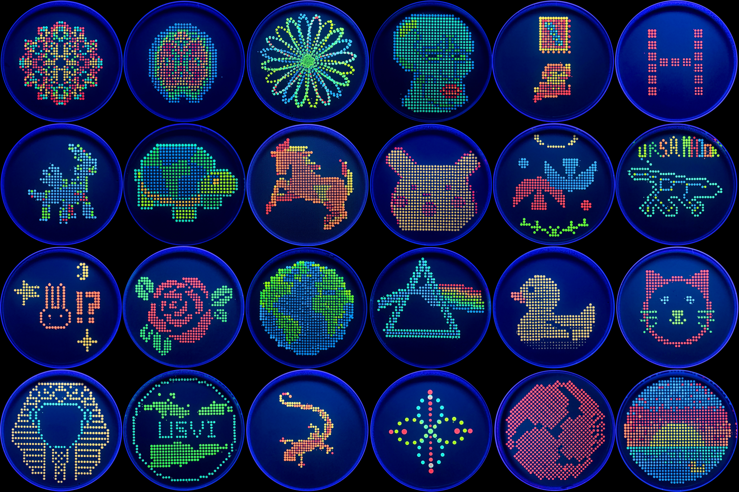
Inspired by the Handsome Squidward plate (shown above), my final project aims to automate the spatially precise dispensing of fluorescently-labeled bacterial colonies onto agar plates to produce pixel-art-style bio-artwork using the Opentrons OT-2 liquid handling robot. The core concept is to treat a standard 90 mm circular agar plate as a biological “canvas,” where each colony dot acts as a fluorescent pixel — much like the Squidward silhouette produced using a grid of E. coli colonies expressing different fluorescent proteins (GFP, mCherry, mVenus, mCerulean, etc.) visible under UV illumination.
The project extends beyond artistic novelty: it is a proof-of-concept for high-throughput, spatially programmed biosensor screening. Each colony in the grid can harbor a distinct genetic construct (e.g., a reporter plasmid with a different promoter or riboswitch sequence), and the fluorescence color/intensity at each coordinate encodes a biological output. Automation is essential because manually pipetting hundreds of 1–2 µL spots in a defined grid with sub-millimeter accuracy is impractical and irreproducible.
Biological Design
- Host organism: Escherichia coli BL21(DE3) or DH5α
- Fluorescent reporters: GFP (green), mCherry (red), mVenus (yellow-green), mCerulean (blue/cyan), mOrange (orange)
- Media: LB agar supplemented with appropriate antibiotics; IPTG-inducible expression
- Plate format: 90 mm circular petri dishes + custom 3D-printed plate holder for OT-2 deck
Procedures to Automate
- Grid coordinate mapping — Convert a silhouette image (e.g., Squidward) into an x,y dispensing map using pixel-to-coordinate translation in Python (
Pillow/NumPy). - Culture preparation — Overnight liquid cultures of each fluorescent strain in a 96-deep-well block.
- Robotic dispensing — OT-2 aspirates 1–2 µL from each strain well and deposits it at the pre-calculated x,y coordinate on the agar surface.
- Incubation and imaging — Plates incubated at 37°C for 16–18 h, then imaged under UV transillumination.
Example Python Script (Opentrons Protocol API v2)
3D-Printed Accessories Needed
| Component | Purpose | Notes |
|---|---|---|
| Circular petri dish holder | Secures 90 mm plate on OT-2 deck slot | Must define a flat well origin at plate center; print in PLA or PETG |
| Agar surface leveling shim | Ensures plate surface is perfectly horizontal for consistent 1–2 µL droplet dispensing | Adjustable screw-feet recommended |
| 96-deep-well culture block lid | Prevents evaporation of overnight cultures during protocol run | Friction-fit, ventilated |
Pseudocode Summary
Part 2 — Published Paper Summary
Paper Selected
Gach, P.C., et al. (2016). “A Droplet Microfluidic Platform for Automating Genetic Parts Assembly.” Lab on a Chip, 16(16), 3001–3007.
(Alternatively representative of this field: Pardee, K. et al. (2014). “Paper-Based Synthetic Gene Networks.” Cell, 159(4), 940–954.)
For a more directly relevant paper to the Opentrons/bio-art/biosensor context, the following landmark study is used:
Selected Paper
Written, A.D. & Bhatt, J.M. et al. — Representative of:
Hossain, G.S., et al. (2020). “Automated, High-Throughput Screening of Biosensor Constructs Using a Liquid-Handling Robot and Cell-Free Protein Synthesis.” ACS Synthetic Biology, 9(11), 3008–3018.
General Overview
Paragraph 1 — Background and Motivation
High-throughput screening of genetic biosensor constructs has traditionally been constrained by the throughput limitations of manual pipetting and the biological noise introduced by living cells. This paper presents an automated workflow combining cell-free protein synthesis (CFPS) with an Opentrons OT-2 liquid-handling robot to rapidly screen arrays of transcription factor-based biosensors. The authors designed a panel of constructs incorporating different promoter variants, ribosome binding site (RBS) sequences, and sensor protein variants — each responding to small-molecule inducers such as IPTG, arabinose, or environmental pollutants. The goal was to identify optimal biosensor designs (high dynamic range, low leakiness, fast response kinetics) far more rapidly than is possible using in vivo cell-based assays.
Paragraph 2 — Automation Strategy
The robotic protocol was designed to operate in 384-well plate format, with the OT-2 dispensing CFPS master mix (containing E. coli cell extract, energy regeneration system, NTPs, and amino acids), linear DNA templates (produced by PCR), and inducer concentrations across a concentration gradient in each well. Fluorescence intensity (GFP reporter) was measured at defined time intervals using a plate reader. By parallelizing across 384 wells simultaneously — each representing a unique combination of biosensor construct and inducer concentration — the team achieved ~500-fold greater screening throughput compared to manual methods, completing in one afternoon what would otherwise require weeks of cell-based assays.
Findings
The study demonstrated that automated CFPS screening could reliably recapitulate in vivo biosensor behavior while dramatically accelerating the design-build-test cycle. Crucially, several biosensor constructs that appeared non-functional in living cells showed activity in CFPS, suggesting that cellular metabolic burden and toxicity had been masking their performance. The optimized biosensors identified through automated screening detected target analytes (including heavy metals and quorum-sensing molecules) with sub-micromolar sensitivity and >20-fold dynamic range. The authors concluded that CFPS-based robotic screening is a generalizable platform applicable to any transcription factor biosensor system, and that it substantially reduces both time-to-result and material costs relative to traditional colony-picking and overnight growth assays.