What are some components in the Phusion High-Fidelity PCR Master Mix and what is their purpose?
Phusion DNA Polymerase: Building enzyme that reads the original DNA and constructs the new copies with high accuracy.
nucleotides
Optimized reaction buffer: A liquid that maintains the perfect chemical environment and pH for the enzyme to work.
MGCL2: Helper molecule (cofactor) that the polymerase needs to function properly.
What are some factors that determine primer annealing temperature during PCR?
Primer Length: Longer primers have more binding area, so they also require higher temperatures.
GC Content: The DNA bases Guanine (G) and Cytosine (C) bind to each other with three chemical bonds, while Adenine (A) and Thymine (T) only use two. Therefore, primers with more Gs and Cs hold on tighter and require a higher temperature.
There are two methods from this class that create linear fragments of DNA: PCR, and restriction enzyme digests. Compare and contrast these two methods, both in terms of protocol as well as when one may be preferable to use over the other.
PCR Protocol: Uses heat cycles to melt DNA apart, lets primers attach, and uses an enzyme to build new copies.
When to use: When you have a tiny amount of DNA and need billions of copies of a very specific segment, or when you want to add custom ends to a DNA sequence.
Restriction Digest Protocol: Mixes DNA with restriction enzymes and incubates them at a steady temperature. The enzymes physically cut the DNA at specific sequences.
When to use: When you want to extract a specific chunk of DNA out of a larger, already-existing piece, or when you want to verify that a DNA sequence is correct by seeing what sizes it cuts into.
How can you ensure that the DNA sequences that you have digested and PCR-ed will be appropriate for Gibson cloning?
Must design the PCR primers so that the ends of DNA pieces overlap. The tail end of piece A must have the exact same sequence (usually 15 to 40 base pairs) as the starting end of piece B. The Gibson mix will chew back one strand of these ends, allowing the matching sequences to find each other and stick together like perfect puzzle pieces.
How does the plasmid DNA enter the E. coli cells during transformation?
Usually through heat shock or electroporation.
Heat Shock (Chemical): The bacteria are treated with chemicals (like calcium) to neutralize their charge, then subjected to a sudden spike in heat. This sudden temperature change creates temporary “pores” or holes in the bacterial wall, allowing the DNA to slip inside.
Electroporation: The bacteria are hit with a quick zap of electricity, which shocks the cell membrane into opening those temporary pores.
Describe another assembly method in detail (such as Golden Gate Assembly)
Golden Gate assembly is a method for joining multiple DNA fragments together in a single tube. It uses special “molecular scissors” called Type IIS restriction enzymes. Unlike normal restriction enzymes that cut exactly where they bind, Type IIS enzymes bind to a recognition sequence but reach over and cut the DNA a few steps away. Because they cut outside their recognition site, they leave behind custom “sticky ends” (overhangs) that you can design to match perfectly with the next piece of DNA. When the matching pieces snap together, an enzyme called ligase glues them shut permanently. Crucially, the original enzyme recognition site is cut off and left behind in this process, meaning the final assembled DNA has no “scars” or unwanted leftover sequences. Because the assembled product can no longer be cut by the enzyme, the cutting and gluing can happen simultaneously in one reaction tube.
Model this assembly method with Benchling or Asimov Kernel!
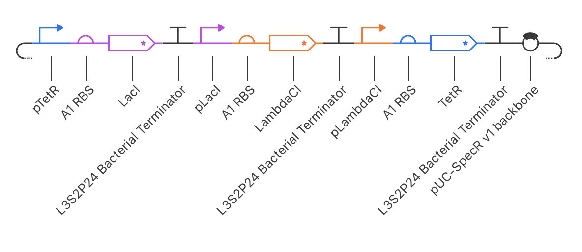
Recreate the Repressilator in that empty Construct by using parts from the Characterized Bacterial Parts repository
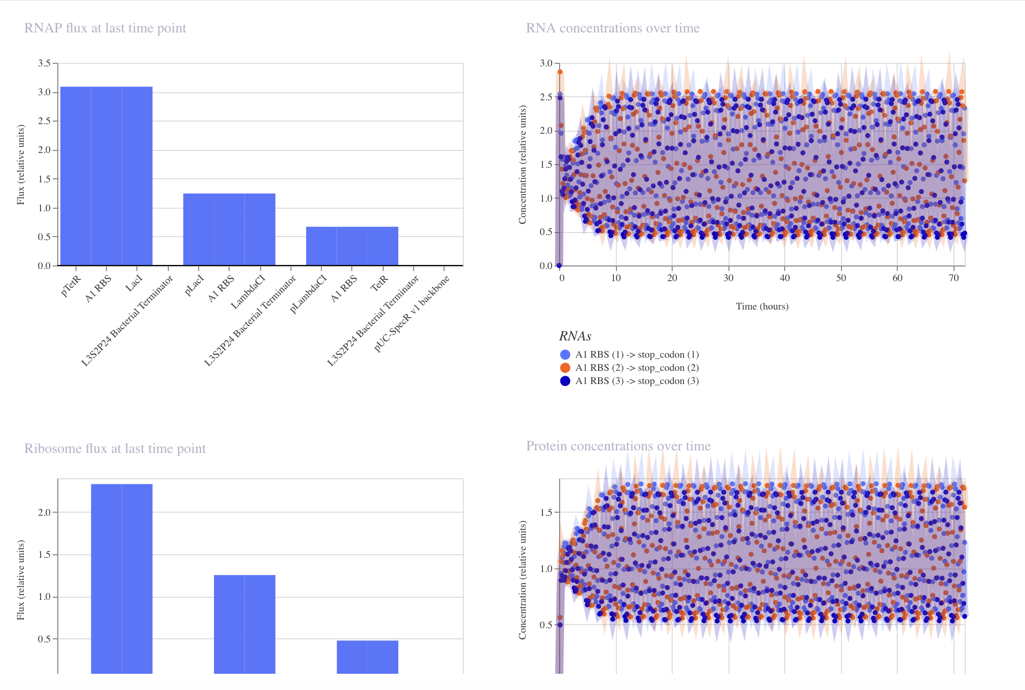
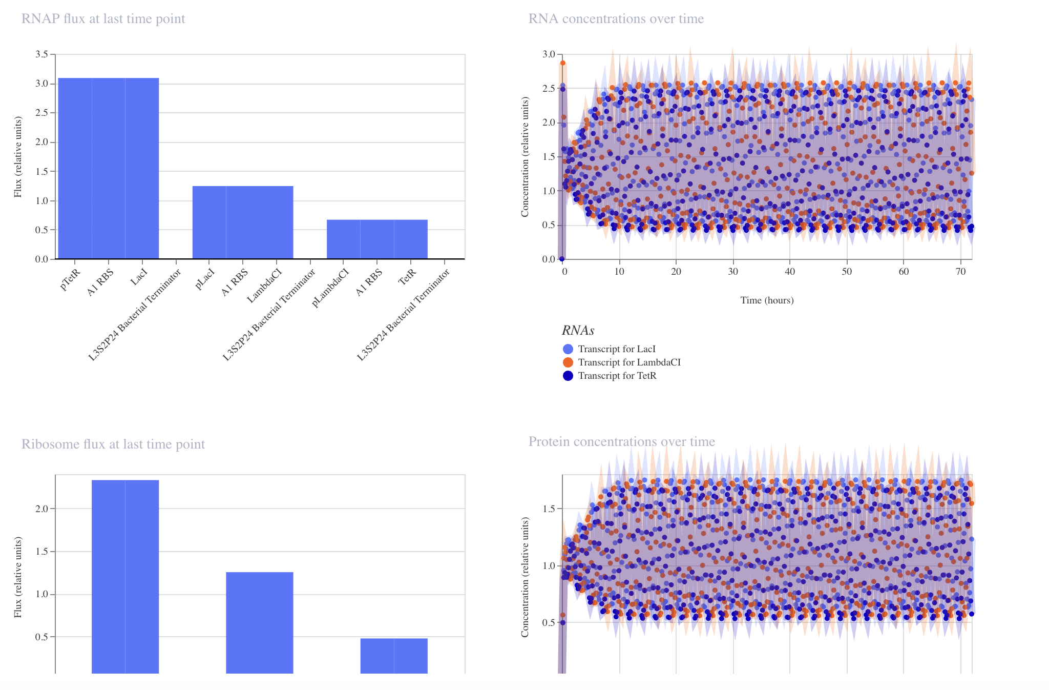
Confirm it works as expected by running the Simulator (“play” button) and compare your results with the Repressilator Construct found in the Bacterial Demos repository
Document all of this work in your Notebook entry - you can copy the glyph image and the simulator graphs, and paste them into your Notebook
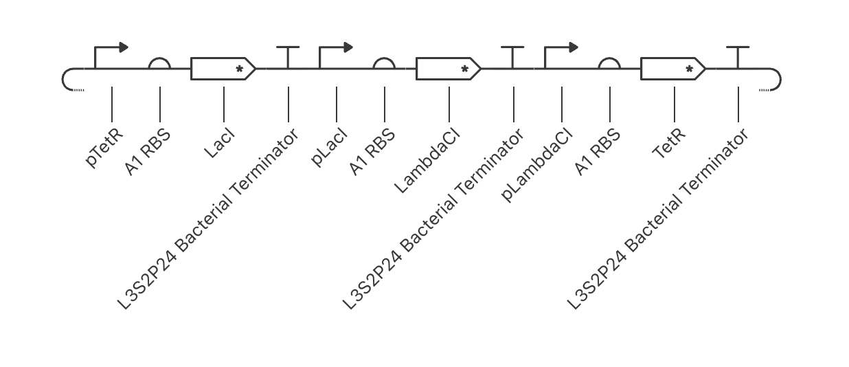
Construct Glyphs
Model — color-coded cassettes, includes pUC-SpecR v1 backboneMy Build — same 3 cassettes, no backbone, monochrome glyphs
Simulation Results
Model — 24h, clean phase separation, transcripts named by repressorMy Build — 72h, oscillation sustained but curves heavily overlapping
Model
My Build
Backbone
pUC-SpecR v1 included
Not added
Duration
24 hours
72 hours
Oscillation
Clear phase separation between curves
Sustained but three curves blur together
RNAP flux pattern
Stepped bars (1.57 / 0.65 / 2.87)
Similar stepped pattern (3.1 / 1.25 / 0.65)
Noise bands
Moderate spread
Wider spread
Build three of your own Constructs using the parts in the Characterized Bacterials Parts Repo
Explain in the Notebook Entry how you think each of the Constructs should function
Run the simulator and share your results in the Notebook Entry
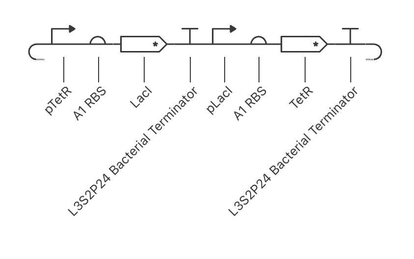
Two cassettes mutually silence each other. The system snaps to one of two stable states — either LacI is high and TetR is low, or vice versa. Acts as a bistable memory switch: once flipped, it holds its state.
No → bistable lock Expect: one protein high, one flat zero
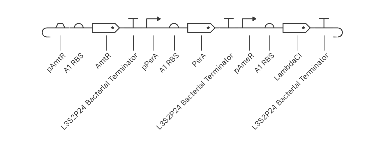
2 — NOR Gate pAmtR → AmtR ⟐ pPsrA → PsrA Both repress pAmeR → LambdaCI
Two input repressors each independently silence the output promoter pAmeR. LambdaCI is only produced when neither AmtR nor PsrA is present — a true NOR logic gate.
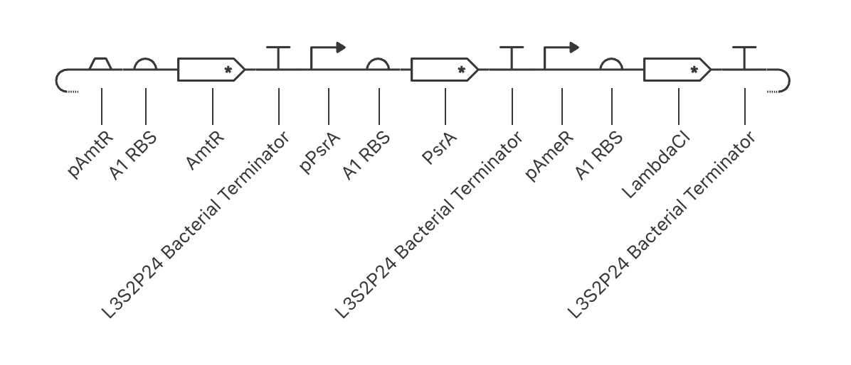
A two-stage repression cascade. When the upstream signal (pAmtR) is active, it silences the chain, keeping output OFF. Remove the signal → repression lifts through both stages → LambdaCI output turns ON.
Signal present → Output OFF Signal removed → Output ON