source: wikimedia commons
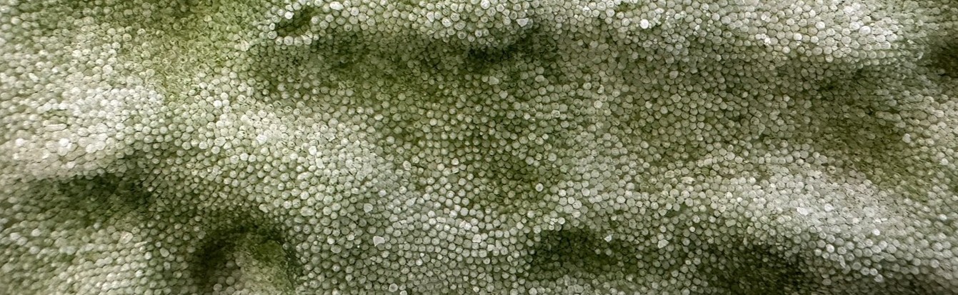
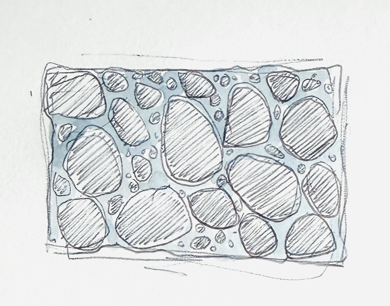
Genetically Engineered Diatoms to Bind with Building Rubble/Waste Building materials like cement and brick are difficult to reuse and natural weathering or active demolition leaves behind tons of waste material that remains under-recycled. In a previous project my team from graduate school developed a porous, bio receptive glass using glass waste and I would like to expand upon that research by bioengineering diatoms into a silica scaffold of cement and glass rubble/frits to fuse these waste materials into a new architectural material. Diatoms are an exciting prospect for architecture for their silica frustules, inherent translucency, and their lacy pore structure. I am curious to see if there would be a way to pattern their silica deposits for enhanced carbon sink and particle processing in urban spaces. It would also be beautiful to see the formation of silica deposits depending on sun patterns on site, filling in the rubble scaffold where there is more direct light. It would also be interesting to potentially engineer the directional strength of a diatom-rubble piece and the lace pattern, playing with the idea of directional bias in architecture more theoretically but also for building methods.
Benchling Gel Art DNA Design Challenge When looking into proteins to explore further, I chose to focus on proteins related to the structure of diatom silica walls. These proteins would be exciting to understand to get a better picture of how diatoms would form the lacy micropatterns for the rubble-diatom material proposal for the final project. While there are a few different proteins key to the biosilica formation, and silaffins drive the lacy patterns and micropore structure.
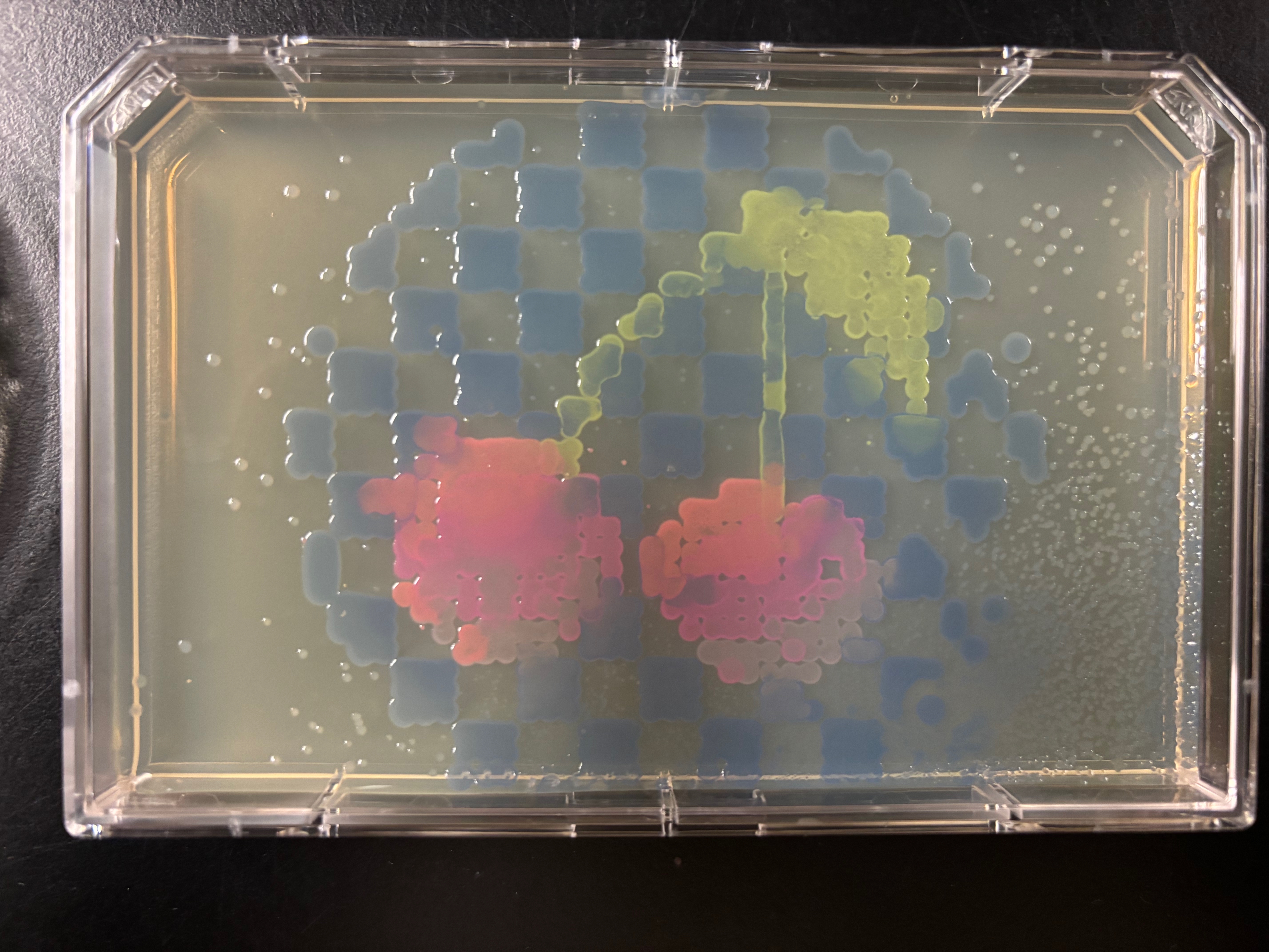
Opentrons Art When creating this GUI art I created an image of cherries with a checkered background to see how common features in traditional drawing would translate to bacterial image creation including shading, regular patterning, thinner and thicker lines, as well as curved and straight forms. I downloaded the python script for the PCR plate system from this simulator.
Conceptual Questions Why do humans eat beef but do not become a cow, eat fish but do not become fish?
As we eat other materials, the proteins and molecules are broken down through our digestive system, leaving us with building blocks to support our cell replication processes.
Why are there only 20 natural amino acids?
These 20 amino acids were created as life was forming, and after the ‘frozen accident’, the proteins that developed at this time seem to have standardized these 20 amino acids. There were more and are more amino acids, however this connection between protein and AA during early evolution created this set of 20.
Intracellular Artificial Neural Networks What advantages do IANNs have over traditional genetic circuits, whose input/output behaviors are Boolean functions?
IANNs allow for less prescriptive circuit method, whereas traditional genetic circuits need to be specified at each step, making it a target specific circuit by finding signalling patterns across a vast dataset. Traditional genetic circuits are limited to simpler action/reaction function simulations to create an mRNA therapy.
General Homework Questions Explain the main advantages of cell-free protein synthesis over traditional in vivo methods, specifically in terms of flexibility and control over experimental variables. Name at least two cases where cell-free expression is more beneficial than cell production.
Because cell-free methods exist without the limitations of maintaining a living cell, they offer greater flexibility in exploring the details of protein synthesis without worrying about a cell membrane or the knock-on impacts of every modification before its more
Based on the older information:
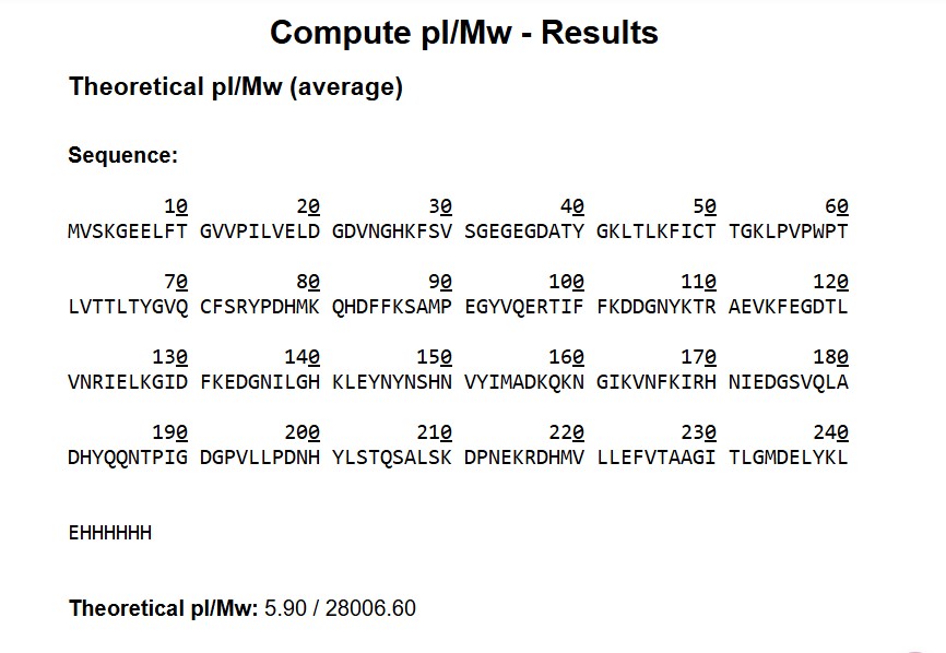
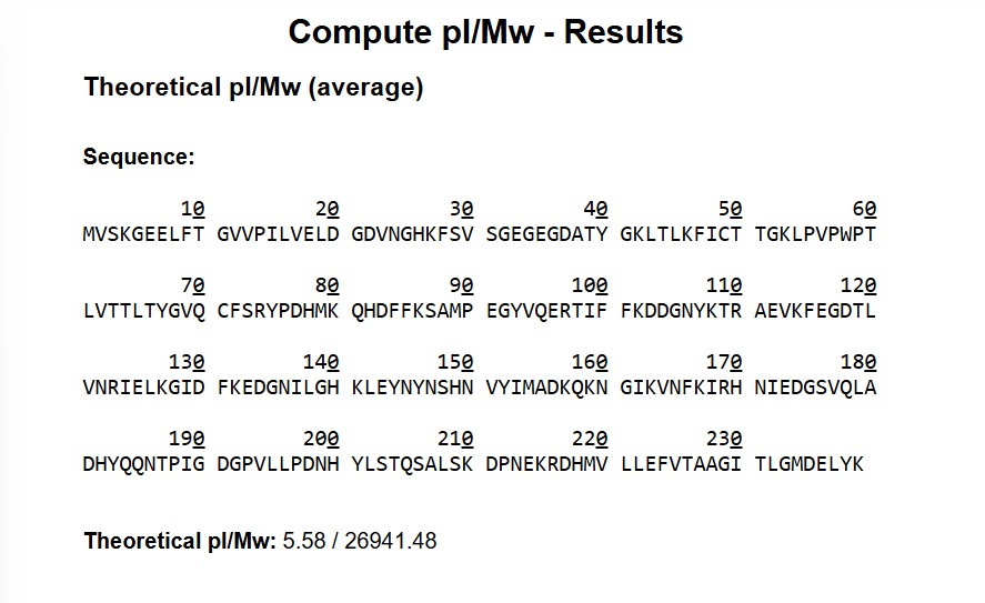
• m/z(n) = 824.1148
• m/z(n+1) = 800.608
• n = 34.0172
MW = (n * m/z(n)) - n
MW = ((34.0172) (824.1148)) - 34.0172
MW = 28,000.06 daltons
Error Rate/Accuracy = ((28006.6 - 28000.06)/28006.6)1000000 = 0.0002341000000 = 233.52ppm
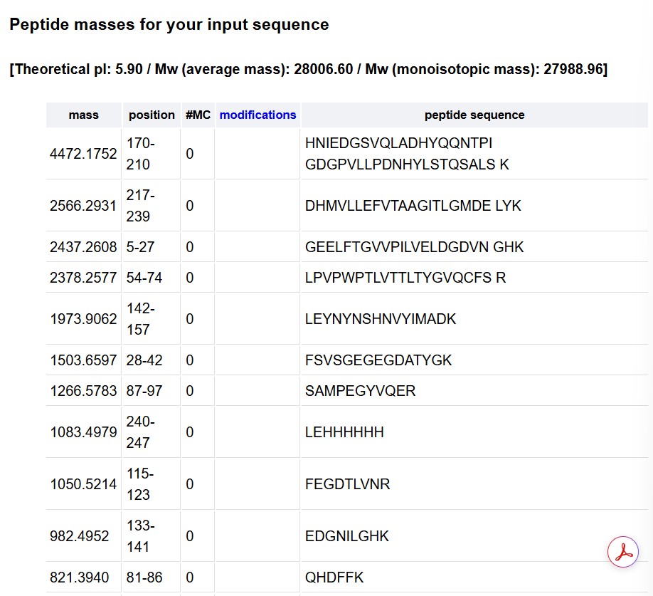
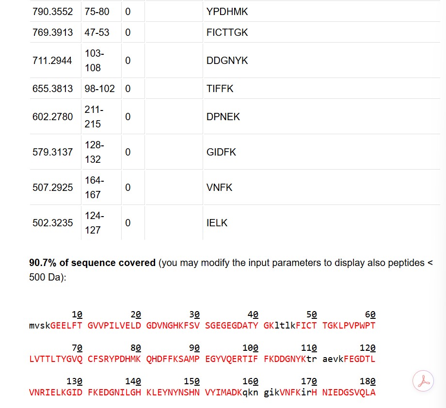
This value is much higher than the 50ppm threshold, indicating an issue with the tested product. Also, I think this calculation seems to actually be for inaccuracy or error rate. The sequence has 20 K’s and 6 R’s for peptide segmentation sites
Part A: Pixel Art Canvas
For the HTGAA pixel artwork, I really enjoyed that the experiment was a playful approach to both a large-scale group project and to cloud-based bio projects. I added a blue dot and a yellow dot to a flower-like design that was absorbed into other designs as the artwork developed, but it was cool to try to predict the interests of the other users and contribute to what I thought the pattern would need to have to be completed.
Subsections of Homework
Week 1: Principles and Practices
source: wikimedia commons
Genetically Engineered Diatoms to Bind with Building Rubble/Waste
Building materials like cement and brick are difficult to reuse and natural weathering or active demolition leaves behind tons of waste material that remains under-recycled. In a previous project my team from graduate school developed a porous, bio receptive glass using glass waste and I would like to expand upon that research by bioengineering diatoms into a silica scaffold of cement and glass rubble/frits to fuse these waste materials into a new architectural material. Diatoms are an exciting prospect for architecture for their silica frustules, inherent translucency, and their lacy pore structure. I am curious to see if there would be a way to pattern their silica deposits for enhanced carbon sink and particle processing in urban spaces. It would also be beautiful to see the formation of silica deposits depending on sun patterns on site, filling in the rubble scaffold where there is more direct light. It would also be interesting to potentially engineer the directional strength of a diatom-rubble piece and the lace pattern, playing with the idea of directional bias in architecture more theoretically but also for building methods.
Goals:
• When speculating such panels, it is important to keep policy which mitigates the impact of the material creation and installation on the surrounding soil and water biodiversity through run off material and resources used to grow the diatom structures. Diatoms can overpower other microorganisms and limits on the volume of diatom production could help reduce the chances of local ecological harm and biological impact.
• Another goal is to secure a non-stress inducing method of cultivating and sourcing the diatoms for this scale of application.
• This material should be lessening the burden of the construction industry.
Concerns:
• Who would have access to this material?
• How does this material impact the local environment?
• Will users receive this material positively and use it? Or will it be demolished or underutilized?
• Will this create excess burden on access to an organism for this scale? What are the political realities of sourcing this material?
• How do diatoms react with materials like cement, brick, and glass? Are there any reactions between materials that can cause issues?
Actions:
• When casting the rubble diatom mixture, create reusable casts/equipment workflows when possible.
• Work directly with ecologists to determine the site for harvesting and to determine if the site for installation is appropriate in case of run-off or other biological interactions.
• One method of mitigating the environmental impact of sourcing and using diatoms at this large scale could be to focus on cultivation from local areas with diatom overgrowth so that this helps cut down on the environmental impact of current unhealthy ecology.
• Lab testing, especially longevity testing, would help clear up uncertainty regarding the impact of the material on site as it weathers and does through periods of high and low growth. This includes the potential toxicity of the rubble, the biological interactions of the diatoms, and how the ,odified diatoms may change over time through mutations and biomass buildup.
• Community workshops/exhibits as user studies to understand how people would interact with the material (biosecurity of sensory interaction), understand whether they would accept it in their built environment, and educate them on the material itself.
Does the option:
Casting
Harvesting
Sourcing
Lab Testing
Community Workshops
Enhance Biosecurity
• By preventing incidents
n/a
***
***
***
***
• By helping respond
n/a
*
*
***
n/a
Protect the environment
• Through sourcing/creation
**
***
***
*
n/a
• During and after installation
*
*
n/a
***
**
Other considerations
• Encourage long term use by stakeholders
n/a
*
*
***
***
Homework Slide Responses
Professor Jacobson:
The error rate for polymerase is 1:10^6. Compared to the length of the human genome of around 3.2Gbp (slide 10), the error rate is minute, occurring perhaps once in one genome. Biology deals with this discrepancy by proofreading the action before it is coded into the DNA.
There are 20 AA and 61 codons that specify amino acids (NHGRI), leading to different ways to code for an average human protein. Some reasons that all of these different codes don’t work for the protein of interest could be due to the physical structure of the protein, and the efficiency of each pathway. (not totally sure of this answer)
Dr.Leproust
The most common method for oligo synthesis is currently a chip based gene synthesis that couples the nucleotide with phosphonamidite. Historically, this has been a solid phase synthesis.
It’s difficult to make oligos longer than 200nt vir direct synthesis because of the surface and primarily the inefficiency of the current state of the art phosphonamidite method.
Source: Yin Y, Arneson R, Yuan Y, Fang S. Long oligos: direct chemical synthesis of genes with up to 1728 nucleotides. Chem Sci. 2024 Dec 18;16(4):1966-1973. doi: 10.1039/d4sc06958g. PMID: 39759933; PMCID: PMC11694485.
You can’t make a 2000bp gene with direct olio synthesis because of the increased error rate in PCR. The new gene pool solution offers a 1 in 3,000 bp error rate which would allow for greater gene lengths to be processed. (slide 39)
Professor George Church
According to the NIH, the nine essential amino acids are histidine, isoleucine, lysine, methionine, phenylalanine, threonine, tryptophan, and valine. This means that the Lysine Contingency is really already a contingency that all animals have upon receiving all nine of these AAs from their diet, not just lysine. (Fictional) dinosaurs are just like you and me, minus 8 more essential amino acids.
Source: Lopez MJ, Mohiuddin SS. Biochemistry, Essential Amino Acids. Updated 2024 Apr 30. In: StatPearls Internet. Treasure Island (FL): StatPearls Publishing; 2025 Jan-. Available from: https://www.ncbi.nlm.nih.gov/books/NBK557845/
Week 2: DNA Read, Write, and Edit
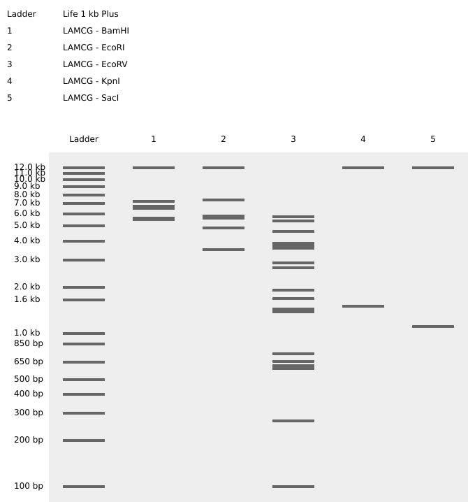
Benchling Gel Art
DNA Design Challenge
When looking into proteins to explore further, I chose to focus on proteins related to the structure of diatom silica walls. These proteins would be exciting to understand to get a better picture of how diatoms would form the lacy micropatterns for the rubble-diatom material proposal for the final project. While there are a few different proteins key to the biosilica formation, and silaffins drive the lacy patterns and micropore structure.
In the NCBI database chose: silaffin protein (Thalassiosira pseudonana CCMP1335)
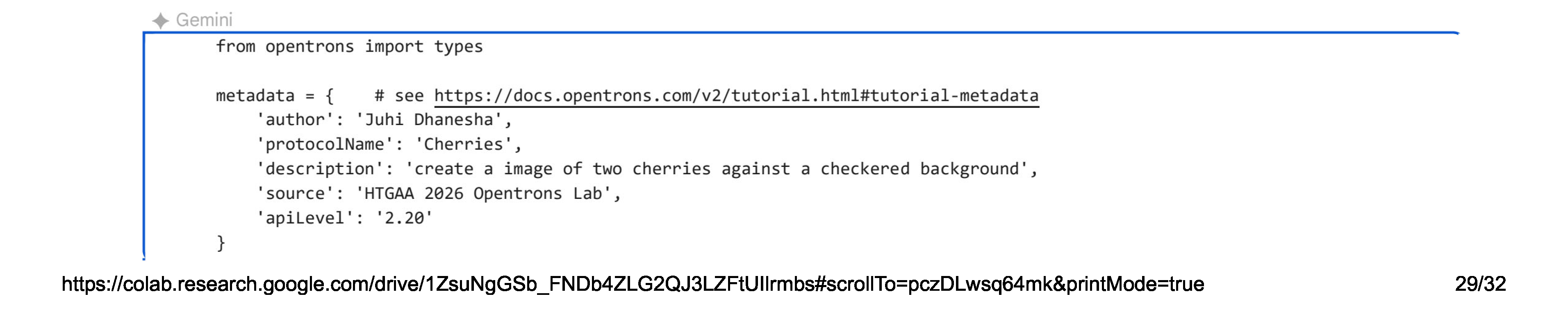
When creating this GUI art I created an image of cherries with a checkered background to see how common features in traditional drawing would translate to bacterial image creation including shading, regular patterning, thinner and thicker lines, as well as curved and straight forms. I downloaded the python script for the PCR plate system from this simulator.
created through Ronan’s Opentrons plate art simulator
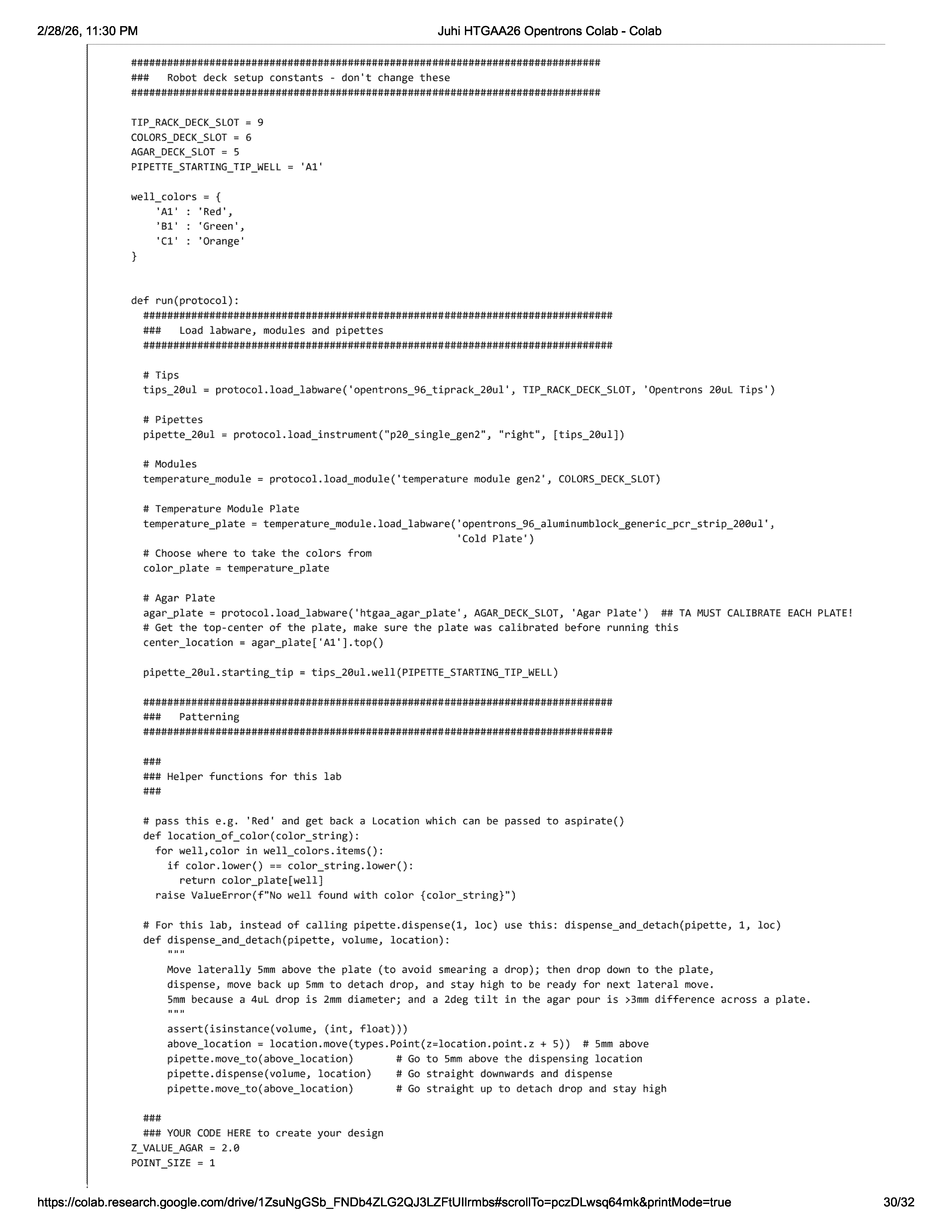
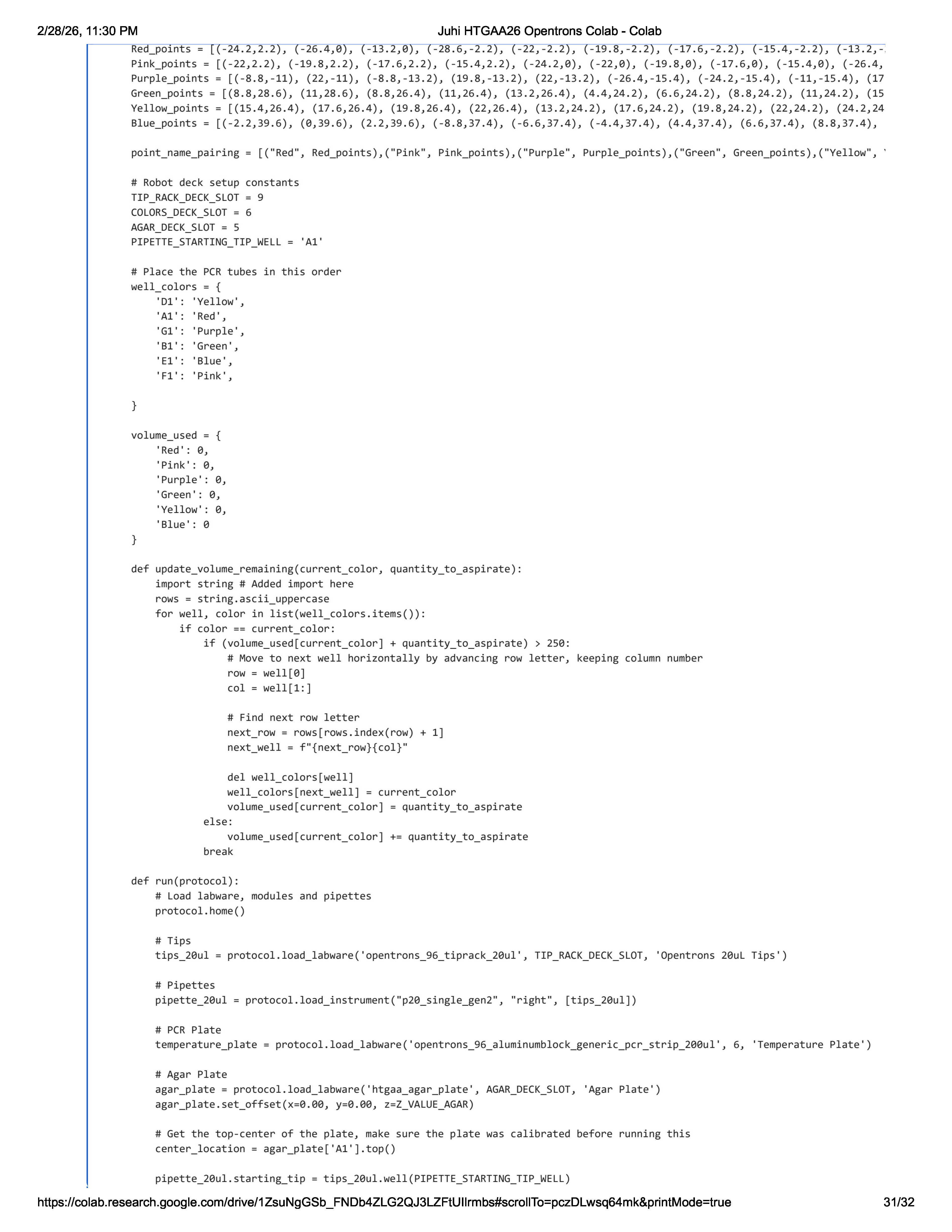
Python Script for Opentrons
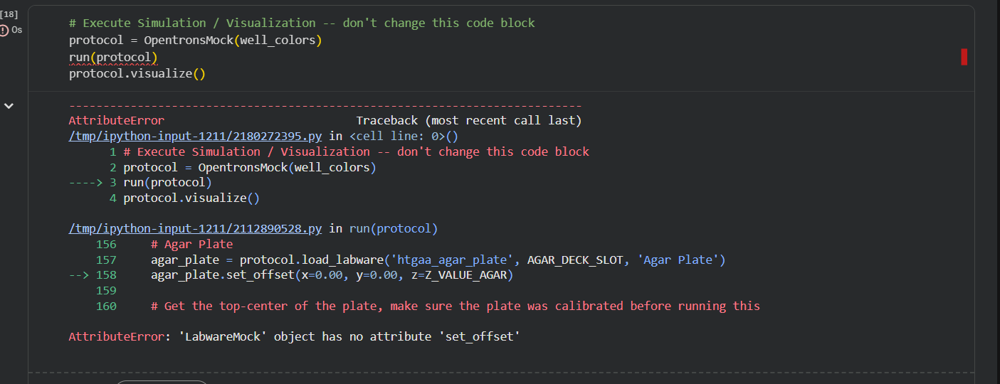
colab work: importing python script and error management
In the colab script I debugged until it was suggesting a bug that could fundamentally change the function for the code to properly converse with the opentrons system. This was about the “set_offset” condition. This error code is not present in the script cell, indicating that the issue is associated with the visualization cell.
Opentrons Plate Art
this lab required lots of troubleshooting, with this plate created after the lab time by the BUGSS TAs Amanda and Joel. Previous plates were unsuccessful due to various trouble shooting issues from the python script, the point size for the asperated droplet, and a potential air bubble in the well.
Opentrons Research and Potential Use of Automated Tools in Final Project
For the final project, if I were to pursue the diatom project idea, would be to use automated systems for cell seeding by scaning contruction rubble and detect the best spots to introduce diatoms into the rubble panel in response to the rubble placement, load bearing support, or areas that make easy bridges between rubble.
Salido et.al used automated technology for diatom identification:
Salido, J.; Sánchez, C.; Ruiz-Santaquiteria, J.; Cristóbal, G.; Blanco, S.; Bueno, G. A Low-Cost Automated Digital Microscopy Platform for Automatic Identification of Diatoms. Appl. Sci. 2020, 10, 6033. https://doi.org/10.3390/app10176033
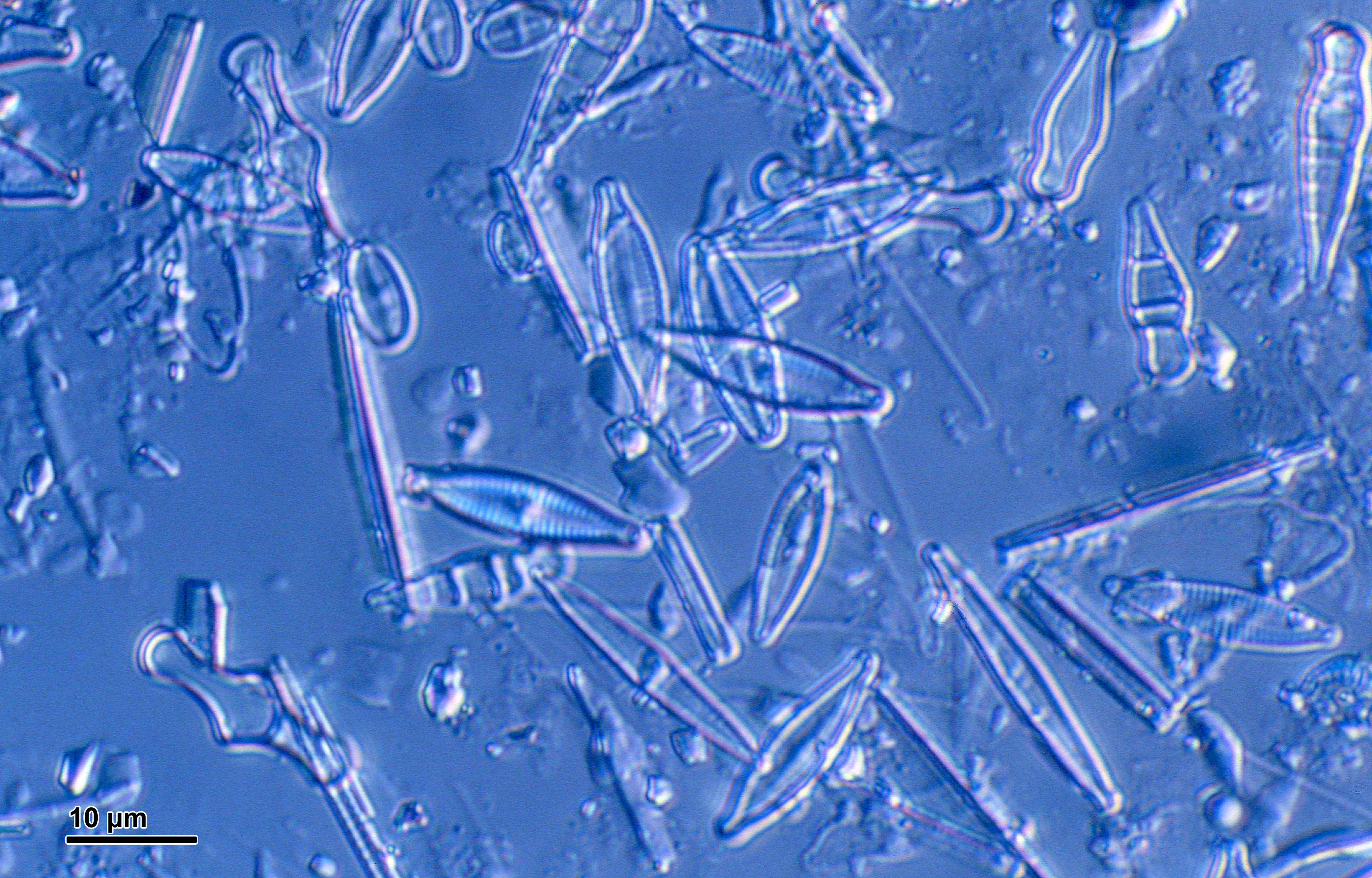
Diatoms are traditionally identified and viewed using LM and SEM imaging, since they are invisible to the naked eye. Different species of diatoms have also remained unidentified due to these limitations. Automated technology and deep neural networks have assisted in the detection, classification, and counting which can be tricky due to the variety of shapes that diatoms may have.
Crit 1 Final Project Ideas
For the final project I would like to look closer at architectural materials and how they can respond to environmental stressors. Some of these ideas are a new perspective on previous ideas of mine, playing with how one concept can be explored through multiple fields.
Project 1: Diatoms and Rubble
This proposal looks into using diatoms to create a new aggregate material out of construction waste (ie cement) and diatoms to create a bioreceptive panel system. It plays with the way that diatoms create silica cell walls, leaving opalescent lacy micropatterns that could bring a new use to materials that are very hard to repurpose.
By using diatoms with rubble, this ‘new’ material would create beautiful translucent seams that could potentially help host other organisms or itself help with nutrient filtration depending on the design application.
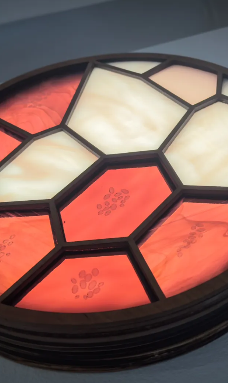
Project 2: Biopigment Stained Glass with Air Quality Indication
Stained glass has a history of storytelling for the masses, usually in churches and other religious spaces. It would be interesting to see if biopigments could:
stain glass without chemical inclusions
react to pollution levels and serve as a visual indicator for air quality in the area.
This research would need to be further refined with the intensity of the color, how sensitive the reaction could be, and how the biofilm pigment could impact its environment in terms of microenvironments, both positive and negative.
Project 3: Brick Extremophiles
Looking back at a previous project where I studied brick walls and reimagined their structure, it would be interesting to see if there are certain prevalent extremophiles present in bricks especially in iron-rich clay areas.
These extremophiles could be further designed to embody carbon and support environmental remediation.
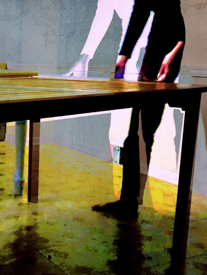
Project 4: Bruising Homes
What if buildings bruised?
This is an idea that I pursued in a filmmaking studio in the past that I would like to unpack through alternative means. A touch-reactive biopigment could become a paint or biofilm that responds to the amount of pressure put onto the surface. Relating back to organisms like touch-me-not mimosa plants that are very sensitive to their environment, a paint like this could become an exploration into the permanence of human action, and document our relationship to our homes. The paint/biofilm would begin as a uniform color, and darken in reaction to pressure.
Week 4: Protein Design Part 1
Conceptual Questions
Why do humans eat beef but do not become a cow, eat fish but do not become fish?
As we eat other materials, the proteins and molecules are broken down through our digestive system, leaving us with building blocks to support our cell replication processes.
Why are there only 20 natural amino acids?
These 20 amino acids were created as life was forming, and after the ‘frozen accident’, the proteins that developed at this time seem to have standardized these 20 amino acids. There were more and are more amino acids, however this connection between protein and AA during early evolution created this set of 20.
Can you make other non-natural amino acids? Design some new amino acids.
Amino acids are primarily composed of hydrogen, oxygen, carbon, and nitrogen and as proven from the beginnings of RNA, there are many non-standard amino acids that could be made in fact many non-natural AAs already exist. In terms of function, I would design an AA that could create florescent tags for injury sites, helping identify issues that are less visible like endometreosis or even to find a bug bite from tics. It could help speed up wound identification and reduce time lost in exploritory tests.
Where did amino acids come from before enzymes that make them, and before life started?
Many natural events have created the primodial soup that led to life as we know it, and the ingredients of AAs seem to have formed in a similar way.
If you make an α-helix using D-amino acids, what handedness (right or left) would you expect?
I would expect righthandedness because ‘D’ typicially refers to right handed molecules and ‘L’ refers to left.
Can you discover additional helices in proteins?
Probably! Given that there is a high percentage of proteins that are still to be discovered and studies, there are probably proteins with additional helices.
Why are most molecular helices right-handed?
DNA is right-handed, but also many aspects of the natural world have a handedness, like the right hand test in physics as well. This seems to be connected back to thermodynamics and create helices that are more stable.
Why do β-sheets tend to aggregate? What is the driving force for β-sheet aggregation?
Their structure is flat, making it easy to pack, and the edges are designed with hydrogen backbones that help form hydrogen bonds with other beta sheets.
Why do many amyloid diseases form β-sheets? Can you use amyloid β-sheets as materials?
Many amyloid diseases happen when proteins misfold, and they form beta sheets because of their thermodynamic stability and their straight hydrogen bond structure allows for tighter folds. They can probably be used to support material tensile strength and rigidity, but I don’t think they classify as materials themselves.
Protein Analysis and Visualization
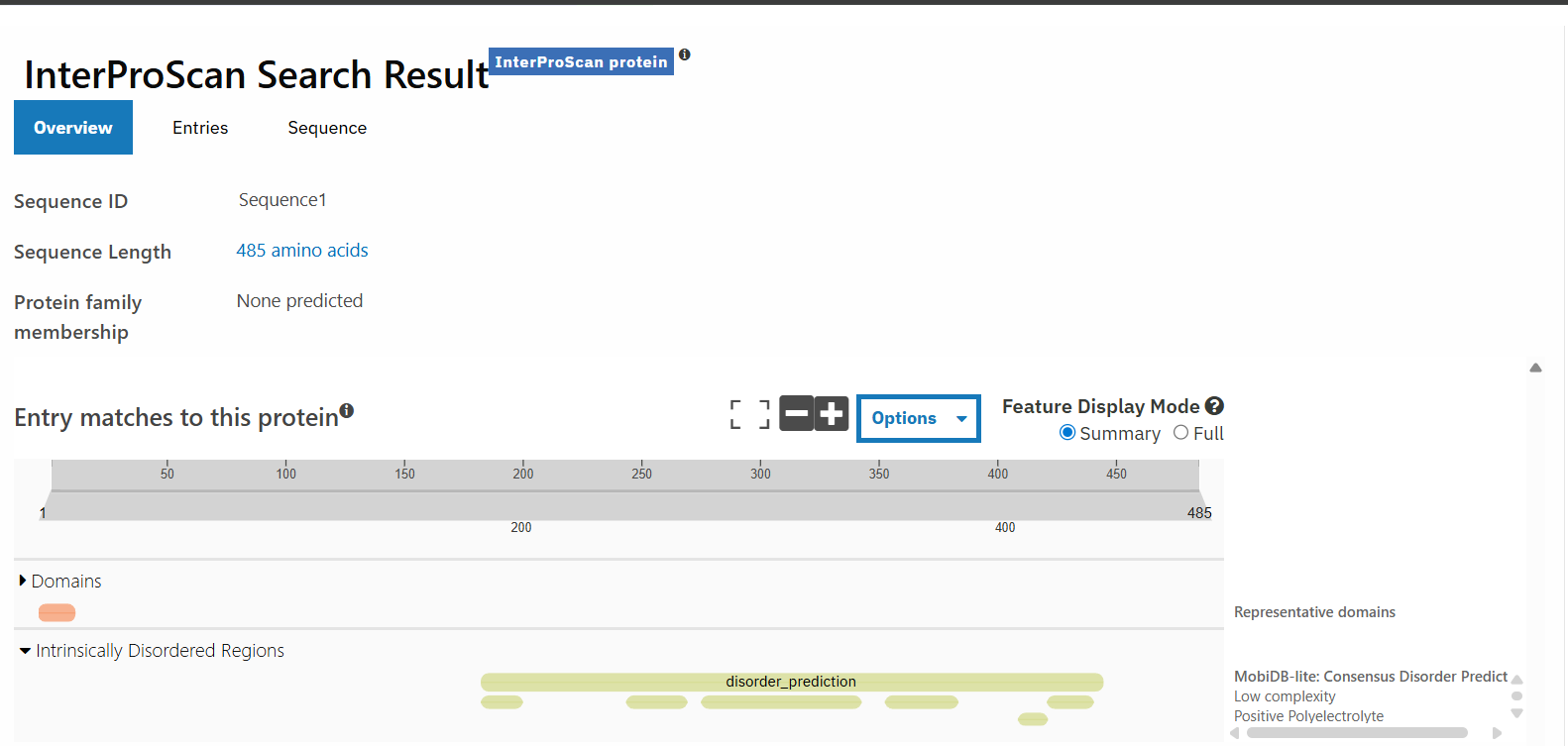
Fro this analysis, I am focusing on silaffin once again, following through from week 2. Silaffin is the protein responsible for the lacy biosilica patterning that diatom frustules form. I am interested in modifying this protein because tuning that pattern could allow for more targeted and intentional interactions between cement rubble and diatom placement for the final project.
silaffin has 485 amino acids, with S appearing most frequently occuring 99 times.
It doesn’t belong to any protein families, which could be because this protein has a very low confidence score and is more rare, leading to less information on its structure and protein family.
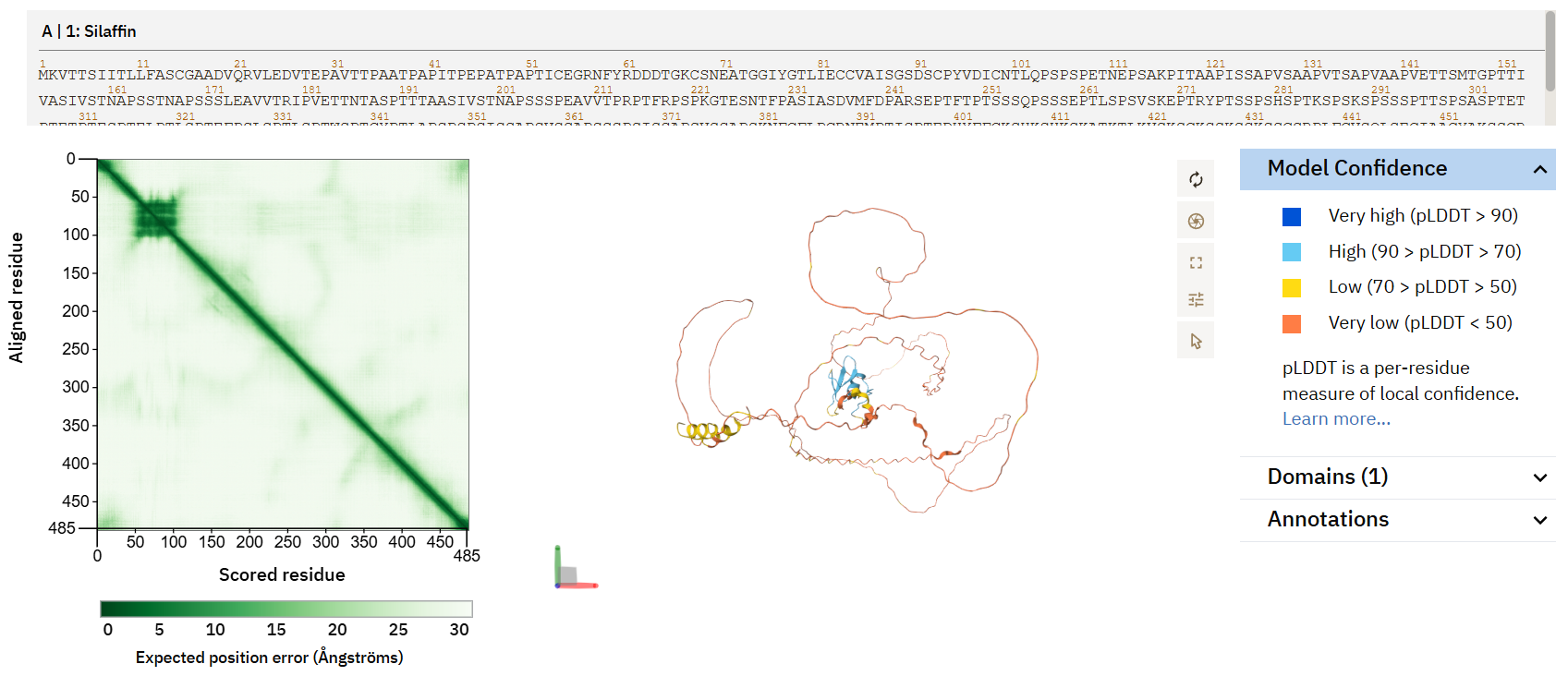
screenshot from uniprot
screenshot from alphafold
With a less documented protein, there is also more likelihood for error. While some areas of the protein with better confidence have a defined beta sheets and alpha helices, most of the protein is modeled with very low confidence.
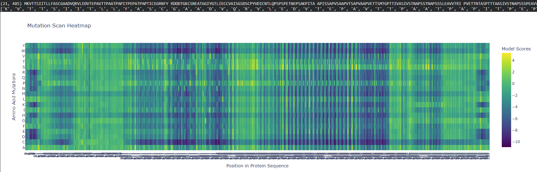
ML Design Tools
the mutation scan shows that amino acids that are most likely to mutate (more yellow in this diagram), are the L,R, and S amino acids. Position 27 is most likely to have a mutation as well.
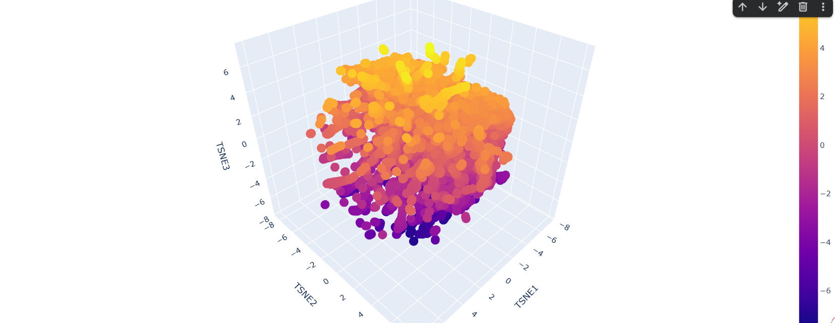
latent space analysis shows concentrated areas. Some of the matches include proteins from organisms ranging from fungi to humans with seemingly low similarity in function. This could be due to the low confidence in the silaffin protein, so the comparative space could be drawing upon very different organisms since there is very little information to respond to.
Week 5: Protein Design Part 2
Week 6: Genetic Circuits
DNA Assembly
What are some components in the Phusion High-Fidelity PCR Master Mix and what is their purpose?
This Master Mix is used to increase the speed and yield of the PCR product, with accurate DNA sequences maintained for DNA replication for testing. The HF buffer variant contains DNA polymerase, deoxynucleotides, buffer, and magnesium dichloride.
What are some factors that determine primer annealing temperature during PCR?
Annealing temperatures are determined by the melting temperature of the selected primers. Some factors that determine the melting point include the number of nucleotides present in the DNA oligo (the short single strand piece of DNA for this experiment).
There are two methods from this class that create linear fragments of DNA: PCR, and restriction enzyme digests. Compare and contrast these two methods, both in terms of protocol as well as when one may be preferable to use over the other.
How can you ensure that the DNA sequences that you have digested and PCR-ed will be appropriate for Gibson cloning?
You can check that the product has the right DNA concentration for the Gibson assembly using the nanodrop after purification.
How does the plasmid DNA enter the E.coli cells during transformation?
The E.coli cells are thermally shocked. First they are kept cold on ice, then heat shocked for a few minutes, before
Describe another assembly method in detail (such as Golden Gate Assembly)
Explain the other method in 5 - 7 sentences plus diagrams (either handmade or online).
Golden Gate Assembly works by
Model this assembly method with Benchling or Asimov Kernel!
Week 7: Neomorphic Circuits
Intracellular Artificial Neural Networks
What advantages do IANNs have over traditional genetic circuits, whose input/output behaviors are Boolean functions?
IANNs allow for less prescriptive circuit method, whereas traditional genetic circuits need to be specified at each step, making it a target specific circuit by finding signalling patterns across a vast dataset. Traditional genetic circuits are limited to simpler action/reaction function simulations to create an mRNA therapy.
Describe a useful application for an IANN; include a detailed description of input/output behavior, as well as any limitations an IANN might face to achieve your goal.
IANNs can be very useful for biomaterials that depend on growth based on the specific conditions of each material batch. For example, if you wanted to make a stained glass using biopigments that stains relative to the colors around it, it could be useful to create a series of relative inputs (ex: high presence of red, medium presence of blue, and no presence of black) that could generate a very specific output (purple gene expression) without manually or preemtively determining color expression. The limitation of the IANN approach is that it has a ‘blackbox’ in reasoning, and doesn’t always present a clear reasoning, unlike traditional genetic circuits. When the reasoning is hidden, it could be making decisions from the wrong signals or not following a genetically safe order of operations, potentially leading to unforseen outcomes.
Below is a diagram depicting an intracellular single-layer perceptron where the X1 input is DNA encoding for the Csy4 endoribonuclease and the X2 input is DNA encoding for a fluorescent protein output whose mRNA is regulated by Csy4. Tx: transcription; Tl: translation. Draw a diagram for an intracellular multilayer perceptron where layer 1 outputs an endoribonuclease that regulates a fluorescent protein output in layer 2.
Fungal Materials
What are some examples of existing fungal materials and what are they used for? What are their advantages and disadvantages over traditional counterparts?
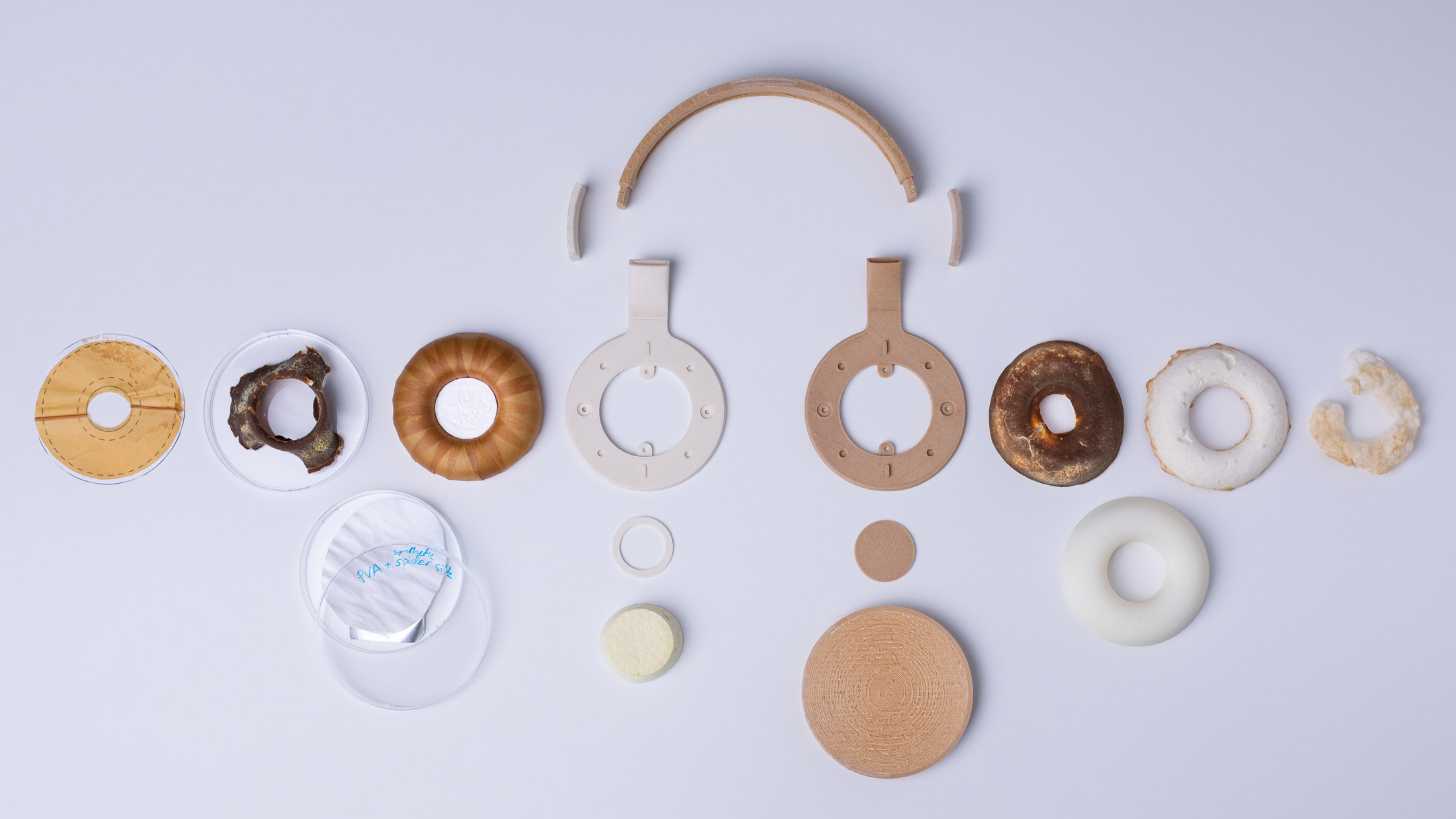
There are headphones designed by Aivan that use mycelium in an experimental synbio headphone design for the leather ear cushion covers. This product is a beautiful example of the multitude of characteristics that bio-based materials like mycelium, silk proteins, and PLA could embody instead of using plastics to make these parts of the headphones. The issue with this product at this time goes partly into user trust and long-term durability of each component. User’s may not feel comfortable having fungus pressed against their ears, and the materials are more susceptible to degredation when exposed to water, heat, sun, etc than a traditional plastic object would be. Headphones are also often tossed into a backpack, worn over earrings, or worn during exercise, leading to a whole range of user habits that could degrade each layer over time. Traditional plastics are also more easy to control and form at this time, when even simple glues may not act as expected with fungal materials over time.
What might you want to genetically engineer fungi to do and why? What are the advantages of doing synthetic biology in fungi as opposed to bacteria?
Fungi feed on simple sugars very well, but this also means that it is easy for mycelium products (before they are fired) to get contaminated during the growth cycle of the fungal products. It would be very useful to have a mycelium product that grows using a specalized sugar source over simple glucose to mitigate the issues of contamination. However this may also create a dependency on a narrow stream of sugar sources that could be used reliably for mycelium fabrication, and potentially limit the accessibility of lo-fi mycelium explorations. Instead it would be cool to have a strain of mycelium with an anti-mold protein, preventing mold from growing in mycelium products whereever mycelium spores are present. Each option would need to be tested for any side effects such changes could have on the mycelium in use. One key advantage of fungi synthetic biology is the speed of growth and visibility of the fungal spores. They are easy to notice by the human eye (larger scale), and still have a high replication rate.
Week 9: Cell-Free Systems
General Homework Questions
Explain the main advantages of cell-free protein synthesis over traditional in vivo methods, specifically in terms of flexibility and control over experimental variables. Name at least two cases where cell-free expression is more beneficial than cell production.
Because cell-free methods exist without the limitations of maintaining a living cell, they offer greater flexibility in exploring the details of protein synthesis without worrying about a cell membrane or the knock-on impacts of every modification before its more
Describe the main components of a cell-free expression system and explain the role of each component.
• lysate: reads the DNA for protein synthesis
• sequence: code for the expression construct
• nuclease free water: the environment for the reaction
• buffers: provides the pH, minerals, etc that are needed for the environment to be right for the reaction to occur
• energy sources from enzymes and substrates: necessary to fuel the transcription and translation for the protein synthesis
Why is energy provision regeneration critical in cell-free systems? Describe a method you could use to ensure continuous ATP supply in your cell-free experiment.
Usually cells have components that can continuously convert energy sources from their environment into ATP, which cell-free systems are not built to do as well. You could build a system of indirect energy sources that get triggered by stages of reactions, creating a more sustained energy supply.
Compare prokaryotic versus eukaryotic cell-free expression systems. Choose a protein to produce in each system and explain why.
The two cell types have a completely different structure, and the way that DNA is folded in these cells is completely different. The protein transcription would be very different, any folding tags, etc.
How would you design a cell-free experiment to optimize the expression of a membrane protein? Discuss the challenges and how you would address them in your setup.
Imagine you observe a low yield of your target protein in a cell-free system. Describe three possible reasons for this and suggest a troubleshooting strategy for each.
Not enough energy source (modify the master mix), the system hasn’t run for long enough (incubate for longer), or maybe the sequence was incorrectly optimized for the lysate system in the protocol (adjust the organism its optimized for) .
Homework Questions from Kate Adamala
Design an example of a useful synthetic minimal cell as follows:
Pick a function and describe it.
a. What would your synthetic cell do? What is the input and what is the output?
It would modify siliffin proteins from diatoms. the input is modified siliffin and the output would be a potential spectral shift between cell free samples
b. Could this function be realized by cell-free Tx/Tl alone, without encapsulation?


c. Could this function be realized by genetically modified natural cell?
Potentially, but the silica cell membrane of the diatoms make them very difficult to open for modification like gibson assembly without killing the cell.
d. Describe the desired outcome of your synthetic cell operation.
Design all components that would need to be part of your synthetic cell.
a. What would be the membrane made of?
b. What would you encapsulate inside? Enzymes, small molecules.
c. Which organism your Tx/Tl system will come from? Is bacterial OK, or do you need a mammalian system for some reason? (hint: for example, if you want to use small molecule modulated promotors, like Tet-ON, you need mammalian)
d. How will your synthetic cell communicate with the environment? (hint: are substrates permeable? or do you need to express the membrane channel?)
Experimental details
a. List all lipids and genes. (bonus: find the specific genes; for example, instead of just saying “small molecule membrane channel” pick the actual gene.)
b. How will you measure the function of your system?
Homework Questions from Peter Nguyen
Freeze-dried cell-free systems can be incorporated into all kinds of materials as biological sensors or as inducible enzymes to modify the material itself or the surrounding environment. Choose one application field — Architecture, Textiles/Fashion, or Robotics — and propose an application using cell-free systems that are functionally integrated into the material. Answer each of these key questions for your proposal pitch:
Write a one-sentence summary pitch sentence describing your concept.
A ‘bruising’ paint that reacts to pressure, creating a lasting impression on a house as it is used more routinely.
How will the idea work, in more detail? Write 3-4 sentences or more.
It would work by creating a biofilm ‘paint’ with pressure-reactive sequences from organisms like mimosa plants. Alternatively the biofilm could use thermoreactive organisms, marked with florescent protein to signal when the temperature is around the range of human skin.
What societal challenge or market need will this address?
This starts a conversation around permanence in a space, and how buildings are seen as more temporary than the people as we move away from being able to afford to own our homes and instead move through rental properties, where our spaces are more defined through the objects than a sense of connection to the walls around us.
In a healthcare sense, this could be useful to highlight surfaces that have been touched, potentially helping with sanitation for sterile rooms if we enter another large-scale health event.
How do you envision addressing the limitation of cell-free reactions (e.g., activation with water, stability, one-time use)?
Walls are not often washed outside of bathrooms and kitchens perhaps, but our hands have moisture, but probably not enough to introduce water into the cell-free system. Pressure on the wall could compress and break micro-scale water cells that are suspended in the biofilm, causing a localized water exposure for the biopigment to react with.
Week 10: Advanced Imaging and Measurement Technology
This value is much higher than the 50ppm threshold, indicating an issue with the tested product. Also, I think this calculation seems to actually be for inaccuracy or error rate.
The sequence has 20 K’s and 6 R’s for peptide segmentation sites
There are 21 labeled peaks, and one unlabeled peak in the chart between 0.5 seconds and 6 seconds.
Compared to the 19 rows for the peptide prediction, the 21 labeled peaks in the chart indicate that there is a slight discrepecy in the threshold between the two software. One observation is that it could down to the two double peaks within two of the larger peaks on the chart, which accounts for the difference of 2 between the rows and peaks. The double peaks could be referring to the same peptide, while each potential peptide in the table is unique.
• m/z(n) = peak 1 = 525.767
• m/z(n-1) = peak 2 = 526.259
delta between peaks = 0.492
z = 1/delta = 2+ charge
Week 11: Bioproduction and Cloud Labs
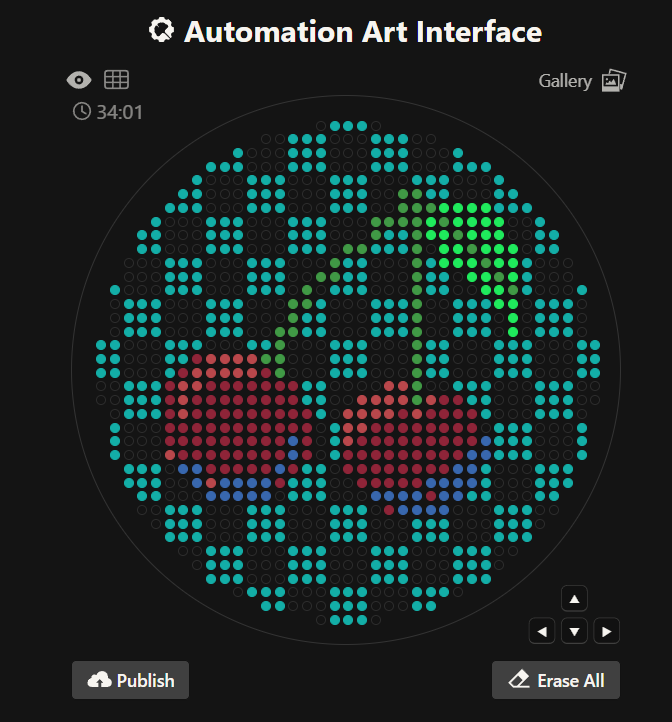
Part A: Pixel Art Canvas
For the HTGAA pixel artwork, I really enjoyed that the experiment was a playful approach to both a large-scale group project and to cloud-based bio projects. I added a blue dot and a yellow dot to a flower-like design that was absorbed into other designs as the artwork developed, but it was cool to try to predict the interests of the other users and contribute to what I thought the pattern would need to have to be completed.
For the future, it could be interesting to have artwork that has space designated per node perhaps to just see the character of each node as well and how they choose to use those limited pixels to connect to the larger HTGAA community.
Part B: Cell-Free Protein Synthesis
Role of each component
• e.coli lysate: this is still necessary to read DNA for protein synthesis
• salts/buffer: maintain the right environment (pH, certain minerals, etc)
• energy/nucleotide system: provide the energy for the synthesis to be conducted
• translation mix: provides base amino acids necessary for synthesis
• additives: supports the systems environment
• backfill: adjust the volume of the reaction to match the experiment needs
The 1 hour and 20 hour incubation master mixes for cell-free systems have some similarities in salts (with some variations) and translation mixes, however the energy system and additives are completely different. Since the 1 hour incubation mix is a PEP-base system, it is composed for immediate energy, while the ribose-based 20 hour incubation mix uses indirect energy sources like NMPs and ribose for more sustained energy sources to match the extended incubation period.
Part C: Global Experiment
sfGFP - an optimized green florescent protein that matures rapidly.
mRFP1 - red florescent with slow maturation and low acid sensitivity
mKO2 - orange florescent with moderate acid sensitivity
mTurquoise2 - cyan florescence that matures rapidly and has very low acid sensitivity
mScarlet_I - red florescent protein with moderate acid sensitivity
Electra2 - basic blue florescence
For my slot (B20), I played with the composition for Electra2, introducing GMP and reducing the nuclease-free water. GMP supports high speed protein production so it could help the synthesis occur faster, and floresce faster.
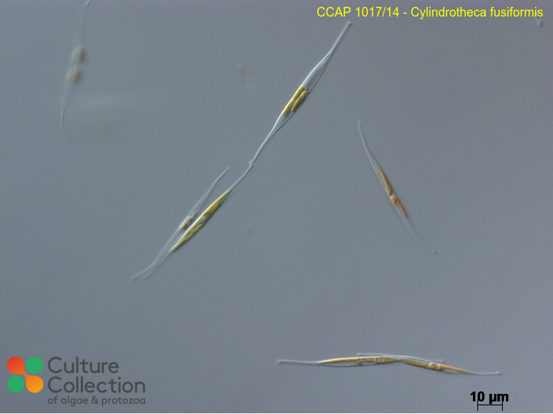
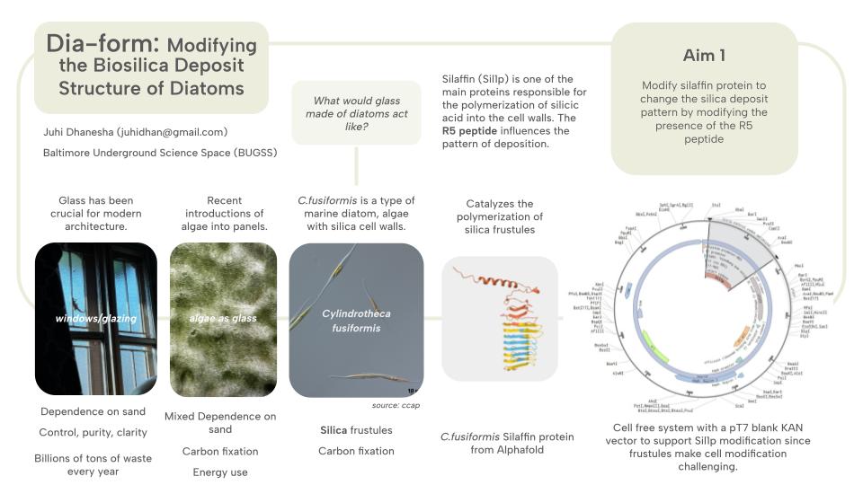
Abstract Clear glass has been crucial to the development of modern architecture, with windows and clear glazing being a major catalyst for indoor living. However this dependence on clear glass has also created a dependence on new material, from a specific and limited sand for glass making, as well as high energy use to fire and float form the sand into glass panes. This project aims at exploring a biological alternative to glass panes, as well as developing the scientific results that could point to future work that uses this biosilica for novel materials, both aggregated with construction waste and as a pure material out of diatom blooms. Diatoms are a type of algae that have silica cell walls called frustules, and these frustules form into intricate lacy, opalescant patterns as the colonies of algae grow. Cylindrotheca fusiformis is a marine diatom species that relies on proteins including silaffins for silicic acid polymerization. By modifying the proteins that are responsible for the diatom structure, this project opens up the mechanical properties of diatoms as a material, where structure is responsible for color expression and for potential material attachment and other characteristics for future projects. Initial work would be conducted in cell-free systems before moving into algal systems once again to work towards creating a ‘glass’ panel with shifting structural properties created through silaffin modifications.
Dia-form: Modifying the Biosilica Deposit Structure of Diatoms
Abstract
Clear glass has been crucial to the development of modern architecture, with windows and clear glazing being a major catalyst for indoor living. However this dependence on clear glass has also created a dependence on new material, from a specific and limited sand for glass making, as well as high energy use to fire and float form the sand into glass panes. This project aims at exploring a biological alternative to glass panes, as well as developing the scientific results that could point to future work that uses this biosilica for novel materials, both aggregated with construction waste and as a pure material out of diatom blooms. Diatoms are a type of algae that have silica cell walls called frustules, and these frustules form into intricate lacy, opalescant patterns as the colonies of algae grow. Cylindrotheca fusiformis is a marine diatom species that relies on proteins including silaffins for silicic acid polymerization. By modifying the proteins that are responsible for the diatom structure, this project opens up the mechanical properties of diatoms as a material, where structure is responsible for color expression and for potential material attachment and other characteristics for future projects. Initial work would be conducted in cell-free systems before moving into algal systems once again to work towards creating a ‘glass’ panel with shifting structural properties created through silaffin modifications.
Project Aims
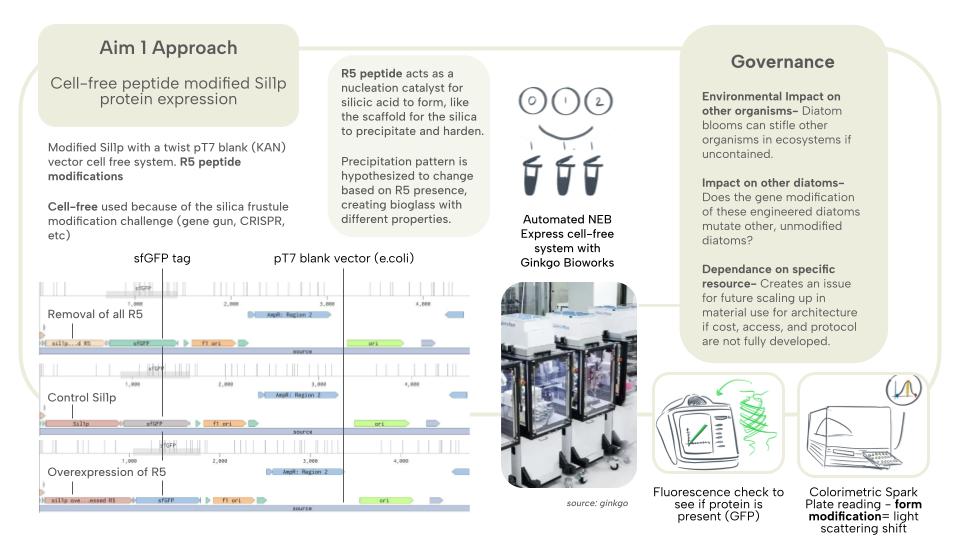
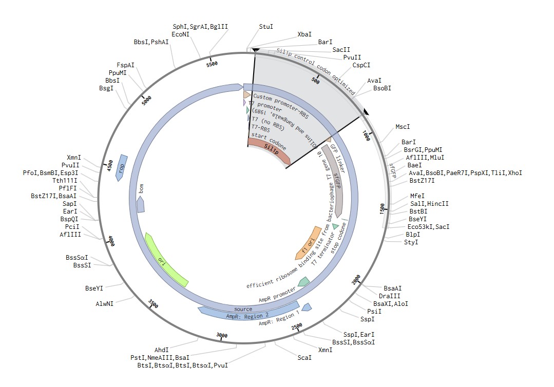
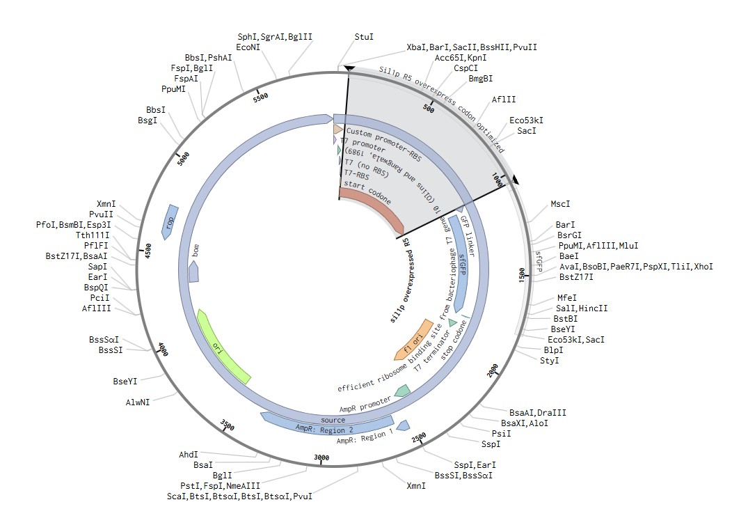
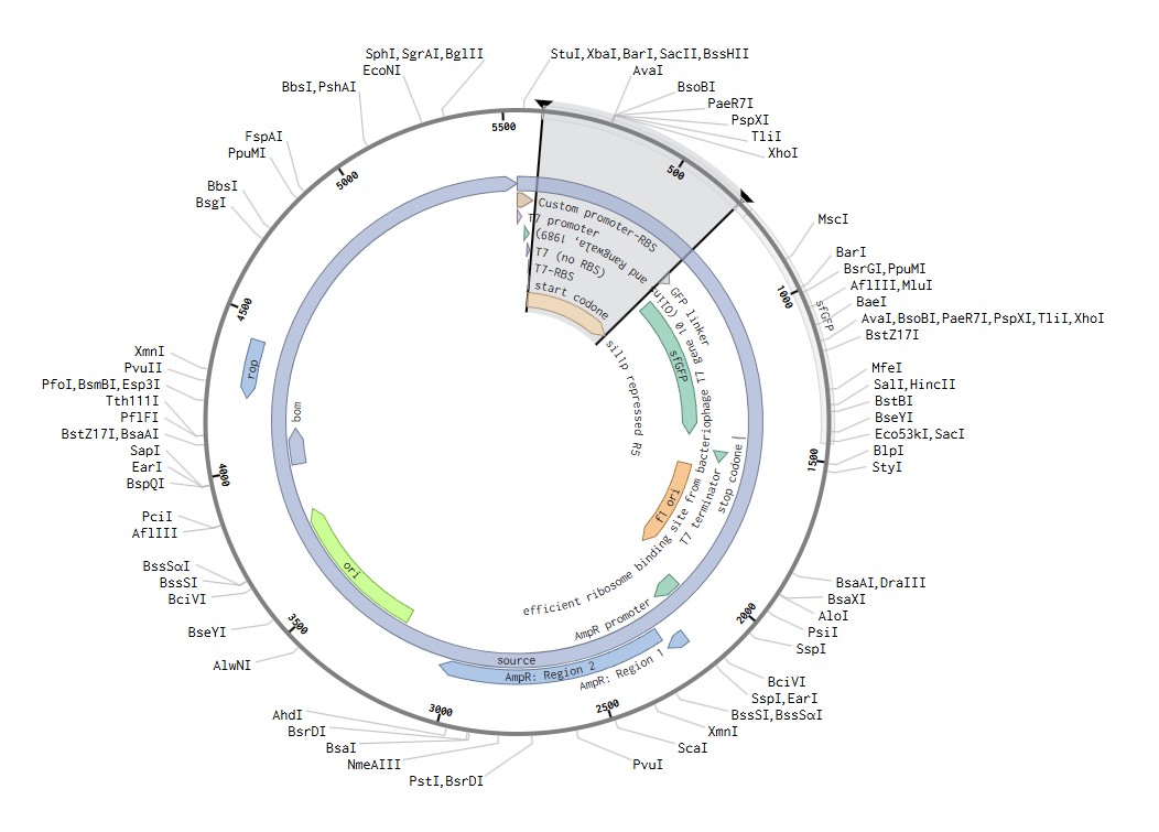
Protein Modification Modify the Sil1p protein of Cylindrotheca fusiformis through cell-free systems. In this aim, I am interested in observing a change of the cell-free system after Sil1p modification from the original sequence. The modification would aim to either overexpress the polymerizing protein, or supress it and see the potential shifts that emerge from those DNA modifications. These shifts would be tested using florescent tagging and a colorimetric assay to check if the system worked, and whether there are notable changes between the modified proteins.
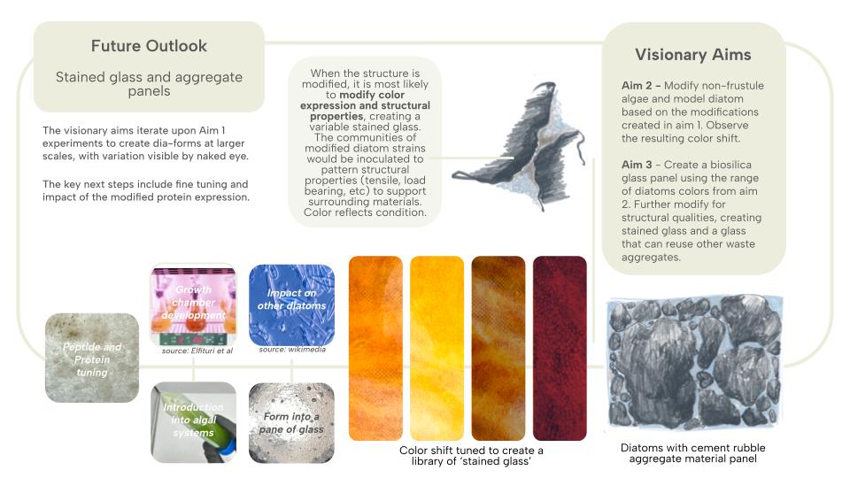
Algae Modification Modify a non-diatom, and non-frustule forming algae and hopefully, C.fusiformis itself, to have the Sil1p protein in place of their traditional cell wall proteins. This would be an exciting yet achievable way to observe the protein in an algae structure, focusing on addressing the challenge of modifying the diatoms without killing the cells.
(OLD AIM 2: Use CRISPR/Cas-9 or the gene gun (most likely CRISPR) to input the modified protein into the T.pseudonona THIS CHANGED AFTER FURTHER DIATOM MODIFICATION RESEARCH.)
Color Shift Modify the TpSil1/2 protein of C.fusiformis to refract light with a visibly different hue through structural modification. The overexpression or knockout of this gene can result in more or less silica deposition, resulting in an altered macropore structure, and thus modifying the light scattering by the physical structure of the frustules.
Rubble Attachment Further modifying the diatom structure to act as a bandaid between two pieces of glass, or two pieces of cement rubble. This takes the structural changes of the silica deposition to a new function, using it to support the use of hard-to-modify architectural waste for new architectural material. Can diatoms from silica patterns that attach onto surrounding objects/surfaces?
Section 3: Background
Diatoms are a unique class of algae that produce silica cell walls called frustules. These frustules have shown up in fossil records, indicating a continued presence of diatoms in our environment over millenia. They are also key organisms for carbon fixation, often looked into for biofuel production. However, by celebrating the unique silica structure of diatoms, this project brings a novel angle of bio-stained glass for architectural applictions to the ways that we may interact with diatoms. Diatoms have been used as aggregates to things like diatomaceous earth, pest control, and for biofuels for their lipid production. Glass production for glazing has created a heavy dependency on specific sand sources that are outsized in a world of skyscrapers. This project proposes an alternative to a sand-derived glass through diatoms. While this project does not suggest that every piece of glass should be made with diatoms from here on out, the production of even a single pane of glass, or single stained glass installation creates a unique condition of a species beyond its natural growning environment and of glass beyond its pure, inert, and uniform state.
Diatoms play an outsized role in oxygen production in the world, (Li et all 2025) producing around 20%, however its silica structure is more of the key focus for this project, with fossil records dating back to the Lower Cretascous Age (https://ucmp.berkeley.edu/chromista/diatoms/diatomfr.html) thanks to their hard silica walls. Kroger et al (1999) identified silaffin as a key protein in the polymerization of silicic acid from aquatic environments into the silica cell walls of diatoms. The R5 peptides were also identified for its repeted occurance within the protein, and its role as the nucleation catalyst for the silicic acid was also identified, acting like scaffolding for the silica to precipitate and harden (Sumper and Brunner 2008), and this peptide plays a key role in the pattern of the silica deposition. From these papers, I focused on the role of a single protein, Sil1p, and the R5 peptide to narrow the experiment to a key singular modification, while Tong et al (2021) speaks towards the importance of the environmental conditions of the silaffin for the growth and attachment, showing that the conditions for growth such as container surface may play a role in shaping the silica deposits beyond the protein modifitions. C.fusiformis is one of the few diatoms that are well studied at this time, and the modification of such a diatom could lead to future experiments into the potential for other modification-resistant organisms to modulate the built environment.
This bioglass approach offers a new way for glass to be considered in the built environment, and the macropore structure gives way for other organisms to occupy these panels in the urban environment. The inherent carbon fixation of the diatoms, and the continued environmental impact of the supported microorganisms like photosynthetic algae, or moss and lichen, can help mitigate the biodiversity depeltion of urban spaces that are often overbuilt in glass. They also provide a softened edge, breaking the flood-prone smooth surfaces with the pore structure. In a synthetic biology context, this project aims to modify a very rigid cell structure, contributing to the body of literature that looks to modifications for organisms that produce calcium and silica-rich structures that could be beneficial for synthetic bones, alternative fuels, and more. This project reframes the inertness of glass, and blurs the boundary between the permanent and temporary materials of the environment. If these panels could be created with living diatoms, their growth patterns would indicate environmental conditions like water stagnation and sun exposure, creating a panel with morphology that is hyper-local, something that architects have been attempting to immitate through computational simulations and programs for decades now, that a large research area of computational design is based off of.
Governance Concerns and Speculated Solutions
Impact on local ecosystems: This design depends on the cultivation of a large quantity of diatoms, which can impact local water ecosystems if not contained in a proper manner.
To address this impact, this project speculates conditions similar to algal production for biofuel, however the unique silica deposits of diatoms make the container a more complex area for research. Aquarium-style tanks would be a good place to start, with wider tubes to account for silica deposits within the filtration system.
Impact on other organisms: If these modified diatoms were to be introduced to an environment through the proposed architectural panels, is there a chance that the mutated diatoms can impact the other organisms in the ecosystems around it?
To address this potential issue, the diatoms will be studied at a petri dish scale tanks with model ecosystem samples, and the other organisms in these sample ecosystems will be tested compared to the control, unmodified diatom ecosystem to see whether there is an impact. Until those tests have a conclusion and beyond, the diatoms can be kiln fired to kill the diatoms in the panel before installation.
Dependence on specific resources: This, like glass, is dependent on a narrow resource, creating issues for future scale up.
This would be addressed through lab-based cultivation and harvesting, creating an initial dependence on a wild organism before creating a contained synthetic growth system to harvest from.
Experimental Design, Techniques, Tools, and Technology
Aim 1’s focus of protein modification began with a study of the silaffin protein and the key peptides that shape it.
reverse translation of sp|Q9SE35|SIL1_CYLFU Silaffin-1 OS=Cylindrotheca fusiformis OX=2853 GN=SIL1 PE=1 SV=1 to a 795 base sequence of most likely codons.
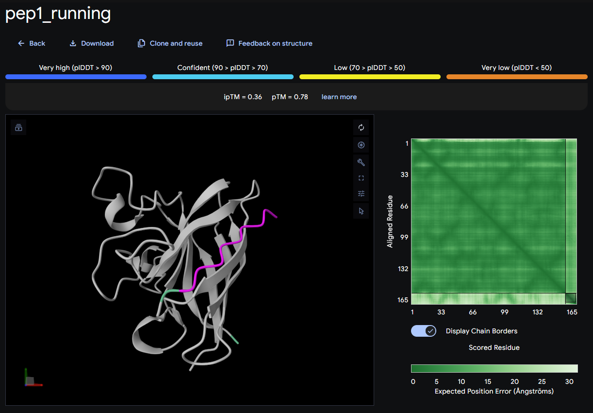
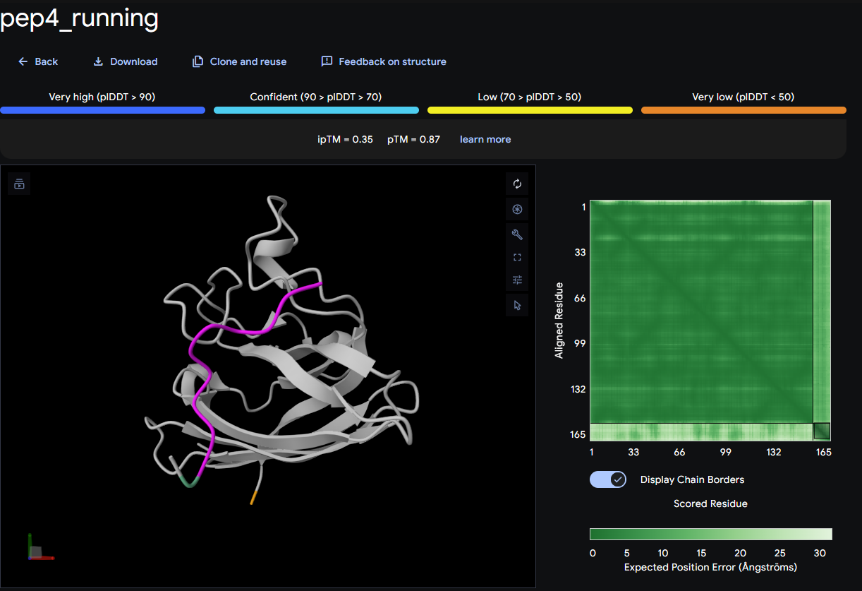
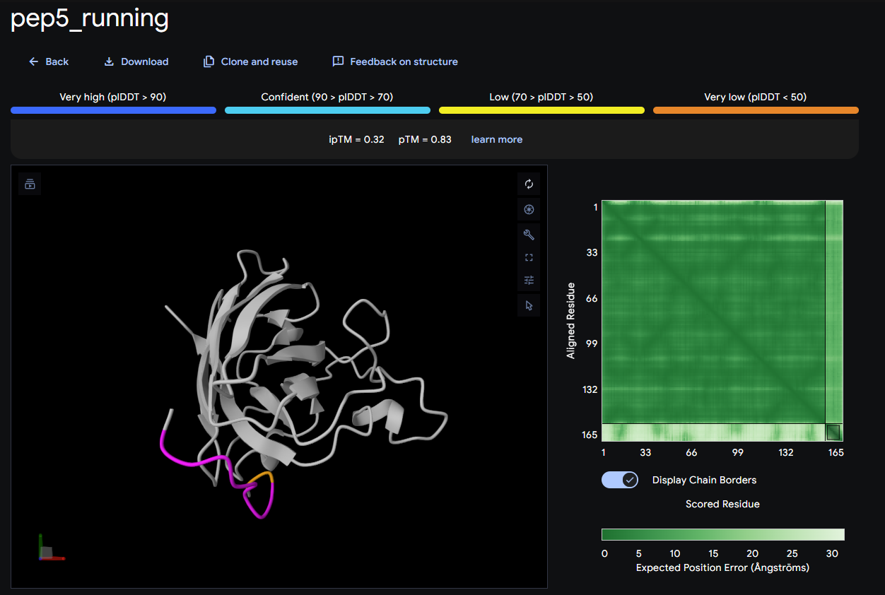
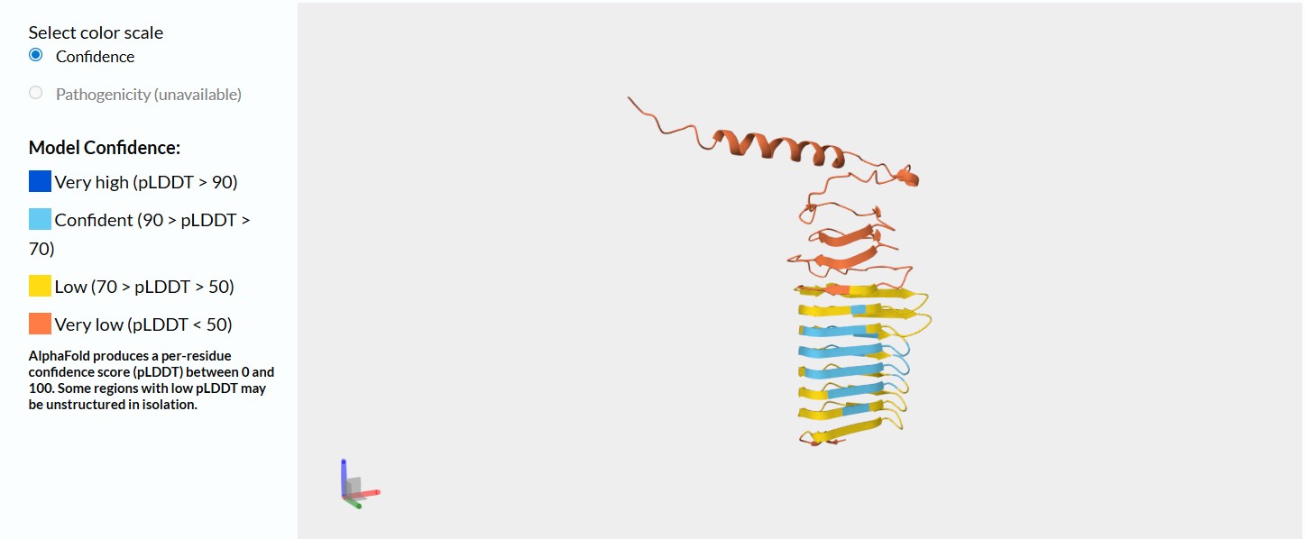
image of the Sil1p protein structure without any modifications
This model on Alphafold demonstrates that this silaffin protein has a much greater structural confidence than the t.pseudonona silaffin protein did. this demonstrates that this diatom is probably more studied than t.pseudonona.
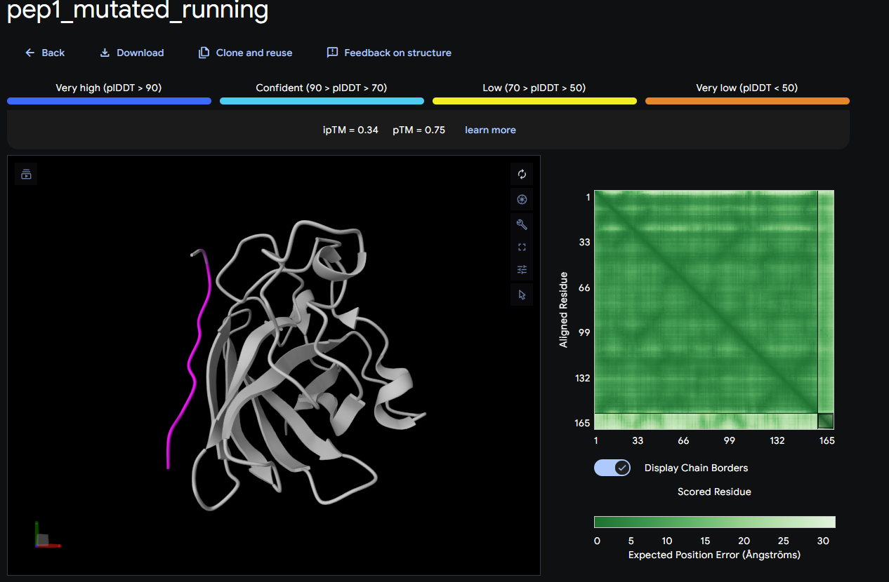
Modify the Sil1p protein R5 peptide
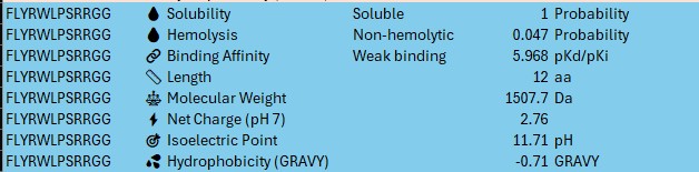
R5 Peptide portion: SSKKSGSYSGSKGSKRRIL (from Claude and double checked through Googling the peptide)
Step 1 — Codon Optimization and Construct Design (Week 1)
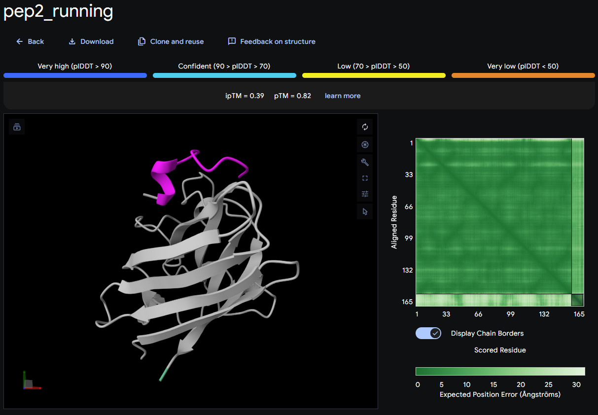
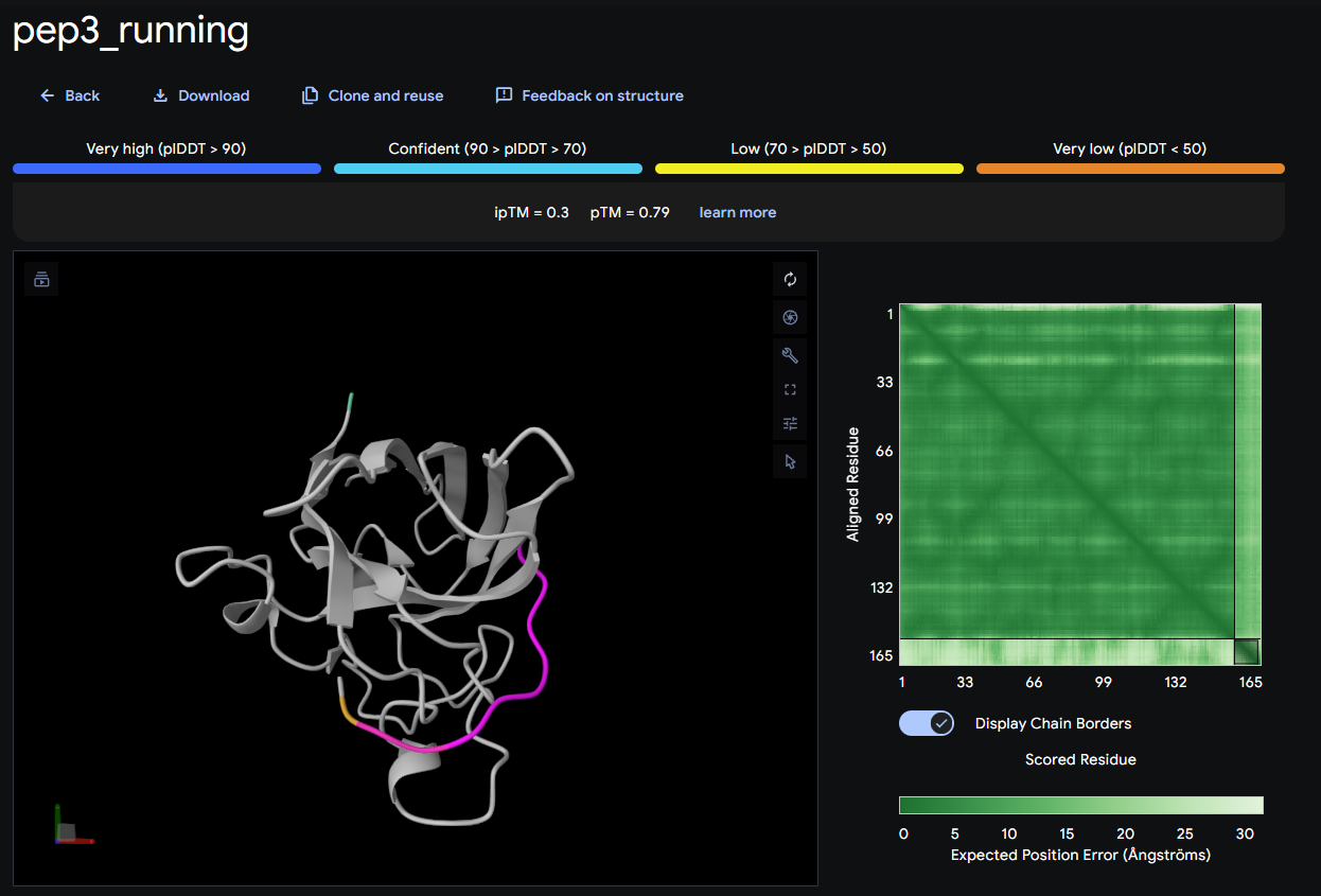
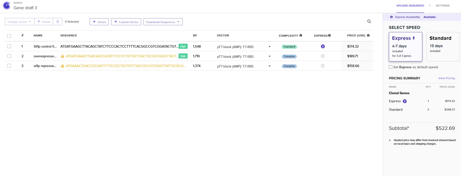
Design three expression constructs encoding: (1) wild-type Sil1p (UniProt O74824), (2) Sil1p-ΔR5 with all five R5 repeat units removed, and (3) Sil1p-2xR5 with the R5 repeat array duplicated from 5 to 10 copies. All constructs include an N-terminal T7 promoter, Shine-Dalgarno sequence, His6-tag, and T7 terminator. Codon-optimize sequences for E. coli BL21 DE3 expression using Twist’s integrated codon optimization tool.
Machine: None (computational design)
Expected result: Three validated construct sequences ready for synthesis ordering
Timeline: Days 1–3
Step 2 — Twist Bioscience DNA Order (Week 1)
Submit all three constructs as whole plasmid synthesis orders to Twist Bioscience using the pTwist-T7 backbone. Select clonal gene synthesis for each construct.
Machine: None (online order)
Expected result: Sequence-verified plasmid DNA delivered within 7–10 business days
Timeline: Days 3–4
Step 3 — Plasmid Receipt and Quality Check (Week 2)
Upon receipt, resuspend lyophilized plasmid DNA per Twist instructions. Verify by Sanger sequencing and gel electrophoresis on a 1% agarose gel.
Machine: ATC Thermal Cycler (for Sanger PCR), gel electrophoresis system
Plate: 96-Armadillo-PCR-AB2396X for PCR setup
Expected result: All three plasmids confirmed sequence-correct
Timeline: Days 10–12
Step 4 — Cell-Free Reaction Setup (Week 2–3)
Using the Echo525 acoustic liquid handler, transfer plasmid DNA (250 ng each) and Ginkgo BL21 DE3 cell-free master mix into a 384-well Echo PP plate in triplicate for each construct plus a no-template negative control. Total reaction volume: 5 µL per well.
Machine: Echo525
Plate: 384-well Echo PP
Expected result: Consistent, low-volume transfers with <5% CV across replicates
Timeline: Day 14
Step 5 — Plate Sealing and Cell-Free Expression (Week 3)
Seal the 384-well plate with the Plateloc using A4s breathable seal to allow gas exchange during expression. Incubate in the Inheco Plate Incubator at 37°C for 4 hours.
Expected result: Protein expression occurs in all construct wells; negative control shows no expression
Timeline: Day 14, hours 0–4
Step 6 — TMOS Silica Precipitation Reaction (Week 3)
Transfer 2 µL of each cell-free reaction to a fresh 384 Greiner black-well clear-bottom plate using the Bravo-384 plate stamp. Using the Multiflo dispenser, add 3 µL of freshly hydrolyzed tetramethyl orthosilicate (TMOS, 1M in 1mM HCl) to each well to initiate silica precipitation.
Machine: Bravo-384, Multiflo
Plate: 384 Greiner black-well clear-bottom
Expected result: Visible silica precipitation in WT and 2xR5 wells within 5–10 minutes; reduced precipitation expected in ΔR5 wells
Timeline: Day 14, hour 5
Step 7 — Mixing and Incubation (Week 3)
Shake the precipitation plate on the BioshakeD3000 at 1,200 rpm for 20 minutes at room temperature to ensure complete silica polymerization.
Machine: BioshakeD3000
Plate: 384 Greiner black-well clear-bottom
Expected result: Complete silica polymerization; visible white precipitate in active wells
Timeline: Day 14, hours 5–5.5
Step 8 — Centrifugation to Pellet Silica (Week 3)
Centrifuge the precipitation plate at 3,000 × g for 10 minutes in the HiG Centrifuge to pellet silica particles.
Machine: HiG Centrifuge
Plate: 384 Greiner black-well clear-bottom
Expected result: Silica pellet visible at well bottom; clear supernatant containing residual free silicic acid
Timeline: Day 14, hour 6
Step 9 — Supernatant Transfer for Colorimetric Assay (Week 3)
Use the Bravo-384 to transfer 4 µL of supernatant from each well into a fresh 384-flat-corning-3640 plate for silicomolybdate assay. Retain the pellet plate for spectrum scanning in Step 11.
Machine: Bravo-384
Plate: 384-flat-corning-3640
Expected result: Clean supernatant transfer without disturbing silica pellets
Timeline: Day 14, hour 6.5
Step 10 — Silicomolybdate Colorimetric Assay (Week 3)
Using the Tempest bulk dispenser, add 1 µL of silicomolybdate reagent (ammonium molybdate in sulfuric acid) to each supernatant well. Incubate 10 minutes at room temperature. Read absorbance at 810 nm on the Spark Plate Reader. Lower absorbance in the supernatant = more silica precipitated by the protein.
Machine: Tempest, Spark Plate Reader
Plate: 384-flat-corning-3640
Expected result: WT and 2xR5 wells show lower A810 than ΔR5 and no-template control, indicating greater silica precipitation
Timeline: Day 14, hours 7–8
Step 11 — Full Spectrum Absorbance Scan of Silica Pellets (Week 3)
Resuspend silica pellets in 5 µL ultrapure water by pipetting. Run a full-spectrum absorbance scan from 400–800 nm on the Spark Plate Reader to capture any optical differences between silica nanoparticles produced by different Sil1p variants.
Machine: Spark Plate Reader
Plate: 384 Greiner black-well clear-bottom
Expected result: Spectral differences between WT, ΔR5, and 2xR5 silica particles; potential blue shift in 2xR5 particles if larger particle size shifts photonic scattering
Timeline: Day 14, hour 8.5
Step 12 — Expression Confirmation: SDS-PAGE (Validation A, Week 3)
Collect 5 µL of cell-free reaction from each construct well before TMOS addition. Load onto SDS-PAGE gel alongside a His-tag protein ladder. Run at 200V for 35 minutes. Stain with Coomassie blue and image.
Step 13 — qPCR Expression Verification (Week 3)
As an orthogonal expression check, extract total RNA from cell-free reactions and run qPCR using primers flanking the His6-tag sequence to confirm transcript levels across all three constructs are comparable.
Machine: CFX Opus qPCR machine
Plate: 96-Armadillo-PCR-AB2396X
Expected result: Similar Ct values across all three constructs, confirming equivalent transcription; any differences in protein yield are post-transcriptional
Timeline: Day 15
Step 14 — Data Analysis and Construct Ranking (Week 4)
Compile colorimetric assay data, full-spectrum scans, SDS-PAGE results, and qPCR data. Calculate silica precipitation efficiency for each construct as: % silica precipitated = (A810 no-template − A810 construct) / A810 no-template × 100. Rank constructs by precipitation efficiency and spectral shift magnitude.
Machine: None (computational analysis)
Expected result: Clear ranking of Sil1p-2xR5 > WT Sil1p > Sil1p-ΔR5 in silica precipitation efficiency
Timeline: Days 16–18
Step 15 — Iteration and Construct Refinement (Week 4–5)
Based on results, design a second round of constructs if needed — e.g., Sil1p-3xR5, Sil1p with randomized repeat spacing, or Sil1p with non-native repeat sequences. Order from Twist Bioscience and re-enter the cell-free pipeline. This iteration loop establishes the quantitative R5 dose-response relationship that forms the foundation for Aim 2 diatom expression work.
Expected result: Refined structure-function map of R5 repeat domains
Timeline: Days 18–25
Results and Quantitative Expectations
Benchling
These cell-free constructs were created to run in the automated lab at Ginkgo. This means that it was created following the NEBExpress protocol, the same one noted in the cell-free lab and one that Ginkgo is familiar with, just with my constructs as the modifier for the experiment.
Technique Checklist (with Claude support)
DNA design and synthesis (Twist Bioscience plasmid synthesis)
The constructs are tagged with sfGFP to floresce if the synthesis was successful, but a colorimetric assay with the spark plate reader would be the best way to see the variation in light scattering that the three different constructs will demonstrate. Since color is a physical structural condition for diatoms, the three modified silica deposit constructs are expected to express color differently and demonstrate a different spectral shift.
These two tests will result in a table that notes whether the sample floresced, and if so, what the spectral reading comes out with. I expect a reading with some green and red, resulting in a brown color, however because it’s in a cell-free system, it may be different since the other parts of the cell system are not present in the same way, however that base variation should be consistent across all three constructs, so the variation is still quantifiable, but may not be representative of the final color expression for Aim 3 as the modified proteins are re-introduced into the diatoms.
This summer, Ronan and I plan to conduct the Aim 1 experiment! I am excited to learn more about the automated lab system and to see if my hypothesis unfolds or if the protein modifications do something unexpected.
• Poulsen, N., Chesley, P.M. & Kröger, N. (2006). Molecular Genetic Manipulation of the Diatom Thalassiosira pseudonana. Journal of Phycology, 42, 1059–1065.
• Tesson B, Lerch SJL, Hildebrand M. Characterization of a New Protein Family Associated With the Silica Deposition Vesicle Membrane Enables Genetic Manipulation of Diatom Silica. Sci Rep. 2017 Oct 18;7(1):13457. doi: 10.1038/s41598-017-13613-8. PMID: 29044150; PMCID: PMC5647440.
Other diatom engineering sources
• Serif, M., Dubois, G., Finoux, AL. et al. One-step generation of multiple gene knock-outs in the diatom Phaeodactylum tricornutum by DNA-free genome editing. Nat Commun 9, 3924 (2018). https://doi.org/10.1038/s41467-018-06378-9
• Li Y, Deng L, Walker EJL, Karas BJ, Mock T. Genetic engineering in diatoms: advances and prospects. Plant J. 2025 Mar;121(6):e70102. doi: 10.1111/tpj.70102. PMID: 40089910; PMCID: PMC11910954.
• Tong, C.Y., Derek, C.J.C. The role of substrates towards marine diatom Cylindrotheca fusiformis adhesion and biofilm development. J Appl Phycol 33, 2845–2862 (2021). https://doi.org/10.1007/s10811-021-02504-1
Budget
Assuming that the lab (in this case, Ginkgo Bioworks), has the plate reader and other equipment needed to conduct the cell-free automated experiment, the price of the Aim 1 experiment is limited to the cost of the Twist order. The cost is fairly high, and for that, Ronan may have only ordered two of the constructs to best balance the cost and time per student on his end at Ginkgo.
Alternative project approaches
Another potential project was to create a touch-reactive biofilm that pigmented with pressure. This could use a system along the lines of the cell-free systems that were discussed in the Week 8 lecture. However for this project, I wanted the color to fade away once the pressure was lessened, however that may not be possible with cell-free systems as they seem to be a one-time reaction.
Alternatively to diatoms, it could also be interesting to use mycelium and genetically modify it to be clear. This uses the strength and potential for mycelium as a building material, binding to architectural waste like glass pieces to create a composite material with the tensile strength and growth rate of mycelium. A clear mycelium would entail a melanin gene knockout, and potentially doing so with a more clear mycelium strain to begin with. This method was developed with the help of CRISPR.
Claude generated information:
SECTION 5: TECHNIQUES, TOOLS, AND TECHNOLOGY
Technique Expansion
1. Cell-Free Protein Synthesis (CFPS)
Cell-free protein synthesis (CFPS) is an in vitro method for producing proteins directly from DNA templates without the use of living cells. The system consists of a cell lysate — in this project, BL21 DE3 lysate prepared at Ginkgo Bioworks — combined with a master mix containing ribosomes, amino acids, energy regeneration components, and RNA polymerase. When a plasmid encoding a T7 promoter-driven gene is added, T7 RNA polymerase transcribes the gene into mRNA, which is then translated by ribosomes present in the lysate into protein. CFPS is particularly powerful for this project because it allows multiple silaffin variants to be expressed and assayed in parallel in a 384-well format within a single day, without the need for bacterial transformation, overnight culture, or IPTG induction — dramatically accelerating the design-build-test cycle for protein engineering.
2. Silicomolybdate Colorimetric Assay
The silicomolybdate assay, also known as the molybdenum blue assay, is a well-established colorimetric method for quantifying free silicic acid (Si(OH)₄) in solution. In the presence of ammonium molybdate under acidic conditions, free silicic acid forms a yellow silicomolybdate complex; upon reduction with ascorbic acid or other reducing agents, this complex turns an intense blue color with peak absorbance at 810 nm. In this project, the assay is applied to the supernatant after silica precipitation — the more silica the Sil1p variant has precipitated from solution, the less free silicic acid remains, and therefore the lower the A810 reading. This indirect measurement elegantly reports on the silica-precipitating activity of each Sil1p variant in a format fully compatible with automated 384-well plate reading on the Spark platform, enabling quantitative comparison of precipitation efficiency across all three constructs simultaneously.
SECTION 6: PROJECT VALIDATION
10a — Validation Choice
Two complementary validation experiments are planned for this project, to be performed based on available lab access and timeline. Validation A (SDS-PAGE) directly confirms that all three Sil1p variants are being produced as proteins of the expected size in the cell-free system, ruling out expression failure as a confound. Validation B (silicomolybdate pilot assay) directly confirms that the wild-type Sil1p is functionally active in precipitating silica from TMOS, and that the ΔR5 truncation reduces this activity — establishing the functional assay and the expected directionality of results before the full 384-well campaign is run.
10b — Validation Protocols
Validation A: SDS-PAGE Expression Confirmation
Collect 5 µL of cell-free reaction from each of the three Sil1p constructs and the no-template control after 4 hours of expression.
Add 5 µL of 2× Laemmli SDS sample buffer to each sample.
Heat samples at 95°C for 5 minutes using the ATC Thermal Cycler.
Load 8 µL of each sample onto a 4–20% gradient SDS-PAGE gel alongside a His-tag protein molecular weight ladder.
Run electrophoresis at 200V for 35 minutes in Tris-glycine SDS running buffer.
Stain gel with InstantBlue Coomassie stain for 30 minutes.
Confirm absence of bands in no-template control lane.
Validation B: Silicomolybdate Pilot Assay
Set up cell-free reactions for WT Sil1p, Sil1p-ΔR5, and no-template control in triplicate in a 96-well plate (25 µL reactions).
Incubate at 37°C for 4 hours in the Inheco Plate Incubator.
Add 5 µL of freshly hydrolyzed TMOS (1M in 1mM HCl) to each well.
Shake on BioshakeD3000 at 1,200 rpm for 20 minutes at room temperature.
Centrifuge at 3,000 × g for 10 minutes in the HiG Centrifuge to pellet silica.
Transfer 20 µL of supernatant to a fresh 96-well flat-bottom plate.
Add 5 µL of silicomolybdate reagent (0.026M ammonium molybdate in 0.1M H₂SO₄) to each well.
Incubate 10 minutes at room temperature.
Add 5 µL of reducing solution (0.1M ascorbic acid) if molybdenum blue endpoint is desired.
Read absorbance at 810 nm on the Spark Plate Reader.
Calculate % silica precipitated relative to no-template control.
Confirm WT Sil1p shows significantly lower A810 than ΔR5 and no-template control (expected: >40% reduction).
10c — Techniques Used
SDS-PAGE (sodium dodecyl sulfate-polyacrylamide gel electrophoresis) separates proteins by molecular weight under denaturing conditions, allowing direct visualization of whether each Sil1p variant has been produced at the correct size and in sufficient quantity by the cell-free system. The silicomolybdate assay is a spectrophotometric technique that quantifies free silicic acid in solution through formation of a chromogenic molybdate complex, providing an indirect but highly sensitive measure of silica precipitation activity. Cell-free protein synthesis leverages the transcription and translation machinery of bacterial lysates to produce proteins from plasmid DNA templates in vitro, enabling rapid, parallel expression of multiple variants without live organism handling. Together, these three techniques — SDS-PAGE, colorimetric spectrophotometry, and cell-free expression — form an integrated validation pipeline that confirms both the production and the functional activity of each engineered Sil1p variant before proceeding to the full high-throughput 384-well campaign.
Troubleshooting
The most likely technical challenge is low or absent silaffin protein expression in the cell-free system, which could result from poor codon optimization, mRNA secondary structure inhibiting translation, or issues with the His6-tag affecting protein folding. If expression is not confirmed by SDS-PAGE, the first corrective step would be to re-optimize the 5’ UTR sequence using the Salis Lab RBS Calculator and reorder a revised construct from Twist Bioscience before repeating the cell-free expression. A second potential challenge is non-specific silica precipitation in the no-template control wells — TMOS is chemically reactive and will self-polymerize under certain pH conditions independently of silaffin; this can be mitigated by carefully controlling TMOS hydrolysis conditions (1mM HCl, room temperature, freshly prepared) and including a TMOS-only control well in every plate. A third limitation is that cell-free silica precipitation may not faithfully recapitulate the in vivo post-translational modifications of native Sil1p — including phosphorylation and polyamine modifications on lysine residues — which are known to enhance biosilicification activity; this is an inherent constraint of the cell-free platform that will need to be addressed in Aim 2 when the work moves into a native diatom expression system. Finally, the full-spectrum scan for structural color differences is exploratory and may not yield interpretable optical signals at the scale of cell-free silica nanoparticles — if this is the case, dynamic light scattering (DLS) would be pursued as an alternative to characterize particle size distributions.
Claude References
Kröger, N., Deutzmann, R., & Sumper, M. (1999). Polycationic peptides from diatom biosilica that direct silica nanosphere formation. Science, 286(5442), 1129–1132.
Sumper, M., & Brunner, E. (2008). Silica biomineralisation in diatoms: the model organism Thalassiosira pseudonana. ChemBioChem, 9(8), 1187–1194.
Kröger, N., Lorenz, S., Brunner, E., & Sumper, M. (2002). Self-assembly of highly phosphorylated silaffins and their function in biosilica morphogenesis. Science, 298(5593), 584–586.
Poulsen, N., Sumper, M., & Kröger, N. (2003). Biosilica formation in diatoms: characterization of native silaffin-2 and its role in silica morphogenesis. PNAS, 100(21), 12075–12080.
Lechner, C. C., & Becker, C. F. (2015). Silaffins in silica biomineralization and biomimetic silica precipitation. Marine Drugs, 13(8), 5297–5333.