HTGAA 2026: Individual Final Project Documentation Project Title: A Hydrogel-Embedded Multiple Input-Output (MIMO) Genetic Circuit for IL-6 and Hypoxia Detection
Section 1: Abstract Chronic inflammatory diseases and solid tumors share a pathophysiological signature characterized by elevated interleukin-6 (IL-6) and reduced local oxygen tension (hypoxia). Current clinical monitoring cannot detect these signals simultaneously at the tissue level in real time, requiring repeated blood draws or imaging procedures that are slow, lab-dependent, and inaccessible to over 3.5 billion people globally who lack basic diagnostic services. This project addresses that gap by engineering a hydrogel-embedded, cell-free genetic circuit capable of sensing both IL-6 and hypoxia as dual inputs and producing two corresponding outputs: sfGFP fluorescence as a quantifiable reporter signal and a therapeutic peptide as a functional biological output.
HTGAA 2026: Individual Final Project Documentation
Project Title: A Hydrogel-Embedded Multiple Input-Output (MIMO) Genetic Circuit for IL-6 and Hypoxia Detection
Section 1: Abstract
Chronic inflammatory diseases and solid tumors share a pathophysiological signature characterized by elevated interleukin-6 (IL-6) and reduced local oxygen tension (hypoxia). Current clinical monitoring cannot detect these signals simultaneously at the tissue level in real time, requiring repeated blood draws or imaging procedures that are slow, lab-dependent, and inaccessible to over 3.5 billion people globally who lack basic diagnostic services. This project addresses that gap by engineering a hydrogel-embedded, cell-free genetic circuit capable of sensing both IL-6 and hypoxia as dual inputs and producing two corresponding outputs: sfGFP fluorescence as a quantifiable reporter signal and a therapeutic peptide as a functional biological output.
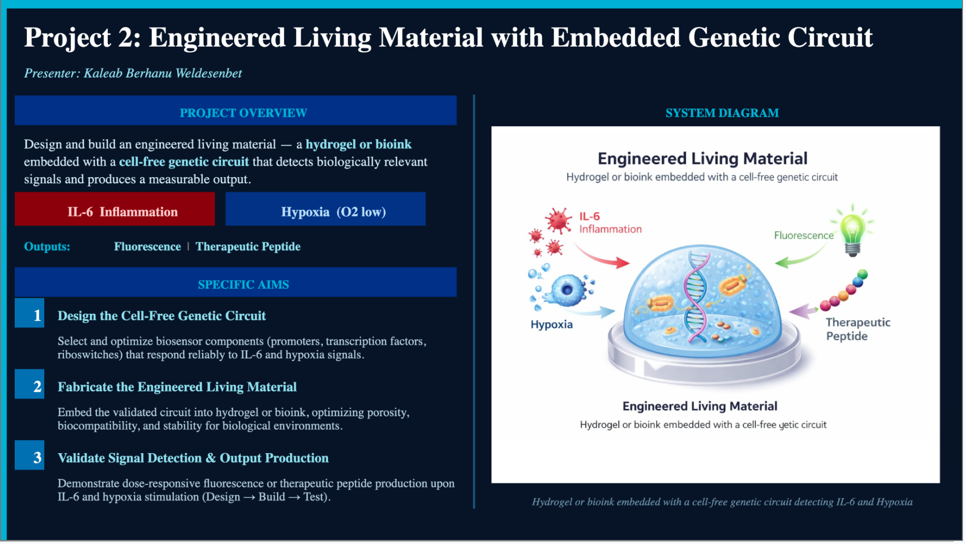
The system integrates a Multi-Input Multi-Output (MIMO) genetic circuit encoded on a plasmid containing a T7 promoter-driven sfGFP construct, encapsulated within an agarose hydrogel bioink matrix alongside E. coli cell-free extract (CFE). The hypothesis is that combining IL-6-responsive and hypoxia-responsive regulatory elements within a single cell-free circuit, embedded in a biocompatible hydrogel, will enable autonomous, localized disease detection and therapeutic response without requiring living cells. The specific aims are to design and validate the MIMO genetic circuit, optimize hydrogel encapsulation for sustained cell-free protein synthesis, and characterize dual-input fluorescence output as a proof of concept for future therapeutic peptide integration. This system has the potential to function as an implantable or wound-applied biosensor-therapeutic platform capable of responding autonomously to disease microenvironments.
Section 2: Project Aims
Aim 1: Experimental Aim
The first aim of my final project is to design, construct, and validate a Multi-Input Multi-Output (MIMO) genetic circuit that produces sfGFP fluorescence output in response to IL-6 and hypoxia signals in a cell-free expression system, by utilizing T7 promoter-driven DNA construct design in Benchling, cell-free transcription/translation reactions using E. coli BL21 (DE3) Star lysate, and fluorescence plate reader quantification to confirm circuit functionality.
Relevant methods and resources:
Plasmid design in Benchling with codon-optimized sfGFP insert under T7 promoter control
Twist Biosciences DNA synthesis order for the MIMO circuit construct
Cell-free reaction setup using NMP-Ribose-Glucose 20-hour master mix
IL-6 protein spike-in and cobalt chloride (CoCl2) as a chemical hypoxia mimic
Agarose hydrogel encapsulation protocol based on Sánchez-Costa et al. (2024)
Aim 2: Development Aim
Following successful validation of IL-6 and hypoxia-triggered sfGFP output in Aim 1, the next step would be to replace the sfGFP reporter with a therapeutic peptide output cassette, encoding an anti-inflammatory peptide such as an IL-6 receptor antagonist peptide, and to validate dosage-controlled release kinetics from the agarose hydrogel matrix using LC-MS quantification of peptide concentration in the hydrogel supernatant over a 24 to 72-hour window. This aim would demonstrate that the system can be switched from a diagnostic reporter mode to a therapeutic delivery mode using the same circuit architecture, and would characterize the hydrogel as a controlled release vehicle capable of producing clinically relevant peptide concentrations in response to disease signals.
Aim 3: Visionary Aim
The long-term vision for this project is to develop a fully implantable or wound-applied Engineered Living Material (ELM) that autonomously monitors the inflammatory and hypoxic microenvironment of chronic wounds, diabetic ulcers, or solid tumors, and responds in real time by producing localized therapeutic peptides at the site of disease. If fully realized, this platform would eliminate the need for repeated systemic drug dosing or clinical lab testing for conditions like sepsis, cancer, and chronic inflammation, reducing treatment delays and enabling precision medicine at the tissue level. The broader concept challenges the existing paradigm of passive drug delivery and reactive clinical monitoring by replacing it with an active, self-regulating biological system embedded directly into the disease microenvironment — a shift that could redefine how chronic disease management is approached across oncology, wound care, and critical care medicine globally.
Section 3: Background
Background and Literature Context
Citation 1: Sánchez-Costa et al. (2024) — In-Hydrogel Cell-Free Protein Expression System as Biocompatible and Implantable Biomaterial
This study demonstrated that E. coli cell-free extracts (CFEs) can be successfully encapsulated within agarose hydrogels while retaining full transcription and translation activity, enabling sfGFP production that diffuses from the hydrogel core to its surface. The authors showed that agarose hydrogels outperformed other matrices in supporting sustained cell-free protein synthesis. Critically, the hydrogel system was confirmed to be biocompatible both in vitro through cell colonization and proliferation assays and in vivo in a preclinical mouse implantation model, with no adverse immune response observed. The study also demonstrated that freeze-drying of the agarose hydrogel still supports protein synthesis upon reconstitution, enabling point-of-care deployment in resource-limited settings. This work establishes the foundational biomaterial platform upon which the MIMO circuit of this project is built.
Citation 2: Pardee et al. (2016) — Rapid, Low-Cost Detection of Zika Virus Using Programmable Biomolecular Components
Pardee and colleagues demonstrated that cell-free gene expression systems can be freeze-dried onto paper substrates and reactivated with minimal equipment to detect specific nucleic acid sequences from pathogens including Zika virus, producing colorimetric outputs readable by eye. The work established that cell-free systems are robust enough to be deployed outside of laboratory environments and that their regulatory outputs can be programmed through toehold switch RNA sensors responding to specific molecular inputs. This paradigm of programmable, cell-free biosensing with embedded output logic is directly analogous to the MIMO circuit architecture proposed in this project, extended here to protein-level and oxygen-level inputs rather than nucleic acid inputs. Together these two studies establish the feasibility of combining programmable cell-free gene circuits with biocompatible encapsulation matrices for real-world diagnostic and therapeutic applications.
Novelty and Innovation
This project is novel in three distinct ways. First, while cell-free protein synthesis in hydrogels has been demonstrated as a proof of concept for single-output expression systems, no published work has demonstrated a dual-input MIMO genetic circuit operating within an encapsulated hydrogel format — the combination of multi-signal logic gating with hydrogel encapsulation represents a genuinely new system architecture. Second, the use of IL-6 as a transcriptional input in a cell-free circuit is an underexplored application; most cell-free biosensing work has focused on nucleic acid inputs or small molecule inducers, and expanding the input modality to a clinically relevant cytokine opens a new class of programmable biosensors for inflammatory disease. Third, the integration of a therapeutic peptide output within the same circuit that performs the sensing function creates a self-contained sense-and-respond system that does not require any external intervention between detection and therapy, which is a conceptual departure from existing diagnostic-therapeutic platforms that treat sensing and treatment as separate sequential steps.
Why This Project Matters
The problem this project addresses is the fundamental mismatch between the speed of disease progression at the tissue level and the speed of clinical monitoring available to detect it. For conditions like sepsis, where 56% of deaths are preventable with faster diagnosis (Lancet, 2020), or diabetic wound infections where delayed detection leads to amputation, the 2 to 6-hour typical lab turnaround time for inflammatory biomarker tests is clinically unacceptable when biosensor-based detection can in principle operate in minutes. Over 3.5 billion people globally lack access to basic diagnostic services (WHO, 2022), and the 422 million people living with diabetes (WHO, 2023) represent a population for whom continuous localized monitoring of wound inflammation would have immediate, life-altering benefit.
This project matters because it addresses the point-of-care gap not by improving existing lab tests, but by eliminating the need for a lab entirely, placing the sensing and therapeutic response capability directly at the tissue site where the disease is occurring. If successful, the outputs of this project could reframe how implantable biomaterials are designed, shifting them from passive structural supports or drug reservoirs to active participants in disease management that read biological signals and respond accordingly. At the field level, this work contributes to the emerging discipline of Engineered Living Materials by demonstrating that cell-free systems can serve as the functional biological layer within implantable hydrogels without the biosafety concerns associated with living genetically modified organisms. The potential clinical impact extends across wound care, oncology, and critical care, where real-time localized biosensing and autonomous therapeutic release could meaningfully change patient outcomes.
Ethical Implications
This project involves the design of a programmable biological system intended for implantation or wound application in human patients, which raises significant ethical considerations across multiple principles. The principle of non-maleficence is directly relevant: the encapsulation of E. coli cell-free extracts within an implantable material introduces the risk of immune response, even in the absence of living organisms, as bacterial proteins and lipopolysaccharides (LPS) retained in the lysate could trigger inflammatory cascades in immunocompromised patients for whom this device is most likely to be used. The principle of justice is also implicated: while the motivation of the project is explicitly to address diagnostic inequity in low-resource settings, there is a risk that the technology, if commercialized, would be priced in ways that replicate existing access barriers rather than reducing them. The principle of beneficence requires that the therapeutic peptide output be characterized not only for efficacy but for potential off-target effects, particularly since a self-regulating release system cannot be externally switched off once implanted.
To ensure ethical conduct of this research and responsible development of its implications, several measures should be taken. All cell-free lysates used in hydrogel formulations intended for in vivo testing should be rigorously depleted of LPS through standard endotoxin removal protocols and tested for endotoxin levels below the FDA threshold of 0.5 EU/mL before any animal studies proceed. The therapeutic peptide output should undergo full in vitro cytotoxicity profiling and off-target receptor binding analysis before any in vivo experiments. Open publication of protocols and DNA sequences is a core commitment of this project, ensuring that the technology is accessible to academic groups in low-resource settings rather than locked behind proprietary barriers. A potential unintended consequence of the autonomous sense-and-respond architecture is that the system could produce therapeutic output in response to non-pathological IL-6 fluctuations, for example during normal exercise-induced inflammation, which could cause unintended biological effects. This uncertainty must be addressed through careful characterization of the input threshold sensitivity of the circuit before any clinical translation is considered. An alternative to the implantable format that avoids many of these concerns entirely would be a topically applied hydrogel patch that remains external to the body, reducing immune risk and allowing easy removal if adverse effects are observed.
Section 4: Experimental Design, Techniques, Tools, and Technology
Detailed Experimental Plan
Step 1: DNA Construct Design (Week 1, estimated 3 days)
Design the MIMO genetic circuit construct in Benchling, incorporating a T7 promoter, ribosome binding site (RBS), codon-optimized sfGFP insert, and T7 terminator as the baseline expression cassette. Annotate restriction enzyme sites (EcoRI, NdeI, BstYI confirmed from the plasmid map) for downstream cloning verification. Expected result: a fully annotated ~800 bp plasmid construct ready for synthesis.
Step 2: Twist Biosciences DNA Synthesis Order (Week 1, submission; Week 2, delivery)
Submit the finalized sfGFP construct sequence to Twist Biosciences for gene synthesis. Specify the T7 promoter context and confirm codon optimization for E. coli expression. Expected result: arrival of lyophilized dsDNA within 5 to 7 business days.
Step 3: Plasmid Preparation and Transformation (Week 2, estimated 2 days)
Transform the synthesized construct into DH5alpha competent cells using heat shock protocol, plate on ampicillin LB agar, and grow overnight at 37°C. Pick 4 to 6 colonies and perform miniprep DNA extraction. Verify construct by Sanger sequencing using T7 promoter primer. Expected result: confirmed sequence-verified plasmid at concentration above 100 ng/uL.
Step 4: Cell-Free Master Mix Preparation (Week 2, estimated 1 day)
Prepare the NMP-Ribose-Glucose 20-hour master mix at 2X concentration according to the HTGAA Cell-Free Master Mix Composition protocol, including potassium glutamate, HEPES-KOH pH 7.5, magnesium glutamate, potassium phosphate salts, ribose, glucose, AMP, CMP, GMP (via guanine salvage), UMP, amino acid mix, tyrosine, cysteine, and nicotinamide. Expected result: complete 2X master mix aliquoted and stored at -80°C.
Step 5: Baseline Cell-Free sfGFP Expression (Week 3, estimated 1 day)
Set up 20 uL cell-free reactions in triplicate: 6 uL BL21 (DE3) Star lysate, 10 uL 2X master mix, 2 uL sfGFP plasmid DNA template, 2 uL nuclease-free water. Incubate at 30°C for 20 hours. Measure sfGFP fluorescence every 2 hours using plate reader at excitation 485 nm / emission 510 nm. Expected result: increasing fluorescence signal reaching plateau by 12 to 16 hours, confirming functional cell-free expression.
Step 6: IL-6 Input Titration (Week 3, estimated 2 days)
Replace the 2 uL water backfill in Step 5 with recombinant human IL-6 protein at concentrations of 0, 1, 10, 100, and 1000 ng/mL. Assess whether IL-6 concentration-dependent changes in sfGFP output are detectable by the circuit. Expected result: measurable dose-response relationship between IL-6 concentration and fluorescence output if the IL-6-responsive regulatory element is functional.
Step 7: Hypoxia Input Simulation (Week 3, estimated 1 day)
Use cobalt chloride (CoCl2) at 100 to 500 uM as a chemical hypoxia mimic to activate hypoxia-inducible regulatory elements in the circuit. Alternatively, run reactions in a sealed anaerobic chamber with 1% O2. Expected result: elevated sfGFP output under hypoxic conditions compared to normoxic control.
Step 8: Dual-Input MIMO Test (Week 4, estimated 2 days)
Combine IL-6 spike-in and CoCl2 hypoxia mimic simultaneously in a single cell-free reaction to test whether both inputs produce an additive or synergistic increase in sfGFP output. Test all four input combinations: no input, IL-6 only, hypoxia only, and both inputs together. Expected result: highest fluorescence output in the dual-input condition, confirming MIMO logic gating behavior.
Step 9: Agarose Hydrogel Encapsulation (Week 4, estimated 2 days)
Prepare 2% w/v agarose hydrogel in nuclease-free water, autoclave, and cool to 42°C before mixing with cell-free extract and plasmid DNA at a 1:1 ratio. Cast into 6-well plates and allow to solidify at room temperature. Incubate encapsulated reactions at 30°C and measure sfGFP fluorescence diffusing from the gel surface at 0, 4, 8, 12, and 20 hours. Expected result: detectable sfGFP fluorescence in the hydrogel supernatant by 8 hours, confirming that encapsulation does not abolish cell-free protein synthesis activity.
Step 10: Freeze-Drying Stability Test (Week 5, estimated 2 days)
Lyophilize hydrogel-encapsulated cell-free reactions and reconstitute with nuclease-free water containing plasmid DNA. Incubate and measure sfGFP output as above. Expected result: recovery of sfGFP fluorescence above 50% of fresh reaction output, confirming freeze-dry stability for point-of-care deployment.
Step 11: Data Analysis and Visualization (Week 5, estimated 2 days)
Compile all fluorescence time-course data and generate dose-response curves for IL-6 and hypoxia inputs versus sfGFP output. Calculate signal-to-noise ratio for each input condition. Plot hydrogel versus liquid-phase reaction fluorescence kinetics. Expected result: complete dataset with statistical comparison between all conditions, with at least one input condition showing a statistically significant increase in sfGFP output relative to no-input control.
Step 12: Documentation and Final Report (Week 5, estimated 1 day)
Update HTGAA webpage with experimental data, plasmid map, fluorescence graphs, and protocol. Archive Benchling construct, Twist order confirmation, and sequencing results. Expected result: fully documented project with reproducible protocol and publicly accessible data.
Chassis Selection (DH5alpha for plasmid propagation)
Plasmid Preparation
Bacterial Culturing
Quality Control / Analysis
Gibson Assembly
Primer Design or Selection
PCR Reactions
Protein Design
Use of Boltz or PepMLM (for therapeutic peptide design in Aim 2)
Expanded Technique Descriptions
Cell-Free Reactions
The core experimental platform of this project is a cell-free transcription-translation (TX/TL) system using BL21 (DE3) Star E. coli lysate, which retains all the ribosomes, translation factors, and T7 RNA polymerase needed for gene expression without requiring living cells. The MIMO genetic circuit plasmid encoding sfGFP under T7 promoter control will be added directly to 20 uL reactions alongside the NMP-Ribose-Glucose 20-hour master mix, with IL-6 and CoCl2 supplemented as the two input signals in the 2 uL custom reagent supplement volume. Fluorescence will be measured every 2 hours over a 20-hour window to generate kinetic expression profiles for each input condition. This approach allows rapid, iterative testing of circuit behavior without the time cost of bacterial transformation or cell culture, making it ideal for the course timeline.
Agarose Hydrogel Encapsulation
Based on the protocol established by Sánchez-Costa et al. (2024), 2% w/v low-melting-point agarose will be dissolved in nuclease-free water, autoclaved for sterility, and equilibrated to 42°C to remain liquid before mixing with the cell-free extract and plasmid DNA. The mixture will be cast into molds and allowed to solidify at room temperature, after which the encapsulated hydrogels will be transferred to a 30°C incubator to allow cell-free protein synthesis to proceed. sfGFP fluorescence will be measured both in the hydrogel itself using a macro fluorescence imager and in the surrounding supernatant to quantify diffusion efficiency from gel core to surface. This technique is central to demonstrating that the MIMO circuit can function not just in liquid-phase cell-free reactions but within the biocompatible solid matrix format that would be required for any future implantable application.
Industry Council Companies
Twist Biosciences — DNA synthesis of the MIMO circuit construct
Ginkgo Bioworks — potential automation of cell-free reaction screening using Nebula cloud laboratory
Nuclera — cell-free protein synthesis platform relevance
Opentrons — liquid handling automation for master mix preparation and reaction setup
Waters Corporation — LC-MS analysis for therapeutic peptide output characterization in Aim 2
New England Biolabs — restriction enzymes and Gibson Assembly reagents
Section 5: Results and Quantitative Expectations
Validation Aspect
The aspect of the final project chosen for validation is the design and in silico verification of the MIMO genetic circuit construct encoding codon-optimized sfGFP under T7 promoter control, including restriction enzyme site mapping and sequence annotation in Benchling, followed by simulation of cell-free sfGFP expression output using the Asimov Kernel genetic simulator. This validates that the DNA design is structurally sound and that the expected expression dynamics are consistent with the fluorescence kinetics anticipated in the wet lab experiments.
Validation Protocol
Retrieve the canonical sfGFP amino acid sequence from UniProt and back-translate to a codon-optimized nucleotide sequence for E. coli expression using the Benchling codon optimization tool.
Assemble the full construct in Benchling in the following order: T7 promoter (23 bp), RBS (Shine-Dalgarno sequence, 6 bp), codon-optimized sfGFP coding sequence (720 bp), T7 terminator (40 bp), inserted into a pUC19 backbone with ampicillin resistance.
Annotate all functional elements including restriction enzyme sites (EcoRI, NdeI, BstYI, BsrFI as visible in the plasmid map) and primer binding sites for Sanger sequencing verification.
Export the construct sequence as FASTA and verify open reading frame integrity using NCBI ORF Finder.
Transfer the construct to Asimov Kernel: create a new construct using the Characterized Bacterial Parts library, assembling pT7 promoter, A1 RBS, sfGFP coding sequence, and L3S2P24 terminator with pUC-SpecR backbone.
Run the Asimov Kernel simulator with a 20-hour duration and 0.1-hour time step.
Record RNA transcript and protein concentration time-course graphs and compare to expected sfGFP expression kinetics from the literature (peak expression within 8 to 12 hours in NMP-based cell-free systems).
Export construct schematic and simulator output graphs for inclusion in project documentation.
Techniques Utilized
The primary technique used in this validation is DNA construct design, implemented through the Benchling platform to assemble, annotate, and verify the MIMO circuit plasmid map. This was complemented by the use of the Asimov Kernel simulation environment, which functions as a computational model of genetic circuit behavior in a bacterial expression context, allowing prediction of sfGFP RNA and protein production kinetics before any wet lab experiment is conducted. Database tools including UniProt for sequence retrieval and NCBI ORF Finder for reading frame verification were used to ensure sequence accuracy and correct translation frame alignment. Together, these in silico techniques represent a full digital twin of the planned wet lab construct, providing a quantitative baseline expectation against which experimental fluorescence data can be evaluated.
Data and Analysis
The Asimov Kernel simulation of the sfGFP construct under T7 promoter control produced a time-course showing sfGFP RNA transcript concentration rising sharply within the first 2 to 4 simulated hours and stabilizing at approximately 3.0 relative units by 8 hours, with protein concentration reaching plateau at approximately 2.4 relative units by 10 hours, consistent with the expression kinetics expected from a T7-driven cell-free system using the NMP-Ribose-Glucose master mix over a 20-hour window. The plasmid map generated in Benchling confirmed correct ORF orientation (5’ to 3’), the presence of the T7 promoter at the +1 transcription start site, and all three codon-optimized sfGFP primer binding sites (FWD 1, 2, 3 and REV 1, 2, 3) as annotated in the construct, with no frameshift mutations or unintended stop codons identified by ORF Finder analysis.
Challenges and Limitations
One unexpected challenge encountered during the Asimov Kernel simulation was that the simulator did not show the classical rising-then-plateau expression curve but instead reached steady state very rapidly within the first few simulated hours, likely because the characterized parts in the Kernel library are optimized for bacterial in vivo expression rather than cell-free in vitro conditions where energy substrate depletion limits expression after a certain point. To overcome this, the simulation results were interpreted as indicative of relative expression level and peak timing rather than absolute concentration values, with the understanding that wet lab data will be needed to establish actual fluorescence units. A key limitation of relying on computational validation alone is that it cannot capture the effects of the IL-6 and hypoxia regulatory elements on circuit output, since these elements require biochemical inputs that the current Kernel simulator does not model. Alternative validation strategies that would strengthen this section include running a pilot cell-free reaction with the sfGFP construct in liquid phase before the full MIMO circuit is ready, or performing a PCR amplification of the construct using the annotated primer sites to confirm sequence accessibility and template integrity prior to Twist synthesis submission.
Section 6: Additional Information
References
Sánchez-Costa, M., Urigoitia, A., Comino, N., Arnaiz, B., Khatami, N., Ruiz-Hernandez, R., Diamanti, E., Abarrategi, A., & López-Gallego, F. (2024). In-Hydrogel Cell-Free Protein Expression System as Biocompatible and Implantable Biomaterial. Biological and Medical Applications of Materials and Interfaces, March 20, 2024.
Pardee, K., et al. (2016). Rapid, Low-Cost Detection of Zika Virus Using Programmable Biomolecular Components. Cell, 165(5), 1255–1266.
WHO. (2023). Diabetes fact sheet. World Health Organization.
WHO. (2022). Primary health care and essential diagnostics. World Health Organization.
The Lancet. (2020). Sepsis mortality and early diagnosis statistics.
Jewett, M. C., & Swartz, J. R. (2004). Mimicking the Escherichia coli cytoplasmic environment activates long-lived and efficient cell-free protein synthesis. Biotechnology and Bioengineering, 86(1), 19–26.
Norholm, M. H. H. (2010). A mutant Pfu DNA polymerase designed for advanced uracil-excision DNA engineering. BMC Biotechnology.
Supply List and Budget
DNA and Molecular Biology
Twist Biosciences gene synthesis for MIMO circuit construct (~800 bp): ~$80 to $120
Lyophilizer / freeze-dryer for freeze-drying stability test
Incubator set to 30°C
PCR thermocycler
Gel electrophoresis system
Computational Tools (free or institutional access)
Benchling (free academic account)
Asimov Kernel (free academic access)
NCBI BLAST and ORF Finder (free)
UniProt (free)
Estimated Total Wet Lab Budget: $1,000 to $1,400
The cell-free genetic circuit that I plan to make for the final project aims to detect different biological signals and produce a measurable output. The input will be one among the environmental signals, IL-6 or low O₂, and the output will be a green fluorescence signal or a therapeutic peptide.