Week 9 HW: cell free systems
Homework Part A: General and Lecturer-Specific Questions
General homework questions
- Explain the main advantages of cell-free protein synthesis over traditional in vivo methods, specifically in terms of flexibility and control over experimental variables. Name at least two cases where cell-free expression is more beneficial than cell production.
Cell-free protein synthesis (CFPS) offers several key advantages over traditional cell-based expression by decoupling transcription and translation from the complexity of living cellular metabolism.
Flexibility is perhaps the most significant advantage. In CFPS, the reaction environment is fully open and accessible — you can add, remove, or modify components at any point during the reaction. There is no cell membrane barrier, no need for transformation or transduction, and no requirement for the protein to be compatible with cellular physiology. You can express toxic proteins, incorporate non-canonical amino acids at specific positions using amber suppressor tRNAs, or test dozens of DNA constructs in parallel in a few hours. The template can be linear PCR product rather than a circular plasmid, eliminating cloning steps entirely and enabling rapid prototyping of genetic parts.
Control over experimental variables is far more precise than in vivo. You can independently tune the concentration of every component — template DNA, ribosomes, chaperones, cofactors, metabolic enzymes — and directly observe how each variable affects expression yield or protein folding. In a living cell, these variables are coupled through complex regulatory networks that are difficult or impossible to decouple.
Two cases where CFPS is clearly superior to cell-based production:
Case 1 Cytotoxic proteins: Proteins that kill or arrest the growth of the host cell (pore-forming toxins, antimicrobial peptides, viral proteins) cannot be produced in sufficient quantities in vivo because expression of the protein kills the production host before enough material accumulates. In CFPS, there is no living cell to kill, so the reaction proceeds unimpeded.
Case 2 Rapid prototyping of genetic circuits and biosensors: When screening large libraries of regulatory elements (promoters, ribosome binding sites, 5’ UTRs) for optimal expression, CFPS allows testing of hundreds of linear DNA constructs in a single day using crude cell extract in microwell format, without the bottleneck of cloning and transformation. This is routinely used in synthetic biology for rapid design-build-test cycles.
- Describe the main components of a cell-free expression system and explain the role of each component.
A complete CFPS system requires six categories of components working in concert:
a) Cell extract (crude lysate): The core of the system, typically prepared by mechanical lysis of E. coli, wheat germ, rabbit reticulocytes, or insect cells, followed by centrifugation to remove cell debris and membranes. The extract provides the entire translational machinery — ribosomes, translation factors (initiation, elongation, release), aminoacyl-tRNA synthetases, tRNAs, chaperones, and the enzymatic machinery for energy regeneration. The quality of the extract is the single most important determinant of system performance.
b) DNA template: The genetic information encoding the protein of interest, provided as a plasmid or linear PCR product under the control of an appropriate promoter (T7, T3, or SP6 for prokaryotic-based systems; or endogenous eukaryotic promoters for eukaryotic extracts). The template concentration must be optimized — too little limits RNA production, too much can titrate out transcription factors.
c) RNA polymerase: In E. coli-based systems, T7 RNA polymerase is typically added exogenously to drive transcription from the T7 promoter with high efficiency and processivity. T7 RNAP is orthogonal to the endogenous E. coli RNAP system, minimizing competition with background transcription.
d) Amino acids: All 20 canonical amino acids must be supplied in excess as substrates for translation. In specialized applications, non-canonical amino acids (ncAAs) can be included alongside an orthogonal aminoacyl-tRNA synthetase/tRNA pair for site-specific incorporation.
e) Energy regeneration system: Transcription and translation are energetically expensive processes requiring continuous supply of ATP, GTP, CTP, and UTP. Since the extract has finite energy reserves that deplete rapidly, an exogenous energy source and regeneration system must be provided (see Question 3 for details).
f) Salts, cofactors, and buffers: Magnesium (Mg²⁺) concentration is particularly critical — it directly affects ribosome stability and enzymatic activity, and must be optimized empirically for each extract batch (typically 4–10 mM free Mg²⁺). Potassium (K⁺) and polyamines (spermidine, putrescine) also significantly affect translation efficiency. The reaction is buffered at physiological pH (~7–7.5 for prokaryotic systems).
- Why is energy provision regeneration critical in cell-free systems? Describe a method you could use to ensure continuous ATP supply in your cell-free experiment.
Translation is one of the most energetically expensive cellular processes: synthesizing a single peptide bond costs approximately 4 ATP equivalents (2 for aminoacylation, 2 for GTP hydrolysis during elongation), meaning that producing a 300-amino acid protein requires ~1,200 ATP equivalents. In a cell-free system, the initial ATP present in the extract is consumed within minutes, and the reaction stalls unless ATP is continuously regenerated.
Without energy regeneration, CFPS reactions typically exhaust their energy supply in 20–40 minutes, severely limiting protein yield. Additionally, accumulation of inorganic phosphate from ATP hydrolysis inhibits key enzymes in the system and eventually halts translation.
Methods for continuous ATP supply:
The most widely used approach in E. coli-based CFPS is the phosphoenolpyruvate (PEP)/pyruvate kinase system: PEP donates its high-energy phosphate group to ADP via pyruvate kinase, regenerating ATP. PEP is added in excess at the start of the reaction, and pyruvate kinase is either present in the crude extract or added exogenously. This system is simple and effective but generates pyruvate as a byproduct, which accumulates and can inhibit the reaction over time.
A more sophisticated alternative is maltose/maltodextrin-based energy regeneration, pioneered by the Jewett lab. Here, maltose or glucose is used as the primary energy source, coupled to endogenous glycolytic and oxidative phosphorylation enzymes retained in the extract. This provides a much larger energy reservoir and significantly extends reaction duration (up to 10 hours versus 1–2 hours with PEP), at much lower cost. The reaction conditions must be carefully tuned to maintain oxygen supply if oxidative phosphorylation is used.
For my own cell-free experiment involving a structurally complex protein requiring longer synthesis times, I would use the maltodextrin system with continuous exchange format: the reaction is performed inside a dialysis membrane against a reservoir containing fresh maltodextrin, amino acids, and cofactors, while allowing inhibitory byproducts to diffuse out. This approach (CECF — Continuous Exchange Cell-Free) routinely achieves yields 10–50× higher than batch reactions.
- Compare prokaryotic versus eukaryotic cell-free expression systems. Choose a protein to produce in each system and explain why.
Protein I would produce in the prokaryotic system — Sfp phosphopantetheinyl transferase: This bacterial enzyme from Bacillus subtilis is widely used in synthetic biology for labeling and assembling nonribosomal peptide synthetases. It requires no glycosylation, folds efficiently in a prokaryotic environment, and is needed in large quantities for in vitro pathway reconstitution. The high yield of E. coli CFPS (~1 mg/mL) and simple setup make it the obvious choice.
Protein I would produce in the eukaryotic system — Erythropoietin (EPO): EPO is a 165-amino acid glycoprotein hormone with three N-linked and one O-linked glycosylation sites that are essential for its biological activity, circulatory half-life, and correct folding. These glycosylation events cannot be recapitulated in an E. coli system. A CHO cell-free system or a glycosylation-competent insect cell extract provides the N-glycosylation machinery (OST complex, glycan processing enzymes) necessary to produce EPO with near-native glycosylation patterns. The lower yield is acceptable because EPO is a high-value therapeutic protein where functional correctness is more important than bulk quantity.
- How would you design a cell-free experiment to optimize the expression of a membrane protein? Discuss the challenges and how you would address them in your setup.
Membrane proteins are notoriously difficult to produce in any expression system because their hydrophobic transmembrane domains tend to aggregate in aqueous solution, and they require a lipid bilayer environment to fold correctly and remain functional. CFPS offers a unique advantage here: the open reaction format allows you to introduce detergents, lipid vesicles, or nanodiscs directly into the reaction mixture, providing a membrane-mimetic environment during synthesis.
Experimental design:
- Choose expression mode: There are three main modes for membrane protein CFPS:
Detergent mode: Add mild non-denaturing detergent (DDM, digitonin, CHAPS) at or just above the critical micelle concentration directly to the reaction. The newly synthesized protein inserts into detergent micelles co-translationally.
Liposome mode: Add pre-formed liposomes (DOPC:DOPG 9:1 molar ratio, ~100 nm diameter by extrusion) to the reaction at 4–8 mg/mL lipid. The protein inserts into the lipid bilayer during synthesis, more closely mimicking the native environment.
Nanodisc mode: Add pre-assembled nanodiscs (empty, without cargo) to the reaction. Nanodiscs are disc-shaped lipid bilayer patches stabilized by membrane scaffold proteins (MSPs). This is currently the most effective approach for producing soluble, functional, and monodisperse membrane proteins in vitro.
I would select nanodisc-coupled CFPS as the primary approach because nanodiscs maintain the protein in a near-native lipid bilayer environment, prevent aggregation, and produce a homogeneous, monodisperse product compatible with downstream structural studies (cryo-EM, NMR) and functional assays.
Optimize nanodisc:lipid:protein ratio: The ratio of empty nanodiscs to the template DNA concentration must be empirically optimized. Too few nanodiscs results in aggregation; too many dilutes the protein below detection. I would perform a 2D titration varying both nanodisc concentration (0.5–5 μM) and template concentration (1–20 nM) in a 96-well format reaction, reading fluorescence from a GFP-fusion tag as a rapid readout of soluble expression.
Supplement with eukaryotic chaperones if needed: Many mammalian membrane proteins (GPCRs, ion channels) require eukaryotic chaperones (HSP70, HSP90, calnexin) for proper folding. If the target is of eukaryotic origin, I would use a wheat germ or CHO cell-free extract rather than E. coli, or supplement the E. coli extract with purified human chaperones.
Assess functional activity: Yield alone is insufficient — the protein must be functional. For a GPCR, I would add the purified nanodisc-reconstituted protein to a binding assay with a fluorescently labeled ligand and verify specific, saturable binding. For an ion channel, I would reconstitute the nanodisc-embedded protein into a lipid bilayer and perform electrophysiology.
Key challenges:
The main challenge is that the hydrophobic transmembrane segments can overwhelm the capacity of the added membranes/detergent, leading to visible precipitation. This is addressed by adding the membrane-mimetic component before starting the reaction, pre-incubating the template with the nanodisc mixture, and optimizing the Mg²⁺ concentration (which affects both ribosome activity and membrane protein insertion kinetics).
- Imagine you observe a low yield of your target protein in a cell-free system. Describe three possible reasons for this and suggest a troubleshooting strategy for each.
Reason 1 — Poor extract quality or activity loss.
The most common cause of low yield is a suboptimal extract preparation. Even small variations in lysis conditions, centrifugation speed, or flash-freezing protocol can dramatically reduce translational activity. The extract may also lose activity during storage due to freeze-thaw cycles or protease degradation.
Troubleshooting strategy: First, run a positive control reaction with a validated template known to express well in your system (GFP-His₆ is standard). If the positive control also shows low yield, the problem is in the extract itself, not the target template. Prepare a fresh extract, verifying the OD₆₀₀ at harvest (0.4–0.6 for E. coli mid-log), lysis efficiency by microscopy (>90% broken cells), and centrifugation protocol (12,000×g, 4°C, 10 min for clearing). Avoid more than 2 freeze-thaw cycles for the extract.
Reason 2 — Suboptimal Mg²⁺ concentration.
Free Mg²⁺ concentration is the single most sensitive variable in CFPS reactions. It affects ribosome assembly and stability, aminoacyl-tRNA synthetase activity, and the activity of the energy regeneration system. The optimal Mg²⁺ concentration varies between extract preparations and between target proteins, and the Mg²⁺ contributed by the template plasmid (as Mg-DNA salt), the energy source, and the added MgCl₂ or Mg-glutamate must all be considered.
Troubleshooting strategy: Perform a systematic Mg²⁺ titration (typically 2–14 mM total Mg²⁺ in 1 mM increments) using a small volume reaction (10 μL) with your target template, measuring yield by fluorescence (if GFP-fused), Western blot, or SDS-PAGE + Coomassie. Identify the Mg²⁺ optimum and use it consistently. This single optimization step often increases yield 5–10-fold.
Reason 3 — mRNA secondary structure or codon usage inhibiting translation.
Strong secondary structures in the 5’ UTR or near the start codon can block ribosome loading and dramatically reduce translation initiation efficiency. Similarly, rare codons in the target sequence can cause ribosome stalling when the cognate tRNA is present at limiting concentration in the extract.
Troubleshooting strategy: Use mRNA structure prediction software (Mfold, RNAfold) to identify stable hairpins near the 5’ end of the mRNA. If secondary structures with ΔG < −5 kcal/mol are present within 15 nucleotides of the start codon, redesign the 5’ UTR spacer sequence or replace it with a well-characterized, highly efficient RBS/5’ UTR from an optimized reference (e.g., UTR1 from the Voigt lab CFPS toolkit). For codon usage, use a codon optimization tool (IDT Codon Optimization Tool, GenScript) to replace rare codons with synonymous common codons, or supplement the reaction with purified rare tRNAs (ArgU, IleX, LeuW, ProL for E. coli rare tRNAs). Additionally, supplementing the reaction with additional tRNA mixture can help if multiple rare codons are present.
Homework question from Peter Nguyen
One-sentence pitch
A smart textile that uses embedded freeze-dried cell-free biosensors to detect harmful environmental toxins (e.g., air pollutants or sweat biomarkers) and respond with a visible color change or enzymatic detoxification.
How will the idea work?
The textile contains small patches or fibers embedded with freeze-dried cell-free synthetic biology circuits, which include DNA templates and transcription–translation machinery. When exposed to moisture (e.g., sweat, rain, or ambient humidity), the system becomes rehydrated and activates gene expression. These programmed circuits detect specific target molecules—such as pollutants, pathogens, or stress biomarkers—and produce a measurable output, such as a color change or fluorescence.
For example, a jacket could detect airborne toxins and trigger a pigment-producing reaction, alerting the wearer in real time. Alternatively, enzyme-producing circuits could actively degrade certain chemicals on the textile surface, creating a functional “self-protective” fabric. Because cell-free systems operate outside living cells, they are safer, faster, and more robust for wearable applications.
What societal challenge or market need will this address?
This concept addresses the growing need for real-time personal environmental monitoring, especially in urban areas with high pollution or in occupations with chemical exposure (e.g., industrial workers, healthcare staff). Traditional detection systems are bulky, expensive, or not wearable, limiting accessibility.
Smart biosensing textiles could provide low-cost, on-the-go diagnostics, enabling early detection of harmful exposure and improving public health outcomes. Additionally, in fashion, there is increasing demand for functional and responsive materials, opening a market for wearable biotech that combines safety, aesthetics, and innovation.
How do you address limitations of cell-free reactions?
1. Activation with water:
Design the textile to exploit natural moisture sources (sweat, humidity, or built-in microfluidic reservoirs). This is already feasible since cell-free systems are commonly activated by rehydration.
2. Stability:
Use freeze-drying (lyophilization) and protective matrices (e.g., polymers or cellulose fibers) to stabilize the biological components for long-term storage at room temperature.
3. One-time use limitation:
Incorporate modular, replaceable patches or layered systems where new reaction zones can be exposed after activation. Alternatively, design garments with multiple independent sensing units distributed across the fabric.
Environmental variability (temperature, humidity):
Engineer protective coatings and optimize reaction kinetics to maintain function across realistic wearable conditions, as current research highlights the need for robustness in real-world environments.
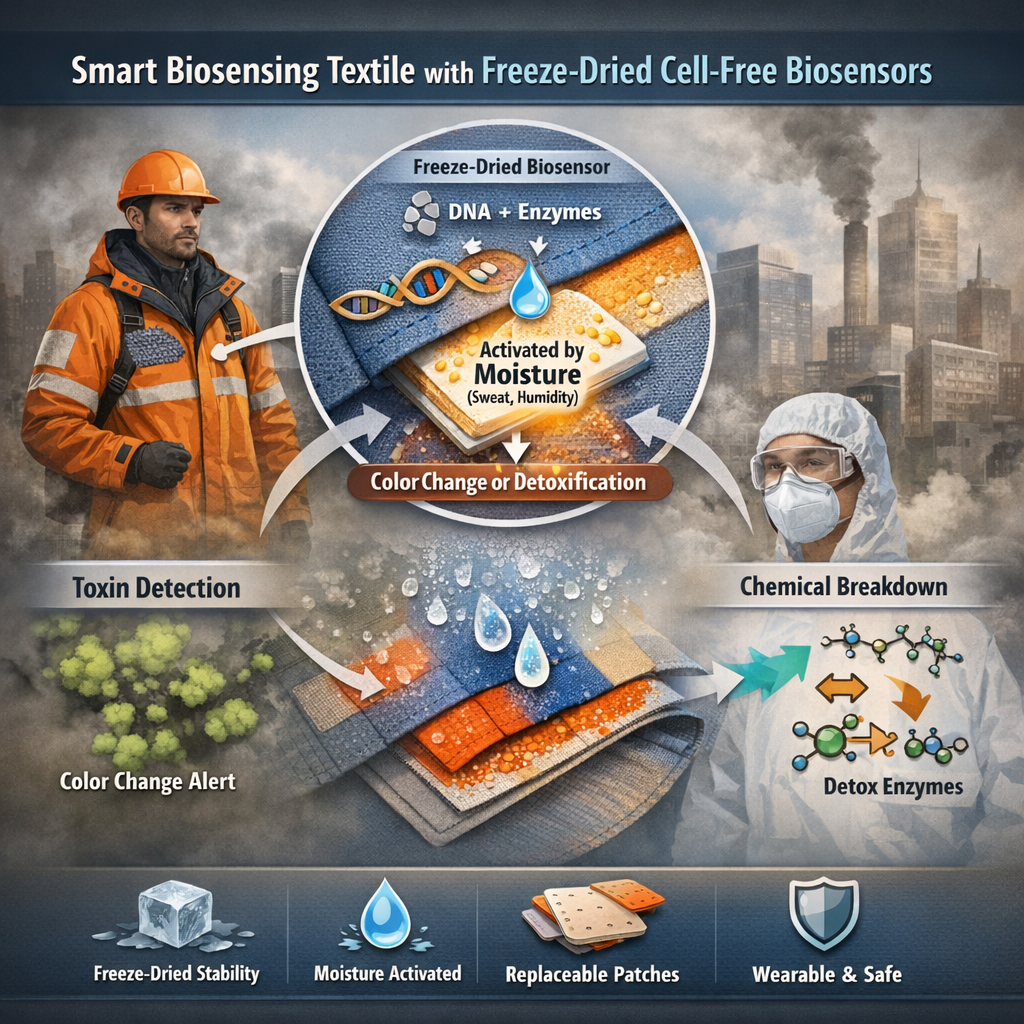 Fig. 1. Image generated with copilot
Homework question from Ally Huang
Long-duration spaceflight exposes astronauts to microgravity and radiation, which can disrupt the human microbiome and increase the risk of infection. Monitoring microbial behavior in space is challenging due to limited equipment and resources. Freeze-dried cell-free systems, such as BioBits®, offer a lightweight and stable alternative for on-demand biological analysis. Understanding how bacteria respond to space conditions is critical for maintaining astronaut health and mission success. This topic is significant because it supports safe human space exploration and advances our ability to perform biotechnology in extreme, resource-limited environments.
Molecular or Genetic Target
Expression of antibiotic resistance genes (e.g., bla gene encoding beta-lactamase) in bacterial DNA samples.
Relevance of Target
Antibiotic resistance genes are a growing concern in confined environments like spacecraft, where microbial populations may adapt differently under microgravity. Studying the expression of these genes helps determine whether space conditions enhance bacterial resistance. By detecting specific resistance-related DNA sequences and expressing them in a cell-free system, we can evaluate potential risks to astronaut health. This approach provides a rapid and safe method to monitor microbial threats without culturing live bacteria, making it highly suitable for space missions.
Hypothesis / Research Goal
I hypothesize that DNA from bacteria exposed to spaceflight conditions will show increased expression of antibiotic resistance genes compared to Earth-based controls. This may occur due to stress responses induced by microgravity and radiation, which can enhance gene regulation mechanisms linked to survival.
My goal is to use the BioBits® cell-free protein expression system to detect and compare the activity of resistance genes in DNA samples. The reasoning is that cell-free systems allow direct measurement of gene expression without requiring live-cell growth, making them ideal for spaceflight. If resistance gene expression is elevated, it would suggest an increased risk of hard-to-treat infections during missions. This knowledge could inform future countermeasures, such as improved sterilization or targeted antibiotic strategies.
Experimental Plan
DNA samples from bacteria grown in space and on Earth (control) will be amplified using the miniPCR® thermal cycler to target resistance genes. The amplified DNA will be added to BioBits® cell-free reactions to express the encoded proteins. Protein expression will be measured using fluorescence output with the P51 Molecular Fluorescence Viewer.
Controls will include reactions without DNA (negative control) and with known resistance gene DNA (positive control). Data collected will include fluorescence intensity, indicating gene expression levels. Results will be compared between space and Earth samples to evaluate differences in resistance gene activity.
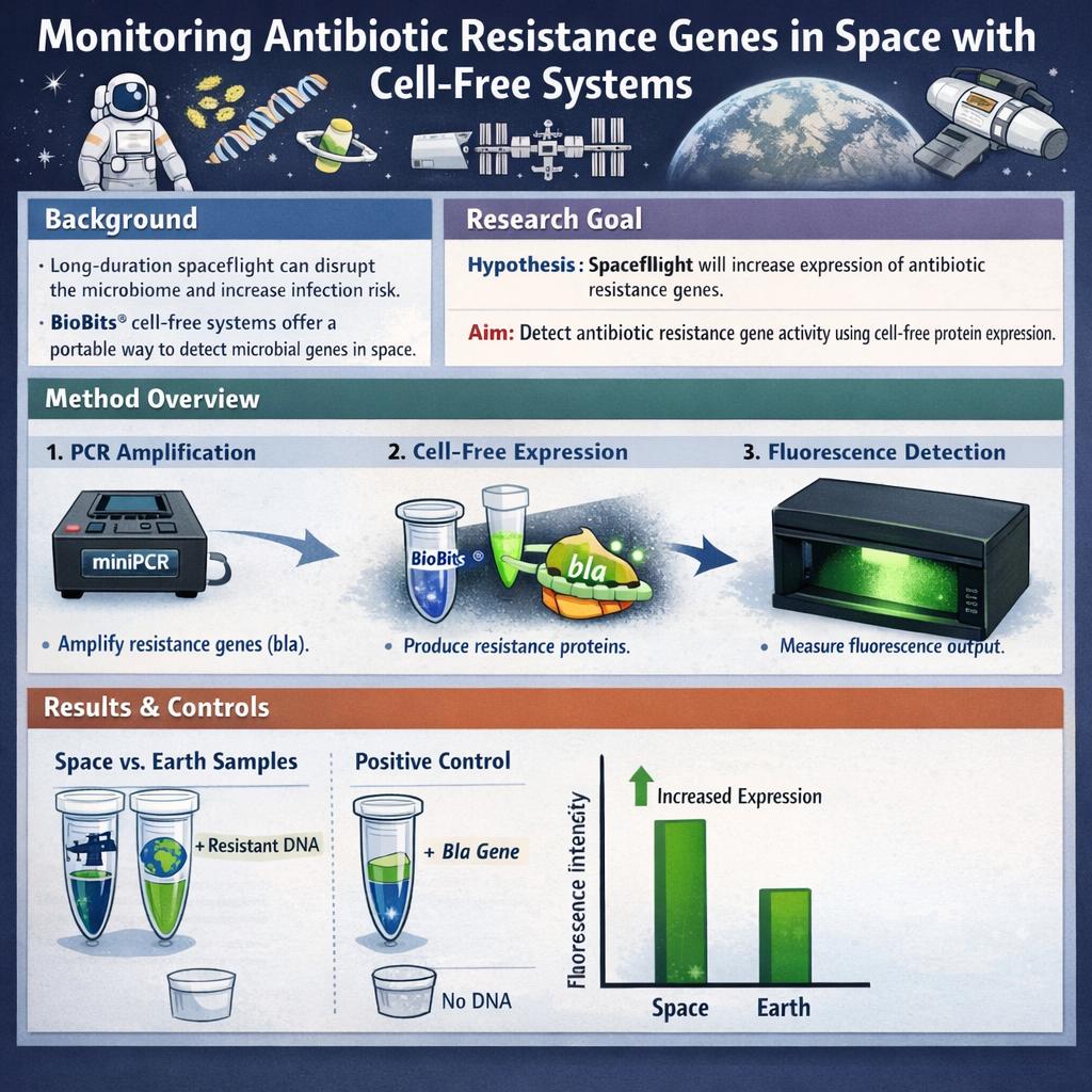 Fig. 2. Image generated with copilot
Homework Part B: Individual Final Project
Put your chosen final project slide in the appropriate slide deck following the instructions on slide 1:
 Note: My final project changed