Optosilence AD: a novel light-activated nanoplatform for precise gene repression
Section 1: Abstract
Alzheimer’s Disease (AD) is characterized by the pathological accumulation of amyloid-beta (Aβ) plaques, primarily derived from the Amyloid Precursor Protein (APP). Traditional gene-editing approaches present risks of irreversible genomic alterations and off-target effects. This project proposes a novel, reversible and multimodal nanotherapeutic strategy based on synthetic biology and nanotechnology. The system utilizes polymeric nanoparticles to deliver a light-activated CRISPRi machinery employing a high-efficiency dCas9-ZIM3(KRAB) repressor, alongside the antioxidant flavonoid Luteolin. By using Near-Infrared (NIR) stimulation for spatially controlled release, this approach enables a reversible and non-cleaving method to downregulate APP expression while mitigating neuroinflammation. This theoretical framework offers a precise, controllable alternative to traditional gene editing, aiming to slow AD progression through a synchronized genetic and chemical intervention.
Section 2: Project Aims
Aim 1: Experimental Aim
The first aim is to design a light-activated CRISPRi system targeting the APP gene by using computational sgRNA design tools (UCSC Genome Browser, CHOPCHOP, Addgene), Benchling for DNA construct assembly and a theoretical transfection and qPCR validation framework in a mammalian neuronal cell model. Specifically, the goal is to design three guide RNAs against the APP promoter TSS-proximal region, construct an in vitro DNA template encoding dCas9-ZIM3(KRAB) mRNA and conceptually integrate this system into PLGA-based polymeric nanoparticles with a NIR photo-responsive release mechanism and luteolin co-encapsulation for neuroprotection.
Aim 2: Development Aim
Building on the theoretical CRISPRi system designed in Aim 1, the next step would be to experimentally validate and optimize the nanoparticle platform in vitro. This includes synthesizing PLGA-PEG nanoparticles functionalized with Angiopep-2 and KLVFF peptides, measuring mRNA encapsulation efficiency and luteolin loading, testing BBB crossing in endothelial cell models and quantifying APP repression and Aβ reduction in human neuroblastoma cell lines (SH-SY5Y) following NIR-triggered cargo release.
Aim 3: Visionary Aim
The final aim is to evaluate the therapeutic potential and safety of the NIR-activated nanoplatform in a complex biological environment using APP/PS1 transgenic mice. By quantifying BBB crossing, APP repression and the reduction of amyloid plaques in a living brain, this stage will provide the necessary evidence to support the long-term vision of establishing a new class of precision nanomedicine. If completed, this clinically translatable platform could shift the paradigm from symptomatic treatment to personalized, reversible epigenetic therapies for Alzheimer’s and other neurodegenerative conditions.
Section 3: Background
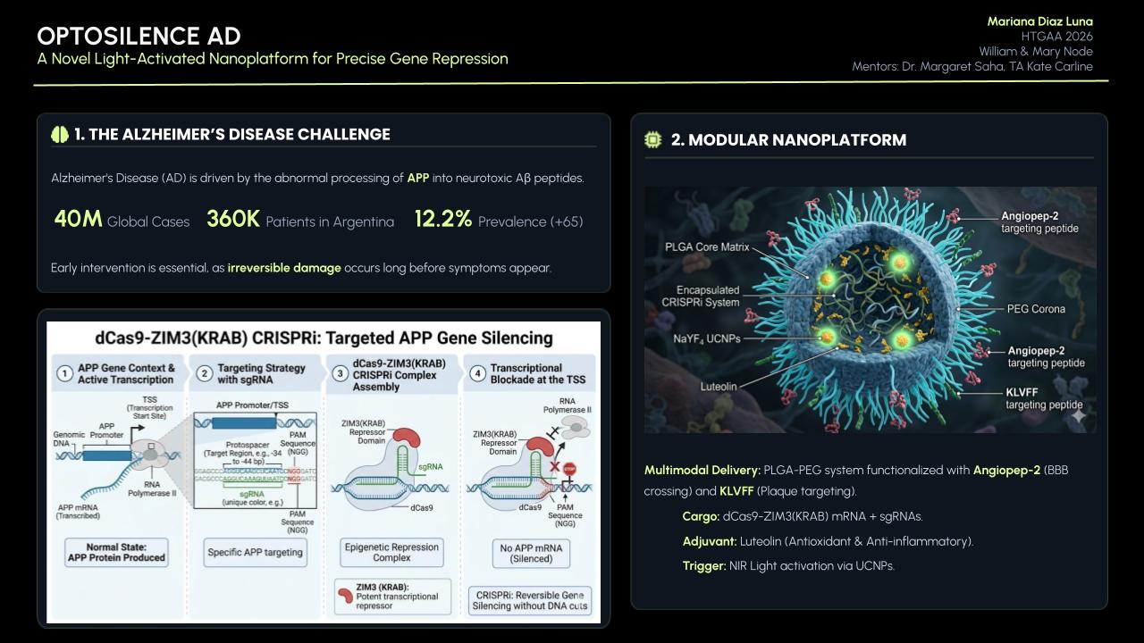
Alzheimer’s disease is the leading cause of dementia worldwide, responsible for 60-80% of dementia cases and affecting an estimated 40 million people globally, a number that continues to rise as populations age (Iosifescu et al., 2023). In Argentina, the prevalence of dementia is estimated at 12.2% in individuals over 65, with approximately 360,000 people living with AD specifically (Iosifescu et al., 2023). At a molecular level, the disease is driven by the abnormal processing of amyloid precursor protein (APP) by β- and γ-secretases, generating neurotoxic Aβ peptides that accumulate into extracellular plaques, triggering tau hyperphosphorylation, synaptic dysfunction and chronic neuroinflammation (Purushothuman et al., 2014). Elevated reactive oxygen species frequently precede Aβ deposition, accelerating neurodegeneration through mitochondrial dysfunction, lipid peroxidation and tau phosphorylation (Purushothuman et al., 2014). By the time symptoms appear, the brain has already sustained irreversible damage, making early, upstream intervention essential.
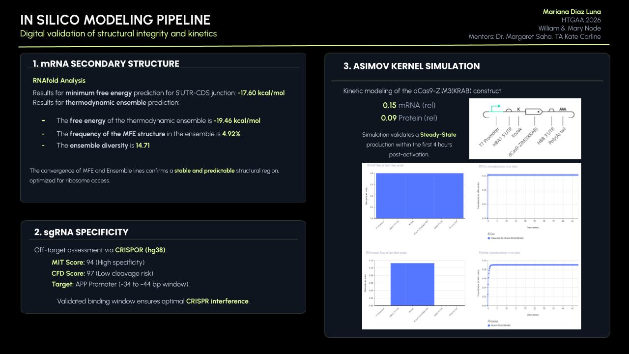
Current pharmacological treatments remain palliative. The blood-brain barrier (BBB) prevents more than 98% of potential therapeutics, including proteins, nucleic acids and antibodies, from reaching the central nervous system, and the most recently approved disease-modifying therapies cost between $27,000 and $34,000 per year per patient, making them inaccessible even in high-income countries (Zhi et al., 2021). Polymeric nanoparticles have emerged as a promising solution: PLGA-based systems functionalized with PEG and targeting ligands have demonstrated improved BBB penetration both in vitro and in vivo (Hoyos-ceballos et al., n.d.), while surface functionalization with Angiopep-2 activates receptor-mediated transcytosis via LRP1, enhancing brain accumulation (Hoyos-ceballos et al., n.d.) and co-functionalization with the KLVFF peptide, enabling selective targeting of amyloid-rich regions (Zhang et al., 2014). Importantly, these platforms can co-deliver multiple agents simultaneously. Abbas et al. (2022) demonstrated that luteolin-loaded nanoparticles significantly improved cognitive function, reduced Aβ aggregation by 50% and decreased neuroinflammatory markers in a sporadic AD mouse model, validating luteolin as a continuous neuroprotective adjunct within this type of platform (Abbas et al., 2022).
Rather than clearing plaques after they form, this project targets APP expression upstream. CRISPRi achieves transcriptional repression without altering the DNA sequence, making silencing reversible and adjustable, which is a critical advantage over traditional CRISPR knockout in post-mitotic neurons (Kristof et al., 2025). The choice of repressor domain determines efficacy: Alerasool et al. (2020) screened 57 KRAB domains and identified ZIM3 as exceptionally potent, demonstrating that the ZIM3(RAB)-dCas9 fusion silences gene expression more efficiently than previously established platforms across multiple human cell lines (Alerasool et al., n.d.). Kristof et al. (2025) further confirmed that ZIM3(KRAB) constructs show lower inter-target variability and consistent performance across cell types, establishing this architecture as the current gold standard for mammalian CRISPRi (Kristof et al., 2025). The system is delivered as synthetic mRNA incorporating a Cap1 structure for immune evasion (Drazkowska et al., 2022), HBA1 and HBB UTRs for stability and translational efficiency (Zarghampoor et al., 2019), and a 120-adenine poly(A) tail (all design choices validated in clinically approved mRNA therapeutics) (Asrani et al., 2018). Finally, upconversion nanoparticles (UCNPs) convert near-infrared light into UV/visible emission to trigger spatially controlled cargo release, adding a layer of precision entirely absent from current AD treatments (Swider et al., 2018).
Novelty and Innovation
This project is novel in three key aspects. First, it applies CRISPRi, a tool primarily used in cancer biology, to neurodegeneration, targeting APP expression before plaques form rather than attempting to clear them afterwards. Second, it combines mRNA-based CRISPRi delivery with NIR-responsive nanoparticles, a pairing not previously explored for APP regulation that introduces spatiotemporal control entirely absent from current AD therapeutics. Third, by integrating reversible gene silencing with simultaneous neuroprotection via luteolin within a single nanoplatform, this project addresses multiple pathological pathways: Aβ production, oxidative stress and neuroinflammation, in a coordinated, multimodal way that goes beyond any single-modality intervention currently available.
Impact
AD represents one of the most pressing unmet medical needs of the 21st century, with a global economic burden projected to rise from $384 billion in 2025 to approximately $1 trillion by 2050 (Iosifescu et al., 2023). The platform proposed here intervenes upstream of Aβ production, targeting the disease before irreversible neuronal damage occurs. If successful, this approach could shift the therapeutic paradigm from symptomatic management to precision epigenetic regulation. Its modular architecture is also adaptable: the same design principles could be applied to other neurodegenerative conditions driven by aberrant gene expression. The societal impact is particularly significant in my country, Argentina, where modifiable risk factors account for 56% of AD risk in Latin America, which is substantially above the global average of 40%, yet access to advanced diagnostics and emerging therapies remain severely limited. Beyond AD, this project demonstrates how synthetic biology tools can be integrated into a single therapeutic platform, advancing the boundaries of precision nanomedicine for complex neurological diseases.
Ethical Implications
The development of a CRISPR-based nanotherapeutic platform for AD raises profound ethical questions from both scientific and social justice perspectives. Even without altering DNA, modulating gene expression in neurons carries risks governed by the principles of non-maleficence and beneficence: off-target transcriptional repression of genes sharing promoter homology with APP could produce unintended neurological consequences. The NIR light trigger further raises justice concerns, as it requires specialized clinical infrastructure accessible only in well-equipped environments. This is especially concerning in Argentina, where over 600,000 people live with dementia and yet the country only established its first National Alzheimer’s Plan in 2026, a striking gap between the scale of the problem and the institutional response to it (Iosifescu et al., 2023). More broadly, dementia is systematically underdiagnosed worldwide, often identified only at late stages due to stigmatization and structural barriers, meaning that any technology requiring expensive infrastructure risks broadening existing inequities rather than addressing them. To ensure ethical conduct, preclinical development must include comprehensive off-target transcriptomic analysis, full nanoparticle immunogenicity and neurotoxicity evaluation and transparent informed consent protocols at the clinical stage. It is also important to acknowledge key uncertainties: NIR light penetration at clinically relevant brain depths has not been validated in humans, and mRNA delivery efficiency may not translate linearly from rodent to human neuronal environments. The cost dimension is equally critical: current disease-modifying therapies for AD already cost between $27,000 and $34,000 per year, and in Argentina, where these drugs remain inaccessible, managing changeable risk factors is our best tool for lowering this disease’s impact (Iosifescu et al., 2023). Affordability and equitable access must therefore be built into the design of this platform ensuring that its benefits can reach the populations who need them most.
Section 4: Experimental Design
Overview of Experimental Steps
- CRISPRi Design and System Validation
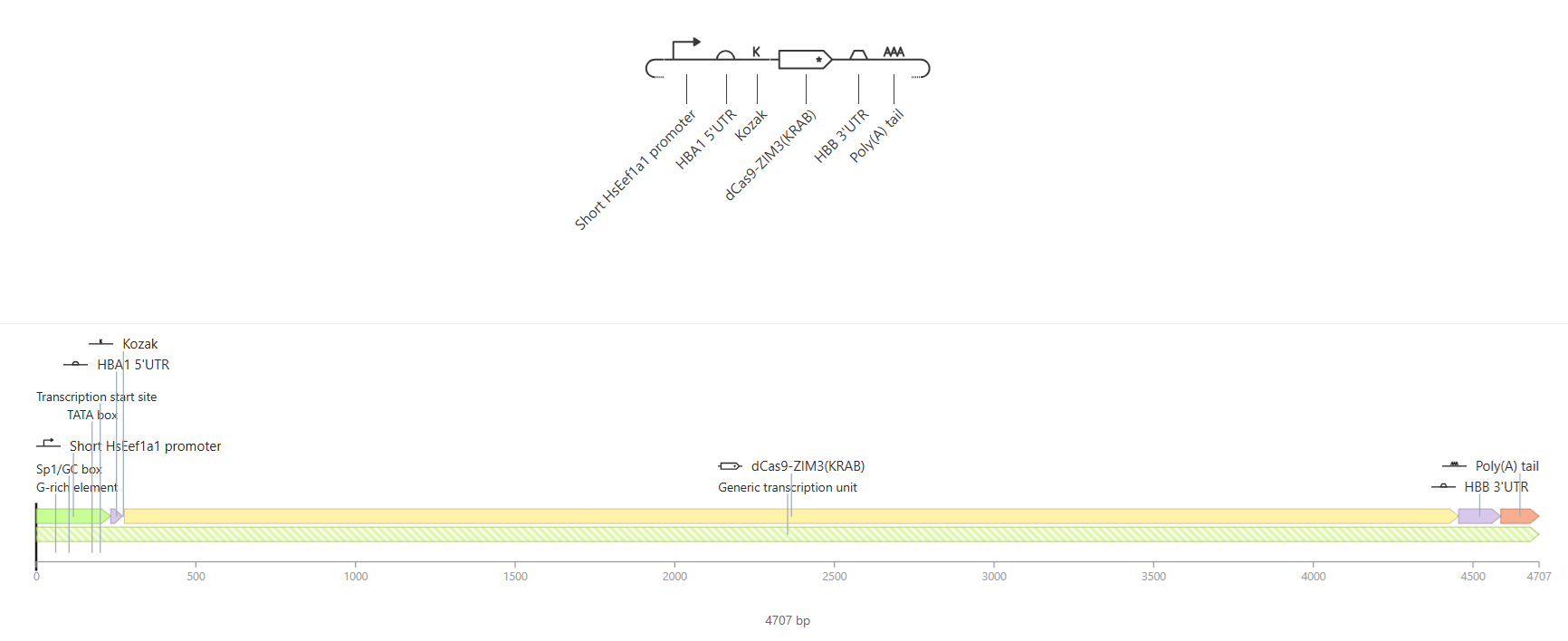
CRISPR and sgRNA design: the APP protein sequence was obtained from UCSC Genome Search. First, the promoter sequence (4000 bp) was identified, and then, the TSS, which was 872 bp distance from the starting point. CHOPCHOP was used to find target sites and determined three candidates with the highest efficiency to become the sgRNA templates. The dCas9-ZIM3(KRAB) construct was designed in Benchling in mRNA format using: 5’ Cap1 – T7 Promoter - 5’ UTR (HBA1) – dCas9-ZIM3(KRAB) – SV40 NLS – 3’ UTR (HBB) – Poly(A) tail, integrated in plasmid for in vitro transcription.
sgRNA cloning and IVT template verification: the four DNA constructs designed in Benchling (pIVT-dCas9-ZIM3(KRAB)-APP and the three sgRNA templates) would be ordered as synthetic linear DNA fragments from Twist Biosciences. Each construct would be verified by Sanger sequencing to confirm sequence integrity before proceeding. We expect the sequences match 100% with the Benchling designs.
- The linear IVT templates would be synthesized by Twist Biosciences and used directly as templates for in vitro transcription using a T7 RNA polymerase kit. For scale-up purposes, these sequences would alternatively be cloned into a T7-based expression plasmid, amplified in DH5α E. coli, and linearized prior to IVT to ensure sufficient template quantity for nanoparticle loading.
In vitro transcription (IVT) of mRNA and sgRNAs: the dCas9-ZIM3(KRAB) IVT template would be transcribed using a T7 RNA polymerase kit (e.g. HiScribe, New England Biolabs). The resulting mRNA would be capped with a Cap1 analog and purified by LiCl precipitation, being a high-selective technique to efficiently recover large mRNA transcripts. The three sgRNA templates would be transcribed separately using the same T7 system. mRNA integrity would be confirmed by gel electrophoresis (expected to see a single clean band at 〜4.5 kb for the mRNA, 〜100 nt for the sgRNas) and nanodrop quantification to determine the final RNA concentration and assess its purity.
Transfection of CRISPRi system into neuronal cell line: the dCas9-ZIM3(KRAB) mRNA and each sgRNA would be co-transfected into SH-SY5Y human neuroblastoma cells (a standard neuronal model for AD research) using a lipid-based transfection reagent (e.g. Lipofectamine MessengerMAX, Thermo Fisher Scientific). Three experimental groups would be established as it follows: (i) dCas9-ZIM3(KRAB) mRNA + APP-targeting sgRNA, (ii) dCas9-ZIM3(KRAB) mRNA + non-targeting control sgRNA, and (iii) untreated cells. We expect to see a successful transfection confirmed by fluorescence microscopy if a reporter (e.g. GFP) is co-transfected, which will be very useful.
qPCR quantification of APP mRNA repression: 48 hours post-transfection, total RNA would be extracted from all three groups using TRIzol (Thermo Fisher Scientific), a reagent used to ensure the immediate inactivation of endogenous RNases and facilitate the selective isolation of high-integrity total RNA. Then, this final RNA would be reverse-transcribed into cDNA. Quantitative PCR (qPCR) with APP-specific primers would be used to quantify APP mRNA levels normalized to a housekeeping gene (like GAPDH) to provide an internal reference for normalization. Expected result: 50-80% reduction in APP mRNA levels in group (i) compared to controls, consistent with published CRISPRi repression efficiency using ZIM3-KRAB (Alerasool et al., n.d.).
Western Blot for APP protein levels: to confirm that transcriptional repression translates to reduced protein levels, total protein would be extracted from all groups and analyzed by Western Blot using an anti-APP antibody. Expected result: reduction in full-length APP protein proportional to mRNA repression observed by qPCR.
ELISA for Aβ secretion: conditioned media from all three groups would be collected and analyzed by sandwich ELISA to quantify secreted Aβ40 and Aβ25 levels, since these two are the primary proteolytic products of APP cleavage and the main constituents of neurotoxic plaques in AD. The expected result would be a significant reduction in Aβ secretion in the CRISPRi-treated group compared to controls, demonstrating that APP repression translates into reduced amyloid production.
- Nanoparticles Synthesis and Characterization
PLGA-PEG nanoparticles would be synthesized by the double emulsion solvent evaporation method. Luteolin would be incorporated into the PLGA matrix during synthesis (through hydrophobic encapsulation). mRNA and sgRNAs would be condensed with protamine sulfate before encapsulation into the aqueous core. We expect to obtain nanoparticles of 100-150 nm diameter with narrow polydispersity index (PDI < 0.2), confirmed by dynamic light scattering (DLS).
Surface functionalization: Angiopep-2 and KLVFF peptides would be conjugated to the PEG terminal via NHS-ester chemistry. UCNPs (NaYF₄:Yb/Er) would be incorporated into the nanoparticle core during assembly. Successful conjugation would be confirmed by zeta potential measurement and FTIR spectroscopy. Expected result: detectable shift in zeta potential after peptide conjugation compared to unfunctionalized nanoparticles.
Encapsulation efficiency and loading capacity: mRNA encapsulation efficiency would be quantified by RiboGreen fluorescence assay, chosen for its sensitivity and specificity compared to traditional UV absorbance, after disrupting nanoparticles with DMSO (Swider et al., 2018). Luteolin loading would be measured by HPLC. It is expected to find a mRNA encapsulation efficiency of >80%, luteolin loading >70%, consistent with reported values for similar PLGA formulations.
NIR-triggered release assay: nanoparticles would be exposed to NIR irradiation defined time intervals and the released cargo quantified over time by RiboGreen fluorescence assay. A non-irradiated parallel group would serve as control. Expected result: significantly higher mRNA release in the NIR-exposed group (>70% release within 2 hours), demonstrating functional photoactivation with minimal leakage in the absence of light, demonstrating functional photoactivation.
- Key parameters remain to be established before clinical translation of this combined system. The field currently faces challenges related to the large number of variables involved (wavelength, power, pulse frequency, session duration and route of administration) making it difficult to develop a standardized therapeutic protocol. In vivo mice studies would therefore specifically aim to determine the optimal NIR parameter for the current system before any clinical translation.
- BBB Crossing and Cellular Uptake
In vitro BBB model - transwell assay: a transwell BBB model would be established using hCMEC/D3 human brain endothelial cells grown to confluence on transwell inserts. Functionalized nanoparticles would be added to the apical (blood-side) compartment and transported to the basolateral (brain-side) compartment measured by fluorescence at 24 and 48 hours. Expected result: Angiopep-2 functionalized nanoparticles showing significantly higher transport efficiency compared to non-functionalized controls, consistent with published LRP1-mediated transcytosis data (Hoyos-ceballos et al., n.d.).
Cellular uptake in SH-SY5Y cells: fluorescently labeled nanoparticles would be incubated with SH-SY5Y cells for 4, 12 and 24 hours. Uptake would be visualized by confocal microscopy and quantified by flow cytometry. Expected result: time-dependent increase in intracellular fluorescence with >70% of cells showing nanoparticle uptake at 24 hours.
Cytotoxicity assessment: cell viability after nanoparticle treatment would be assessed by MTT assay in SH-SY5Y cells at multiple nanoparticle concentrations, ensuring that the delivery system is non-toxic at the proposed therapeutic concentrations. Expected result: >85% cell viability at therapeutically relevant concentrations, confirming the biocompatibility of the PLGA-based formulation.
- Integrated System Evaluation
Combined nanoparticle-CRISPRi treatment with NIR activation: SHSY5Y cells would be treated with the complete nanoparticle system (encapsulating dCas9-ZIM3(KRAB) mRNA + sgRNAs + luteolin + UCNPs), followed by NIR irradiation. APP mRNA levels, APP protein and Aβ secretion would be measured at 48 and 72 hours’ post-treatment. A non-irradiated control group would assess baseline leakage of cargo. It would be expected to see a NIR-activated group showing 50-80% APP repression and significant reduction in Aβ secretion, while the non-irradiated group shows minimal gene silencing, demonstrating successful light-controlled CRISPRi activation.
- Luteolin neuroprotection assessment: to evaluate the antioxidant components, cells would be pre-treated with Aβ42 oligomers to induce oxidative stress, then treated with nanoparticles. ROS levels would be measured by DCFH-DA fluorescence assay and cell viability via MTT. Expected result: nanoparticle-treated cells showing significantly lower ROS levels and higher viability compared to cells treated with Aβ42 alone, confirming the neuroprotective contribution of luteolin.
- In vivo Validation in Mouse Model
Animal model section: the most appropriate model would be the APP/PS1 transgenic mouse, which overexpresses mutant human APP and presenilin-1, developing amyloid plaques and cognitive deficits by 6 months of age (Purushothuman et al., 2014). This model is the most widely used for preclinical AD therapeutic testing and would allow direct evaluation of both APP repression and Aβ plaque reduction.
Nanoparticle administration route: nanoparticles would be administered via intravenous (IV) tail vein injection, which is the standard route for systemic nanoparticle delivery. This route allows the nanoparticles to circulate through the bloodstream and reach the brain via the Angiopep-2 mediated LRP1 transcytosis mechanism designed in this project. A dosing regimen would be tested to determine the number of injections, doses and days. NIR irradiation would be applied transcranially after each injection to trigger CRISPRi activation, according to previous evaluation.
APP mRNA and protein quantification in brain tissue: at the end of the treatment period, mice would be sacrificed and bran tissue collected. APP mRNA levels in the hippocampus and cortex, which are the most affected regions in AD, would be quantified by qPCR. APP protein levels would be assessed by Western Blot. The expected results would be 40-60% reduction in APP mRNA and protein in treated mice compared to untreated APP/PS1 controls.
Amyloid plaque quantification by immunohistochemistry: brain sections would be stained with anti-Aβ antibody and Thioflavin S to visualize and quantify amyloid plaque burden. Plaque number and area would be compared between treated and untreated groups. The expected result would be a significant reduction in plaque burden in the proposed areas of treated mice, proportional to the reduction in APP expression.
Oxidative stress and neuroinflammation markers: brain homogenates would be analyzed for ROS levels, lipid peroxidation and pro-inflammatory cytokines by ELISA. Expected result: significantly lower oxidative stress markers and neuroinflammatory cytokines in nanoparticle-treated mice compared to untreated controls, reflecting the neuroprotective contribution of luteolin.
Safety and biodistribution assessment: to evaluate the safety profile of the nanoparticle platform, major organs like liver, kidneys, spleen, lung, heart and brain would be collected and analyzed by H&E histology. Blood biochemistry panels would assess liver and kidney function. Expected result: no significant histopathological changes or organ toxicity at the therapeutic dose, consistent with the known biocompatibility of PLGA-based nanoparticles.
- Clinical Translation and Regulatory Pathway
Following successful validation in APP/PS1 mouse models, the project would transition into formal clinical trials to evaluate safety and efficacy in humans. Phase I would focus on the safety and tolerability of the NIR-activated nanoplaform delivering synthetic mRNA, monitoring for potential immunogenicity or off-target effects. Phase II would aim to establish the optimal NIR dosing parameters and preliminary efficacy in reducing Aβ biomarkers in patients with early-stage AD. Finally, Phase III trials would be required to demonstrate significant cognitive improvement and long-term stability of the CRISPRi-mediated APP repression.
Techniques proposed for this project
Pipetting
Lab Safety
Bioethical Considerations
DNA Sequencing
DNA Construct Design
Gel Electrophoresis
Databases (NCBI, Ensembl, UCSC Genome Browser)
Benchling
Designing a Twist Order (if performing in vitro)
CRISPR/Cas9
Plasmid Preparation
Quality Control/Analysis
PCR Reactions
Protein Design
Cell Free Reactions
The most important technique (and the first one used) was Benchling for designing and annotating all four DNA constructs in this project. The dCas9-ZIM3(KRAB) IVT template was assembled by sequentially incorporating the T7 promoter, the HBA1 5’UTR, the dCas9-ZIM3(KRAB) coding sequence (extracted from Addgene plasmid #154472 using SnapGene Viewer), the 2x SV40 NLS, the HBB 3’UTR and the 120-adenine poly(A) tail. Each element was individually annotated with color-coded features to facilitate visualization and verification of the construct architecture. The three sgRA templates were designed following the same workflow, with T7 promoter, a 20-nt spacer targeting the APP promoter and the SpCas9 scaffold sequence. Next, the sgRNA design using CHOPCHOP and UCSC Genome Browser was the required technique. The design of the three APP-targeting sgRNAs combined the use of multiple bioinformatics databases and tools in an integrated workflow. First, the UCSC Genome Browser (hg38 assembly) was used to locate the transcription start site (TSS) of the APP gene isoform 695, the dominant neuronal isoform. Because APP is encoded on the minus strand of chromosome 21, the “Get DNA” tool was used with the reverse complement option to extract the correctly oriented promoter sequence (〜4000 bp around the TSS). This sequence was then submitted to CHOPCHOP in repression mode with SpCas9/NGG, which predicted and ranked all possible guide RNAs based on their efficiency score, distance to the TSS and off-target profile. The three final candidates were selected because they all fall within the -34 to -44 bp window relative to the TSS (within the optimal zone for CRISPRi-mediated transcriptional repression) and have efficiency scores above 56, minimal off-targets and GC content within the recommended 40-70% range.
Section 5: Results & Quantitative Expectations
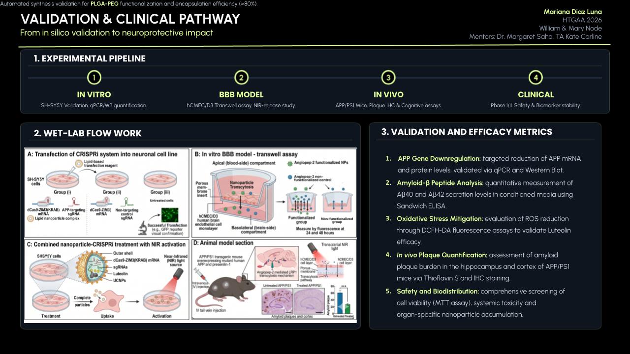
Three complementary computational validations were performed to support the theoretical framework of this project. First, the specificity of the three APP-targeting sgRNAs was evaluated using CRISPOR to assess off-target risk across the human genome. Second, the structural viability of the mRNA construct was analyzed using RNAfold to determine whether the 5’UTR-CDS junction adopts a conformation compatible with efficient ribosomal access. Third, the full dCas9-ZIM3(KRAB) IVT construct was modeled in Asimov Kernel to simulate transcription and translation dynamics and confirm that this design produces functional protein output.
Detailed protocol of validation
i. Validation 1: sgRNA off-target analysis with CRISPOR
a. The three sgRNA spacer sequences previously designed using CHOPCHOP were submitted individually to CRISPOR (https://crispor.gi.ucsc.edu/) using the human genome assembly hg38 and SpCas9/NGG as the PAM.

b. For each guide, the MIT Specificity Score and the CFD (Cutting Frequency Determination) Score were recorded as measures of off-target risk.

c. The off-target table was examined for each guide, counting predicted off-target sites at 0,1, 2, 3 and 4 mismatches, with particular attention to exonic off-targets, which represent the highest functional risk.

d. The genomic position of each guide was compared against the APP transcription start site (TSS) to confirm that all three fall within the -50 and +300 bp window required for effective CRISPRi-mediated transcriptional repression.

e. Efficiency scores (out-of-frame and Lindel predictions) were noted as relative indicators, since dCas9 does not cleave DNA, these values were interpreted as indicators of guide binding efficiency rather than cutting outcomes.
ii. Validation 2: mRNA secondary structure analysis with RNAfold
a. The sequence of the HBA1 5’UTR followed by the first 50 nucleotides of the dCas9 coding sequence was analyzed with the RNAfold web server (ViennaRNA package) using default parameters.

b. The minimum free energy (MFE) structure, thermodynamic ensemble energy, MFE frequency and ensemble diversity were recorded.

c. The resulting dot-bracket structure, mountain plot and base-pair probability coloring were examined to evaluate the accessibility of the 5’ end of the transcript for ribosome engagement.
iii. Validation 3: full construct simulation in Asimov Kernel
a. The complete IVT construct (T7 promoter – HBA1 5’UTR – Kozak – dCas9-ZIM3(KRAB) – HBB 3’ UTR – Poly(A) tail) was assembled and annotated in Asimov Kernel.

b. A simulation was run over a 47-hour time window, generating RNAP flux, ribosome flux, RNA concentration over time and protein concentration over time.

c. Output graphs were examined to confirm uniform transcriptional flux, appropriate engagement at the 5’UTR and CDS, and sustained protein accumulation.
- Synthetic biology techniques utilized
This validation integrated four different synthetic biology approaches as mentioned. Bioinformatics-based sgRNA specificity analysis through CRISPOR constitutes a standard pre-experimental quality control step in CRISPR design, equivalent to computational off-target screening done before any guide RNA is synthesized or tested in cells. RNA secondary structure prediction using RNAfold applies thermodynamic modeling to evaluate if a given mRNA sequence is translationally accessible, a technique commonly used in mRNA therapeutic design to avoid inhibitory structures near the ribosome binding site. Computational construct modeling in Asimov Kernel represents an in silico cell-free assay, simulating the kinetics of transcription and translation from a defined generic part architecture and allowing prediction of RNA and protein output without requiring physical synthesis.
- Data representation and analysis
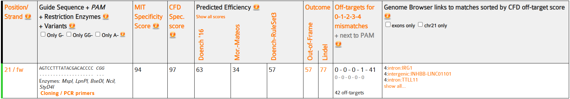
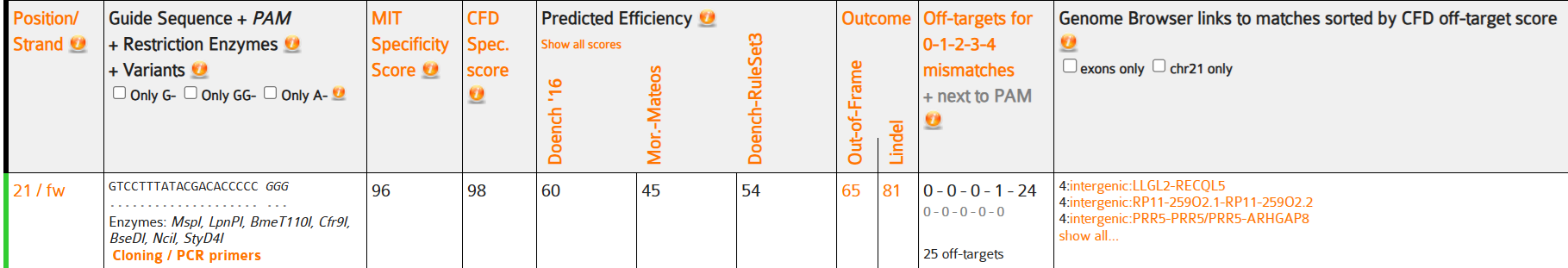
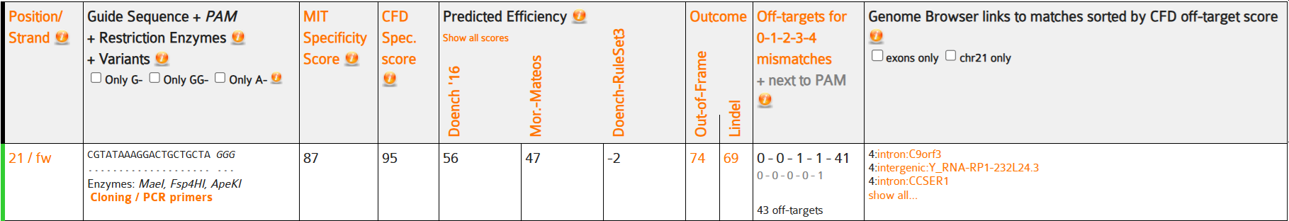
i. Validation 1 – CRISPOR off-target results
All three sgRNAs were confirmed to target the APP promoter within the optimal CRISPRi window relative to the TSS:
sgRNA #1
sgRNA #2
sgRNA #3
sgRNA-1 showed excellent specificity with an MIT score of 94 and a CFD score of 97, showing very low genome-wide off-target risk. The single off-target site at 3 mismatches and 41 sites at 4 mismatches are not located in exonic regions, representing slight functional risk. This system uses catalytically dead dCas9, so the predicted cutting efficiency scores such as out-of-frame: 57% and Lindel: 77% serve only as relative indicators of guide binding strength rather than cleavage outcomes.
ii. Validation 2: RNAfold structural analysis
The RNAfold analysis of the HBA1 5’UTR and the first 50 nt of dCas9 CDS showed the following results:
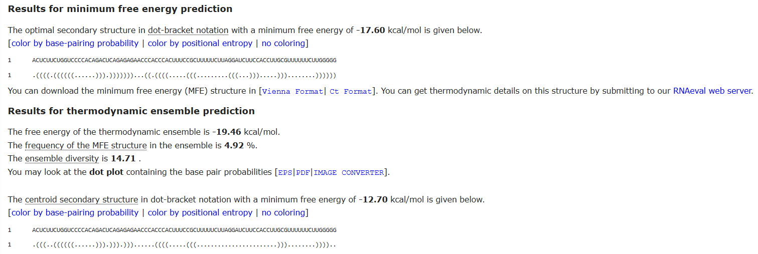
The MFE of -17.60 kcal/mol represents an acceptable range for a fragment of approximately 85 nt. This value is not excessively negative (which would indicate a rigid structure capable of disabling the ribosome binding site), supporting the conclusion that the 5’UTR-CDS junction is translationally permissive. The low MFE frequency of 4.92% indicates that the molecule is highly dynamic and spends the majority of time in alternative conformations, facilitating ribosomal scanning.
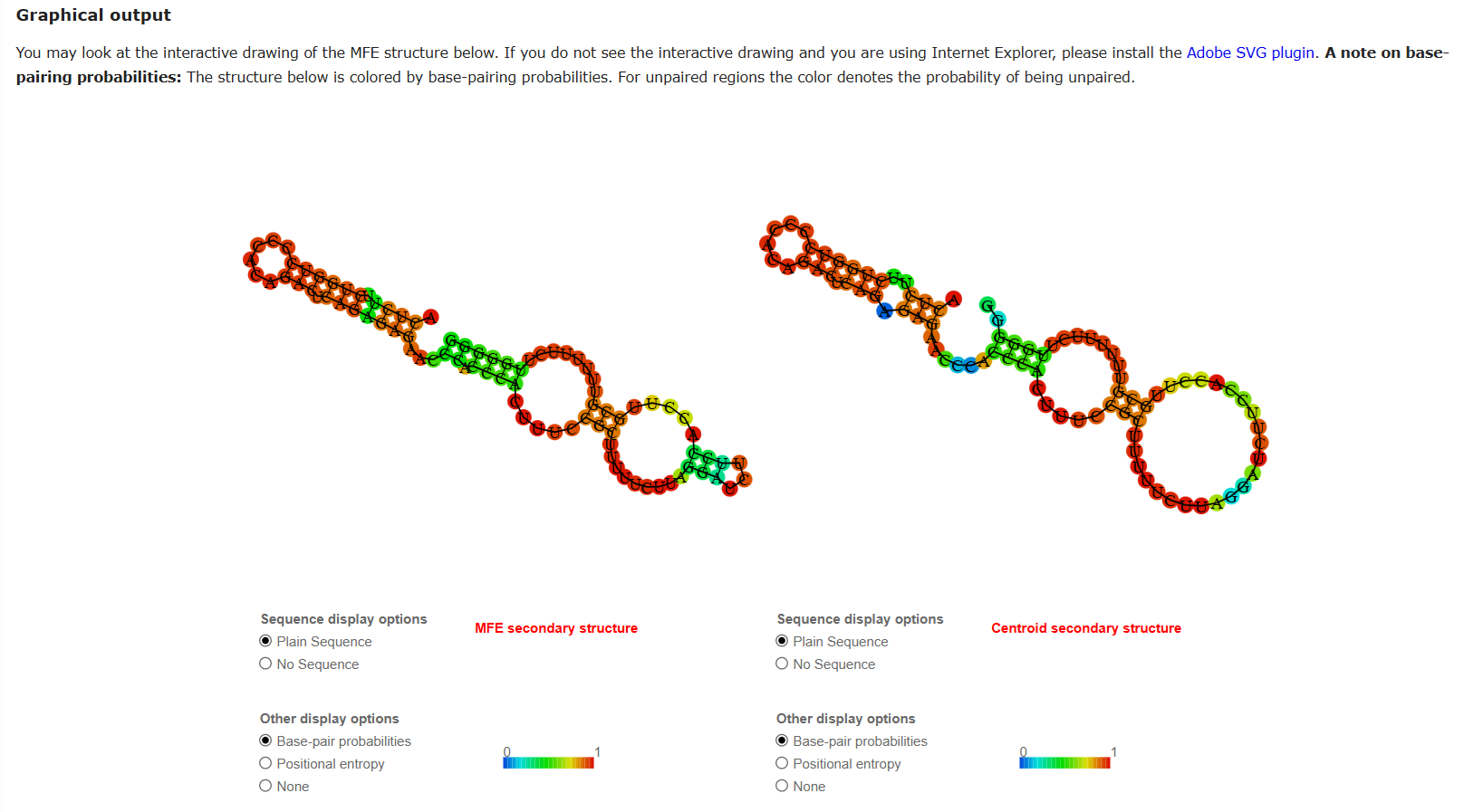
The prominent hairpin observed at the 5’ end of the HBA1 region is a known structural feature of globin UTRs, specifically evolved to protect the mRNA from nuclease degradation without obstructing translation initiation.
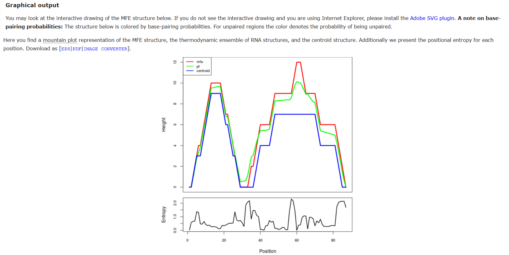
In the mountain plot, convergence of the MFE and ensemble lines at this first helix confirms it is the most thermodynamically stable and structurally predictable region of the construct.
iii. Validation 3: Asimov Kernel simulation This simulation confirmed transcriptional and translational functionality of the full construct across all four output metrics.
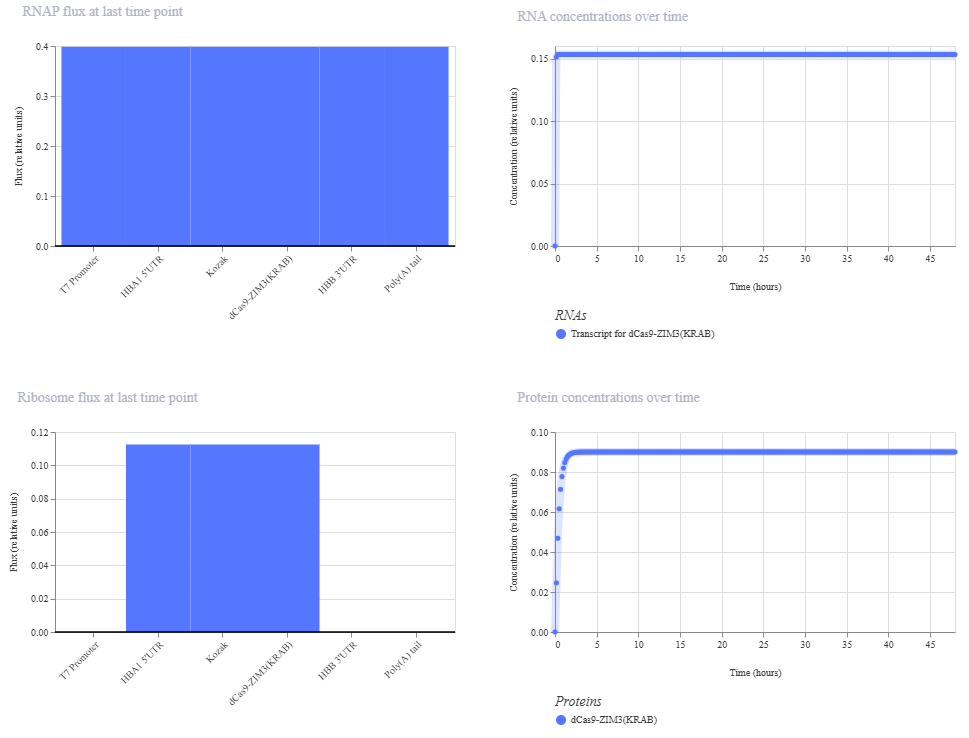
RNAP flux was uniform at ~0.4 relative units across all annotated elements, consistent with the highly processive and non-pausing nature of T7 RNA polymerase. Ribosome flux was appropriately localized to the HBA1 5’UTR and dCas9-ZIM3(KRAB) CDS regions (~0.11 relative units), with no flux detected at the promoter, 3’UTR or Poly(A) tail reflecting correct translational architecture. The dCas9-ZIM3(KRAB) protein accumulated rapidly within the first 2-3 hours and plateaued at ~0.09 relative units, demonstrating sustained expression dynamics consistent with strong mRNA stability given by the HBB 3’UTR and the Poly(A) tail.
- The primary challenge during validation was the use of Asimov Kernel for construct modeling. In the initial attempts to assemble the IVT construct, the platform’s default parts library did not contain several of the regulatory elements I needed for this design at the exact specifications described in the literature. This required learning to manually define and add custom genetic parts to my repository. Through iterative testing, each element was individually added and configured until the complete construct was successfully validated by the platform. Although the results obtained across all three computational validations were consistent with the theoretical expectations, I must say that in silico analyses are extremely limited in their predictive power. Computational tools operate under simplified models that cannot fully represent the complexity of a living biological system –variables such as intracellular mRNA stability, nanoparticle uptake efficiency, blood-brain barrier crossing and the immunological environment of the brain cannot be meaningfully captured by these platforms. Therefore, while these validations provide a strong theoretical foundation and support this design, experimental confirmation through in vitro cell-based assays and in vivo testing in an appropriate animal model would be essential to determine whether the system performs as intended under real biological conditions.
Section 6: Bibliography and References
Abbas, H., Sayed, N. S. El, Abdel, N., Abou, H., Gaafar, P. M. E., Mousa, M. R., Fayez, A. M., & Elsheikh, M. A. (2022). Novel Luteolin-Loaded Chitosan Decorated Nanoparticles for Brain-Targeting Delivery in a Sporadic Alzheimer ’ s Disease Mouse Model : Focus on Antioxidant , Anti-Inflammatory , and Amyloidogenic Pathways. 1–26.
Alerasool, N., Segal, D., Lee, H., & Taipale, M. (n.d.). An efficient KRAB domain for CRISPRi applications in human cells. Nature Methods. https://doi.org/10.1038/s41592-020-0966-x
Asrani, K. H., Farelli, J. D., Stahley, M. R., Miller, R. L., Cheng, C. J., Subramanian, R. R., Brown, J. M., Asrani, K. H., Farelli, J. D., Stahley, M. R., Miller, R. L., Cheng, C. J., Subramanian, R. R., & Brown, J. M. (2018). Optimization of mRNA untranslated regions for improved expression of therapeutic mRNA. 6286. https://doi.org/10.1080/15476286.2018.1450054
Drazkowska, K., Tomecki, R., Warminski, M., Baran, N., Cysewski, D., Kasprzyk, R., Kowalska, J., Jemielity, J., & Sikorski, P. J. (2022). 2 -O-Methylation of the second transcribed nucleotide within the mRNA 5 cap impacts the protein production level in a cell-specific manner and contributes to RNA immune evasion. 50(16), 9051–9071.
Hoyos-ceballos, G. P., Ruozi, B., Ottonelli, I., Ros, F. Da, Vandelli, M. A., Forni, F., Daini, E., Vilella, A., Zoli, M., Tosi, G., Duskey, J. T., & Betty, L. L. (n.d.). PLGA-PEG-Ang – 2 Nanoparticles for Blood – Brain Barrier Crossing : Proof-of-Concept Study. 2, 1–11.
Iosifescu, D. V, Song, X., Gersten, M. B., Adib, A., Cho, Y., Collins, K. M., Yates, K. F., Hurtado-puerto, A. M., Mceachern, K. M., Osorio, R. S., & Cassano, P. (2023). Protocol Report on the Transcranial Photobiomodulation for Alzheimer ’ s Disease ( TRAP-AD ) Study. 1–16.
Kristof, A., Karunakaran, K., Allen, C., Mizote, P., Briggs, S., Jian, Z., Nash, P., & Blazeck, J. (2025). Engineering novel CRISPRi repressors for highly efficient mammalian gene regulation.
Purushothuman, S., Johnstone, D. M., Nandasena, C., Mitrofanis, J., & Stone, J. (2014). Photobiomodulation with near infrared light mitigates Alzheimer ’ s disease-related pathology in cerebral cortex – evidence from two transgenic mouse models. 1–13.
Swider, E., Koshkina, O., Tel, J., Cruz, L. J., Vries, I. J. M. De, & Srinivas, M. (2018). Acta Biomaterialia Customizing poly ( lactic- co -glycolic acid ) particles for biomedical applications. Acta Biomaterialia, 73, 38–51. https://doi.org/10.1016/j.actbio.2018.04.006
Zarghampoor, F., Azarpira, N., Khatami, S. R., Behzad-behbahani, A., & Foroughmand, A. M. (2019). PT. Gene. https://doi.org/10.1016/j.gene.2019.05.008
Zhang, C., Wan, X., Zheng, X., Shao, X., & Liu, Q. (2014). Biomaterials Dual-functional nanoparticles targeting amyloid plaques in the brains of Alzheimer ’ s disease mice. Biomaterials, 35(1), 456–465. https://doi.org/10.1016/j.biomaterials.2013.09.063
Zhi, K., Raji, B., Nookala, A. R., Khan, M. M., Nguyen, X. H., Sakshi, S., Pourmotabbed, T., Yallapu, M. M., Kochat, H., Tadrous, E., Pernell, S., & Kumar, S. (2021). PLGA Nanoparticle-Based Formulations to Cross the Blood – Brain Barrier for Drug Delivery : From R & D to cGMP. 1–17.
Supply list and budget needed for this project
Disclaimer: if a product has no price, it’s because it’s not available for my country.
- DNA design and synthesis
- Twist Biosciences – synthetic linear DNA fragments (x4 constructs: pIVT-dCas9-ZIM3(KRAB) + 3 sgRNA templates
- Cloning and Bacterial Culture
Expression vector, e.g. pUC57 (AddGene Plasmid #51132) $94
Restriction enzymes –EcoRI and XhoI- 〜$120 each, Thermo Fisher
T4 DNA ligase: $380 Thermo Fisher
Competent cells DH5α: $276 Thermo Fisher
LB agar plates with ampicillin (Product L5667 Sigma-Aldrich)
QIAprep Spin Miniprep Kit (50) (Qiagen): $128
HiSpeed Plasmid Kits (Qiagen): $421
Sanger sequencing
General lab material
- mRNA synthesis (IVT)
HiScribe T7 High Yield RNA Synthesis Kit (New England Biolabs)
N1-methylpseudouridine-5’-triphosphate (m1ΨTP) — modified UTP for immune evasion (New England Biolabs)
Vaccinia Capping System (New England Biolabs)
mRNA Cap 2’-O-me (New England Biolabs)
DNase I (Thermo Fisher): $120.94
GeneJET RNA Cleanup and Concentration Micro Kit (50) (Thermo Fisher): $333.23
RNaseZap (250 ml) (Thermo Fisher): $157.29
RNase free water (500 ml) (Thermo Fisher): $68.75
- Nanoparticle synthesis
Carboxylic acid-poly(ethylene glycol)-b-poly(lactide-co-glycolide) (PLGA-PEG-COOH) (Sigma Aldrich): $478
Luteolin (>98% -powder) (Sigma Aldrich): $192
Upconversion Nanoparticles (UCNPs) (Sigma Aldrich) –this product specifies “green light” emission, so this system will be activated by the visible (green) spectrum. $492
Protamine Sulfate (5 g) (Sigma Aldrich): $145
Angiopep-2 hydrochloride (MedChemExpress): $280
Β-Amyloid peptide (KLVFF peptide) (16-20) (MedChemExpress): $100
EDC (1-ethyl-3-(3-dimethylaminopropyl)carbodiimide hydrochloride) Premium Grade (Thermo Fisher): $202.50
NHS (N-hydroxysuccinimide) (Thermo Fisher): $127.58
Estimated Subtotal: ~$4300
More things will be necessary for next steps: Cell Culture (In vitro), Validation Assays, In vivo Instance including Mouse Model and Equipment Access like Dynamic Light Scattering, Confocal microscopy, HPLC, qPCR machine, Sonicator, Ultracentrifuge, Biosafety Cabinet, Nanodrop Spectrophotometer.