Individual Final Project
Melanin-based light-recording bioink/biomaterial
Designing a MelC2-Based Cell-Free Module for Programmable Melanin Bioink
Reframing pigmentation from static dyeing to a programmable chemical state evolution, enabling materials that encode environmental history

Important links:
| Resource | Link |
|---|---|
| Final presentation slides | CL Final Project Slide Deck |
| Final pTwist_MelC2_T7_TXTL_6xHis construct | Benchling |
| Twist Order for my Final Project: MelC2_T7_TXTL_6xHis_expression_cassette | Benchling and Twist (Nodes) Document |
| Cell-free master mix plan - 8 planned reactions | My Week 11 HW Documentation |
SECTION 1 - ABSTRACT
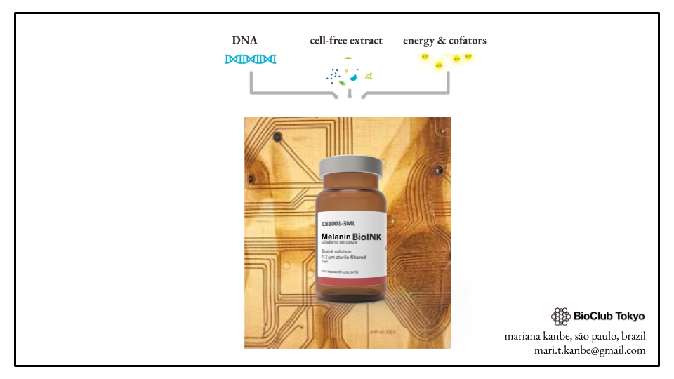
Melanin is a chemically heterogeneous dark biopolymer known for broadband UV-visible optical absorption, photoprotective behavior, photothermal conversion, redox activity, and long-term optical stability. These properties make melanin a compelling biological route to functional color: a pigment chemistry that can absorb and dissipate radiation, preserve optical traces, buffer oxidative stress, and interface with biological or electronic systems. This project proposes controlling melanin-forming chemistry in a synthetic biology system to develop a programmable bioink for engineered biomaterials. The broader vision is to create materials that combine biosensing and functional response: recording environmental inputs such as light, ionizing radiation, or oxidative stress through measurable optical change, while also enabling properties such as UV or radiation protection, photothermal conversion, antioxidant behavior, and bioelectronic interfacing. Depending on concentration, matrix composition, and material format, this melanin-based bioink could be explored for responsive textiles, UV-protective coatings, architectural and design surfaces, tattoo-like dermal pigments, space-oriented materials, bioelectronic interfaces, and localized radioprotective biomaterials. To move toward this goal, this project aims to design a first genetic module that generates measurable melanin-like optical changes in a controlled cell-free system, then use it as a foundation for future integration into engineered biomaterials such as bacterial cellulose. The central hypothesis is that a codon-optimized Streptomyces antibioticus MelC2 tyrosinase construct can provide a tractable route toward cell-free melanin-like pigment formation, with output shaped by tyrosinase activity, substrate availability, copper cofactor loading, oxygen, pH, redox state, and polymerization chemistry. During HTGAA 2026, I designed a MelC2 expression cassette for TX-TL / E. coli use and designed a validation workflow.
SECTION 2: PROJECT AIMS
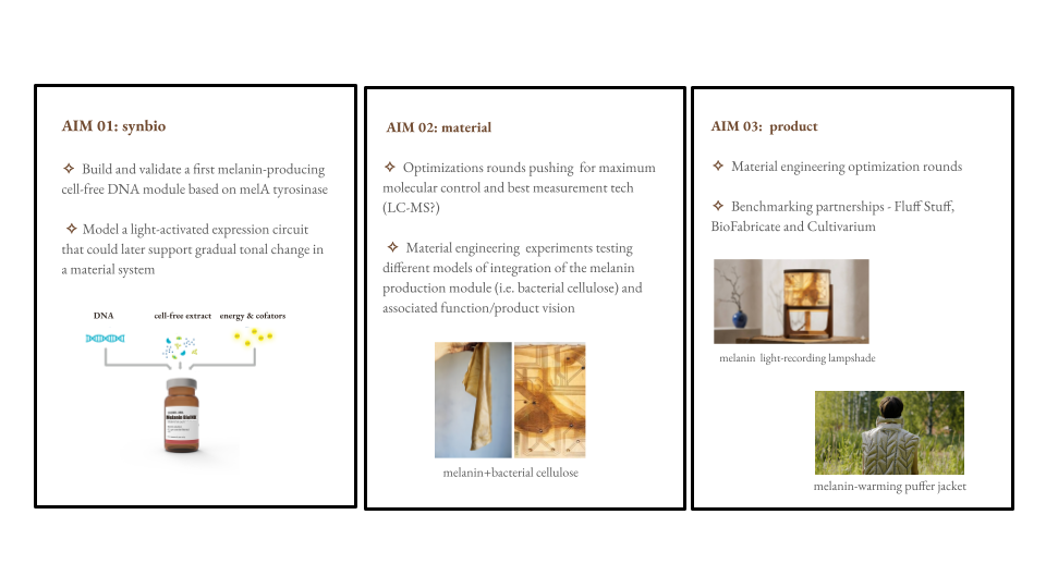
Aim 1: Experimental Aim
Build and validate a first MelC2-based cell-free melanin module
The first aim of this project is to design a codon-optimized Streptomyces antibioticus MelC2 tyrosinase expression cassette for TX-TL / E. coli use and test whether it can generate measurable melanin-like optical changes in a controlled cell-free system. This aim uses DNA design, Benchling assembly, Twist synthesis, fluorescent protein controls, visible darkening, OD 400-500 nm absorbance, SDS-PAGE, and future LC-MS analysis to distinguish protein expression, enzymatic activity, pigment accumulation, and downstream oxidation chemistry.
Aim 2: Development Aim
Optimize the chemical and optical behavior of the melanin-forming system
After validating the first module, the next aim is to optimize the reaction conditions that shape pigment output, including L-tyrosine concentration, copper availability, pH buffering, oxygen exposure, magnesium, incubation time, and reporter choice. This aim will help determine whether the system can be tuned for stronger pigment formation, cleaner optical readouts, and more predictable color response before integration into a material matrix.
Aim 3: Visionary Aim
Develop programmable melanin bioinks for exposure-recording and functional biomaterials
The long-term aim is to integrate the optimized melanin-forming module into bacterial cellulose or other biomaterials to create bio-based surfaces that can both record environmental exposure and respond functionally. If successful, this could support responsive textiles, UV-protective coatings, design surfaces, tattoo-like dermal pigments, bioelectronic interfaces, space-oriented materials, and localized radioprotective biomaterials.
SECTION 3: BACKGROUND
3.1. Peer-reviewed research citations
Melanin is relevant to this project because its material properties extend beyond visible pigmentation. Menichetti et al. 2025 describe melanin photoprotection as a combination of broadband light extinction and antioxidant activity, supporting the idea that melanin-based materials could pair optical response with protection against light-induced damage. Dadachova and Casadevall 2009 further show that melanin changes how biological systems interact with ionizing radiation, with melanized fungi displaying radioprotective behavior and altered electronic properties under radiation exposure. Together, these studies support the central premise of this project: melanin can be treated not only as a pigment, but as a functional material chemistry for exposure-responsive systems.
This material potential has already been explored in several application directions relevant to the proposed bioink. In space-oriented materials, Cordero et al. 2025 showed that fungal melanin-polymer biocomposites exposed to low Earth orbit conditions had improved structural stability and radiation-shielding potential. In photothermal and bioelectronic materials, Yue and Zhao, 2021 review how melanin-like materials can convert absorbed optical energy into heat and support sensor or interface applications through redox activity and mixed ionic/electronic behavior. At the skin interface, Park et al. 2024 developed electroactive melanin tattoo inks using naturally derived melanin nanoparticles to reduce skin impedance, suggesting that melanin-based pigments may be useful for dermal bioelectronic interfaces as well as coloration.
The bioink and textile direction also has direct precedent. Walker et al. 2024 engineered cellulose-producing Komagataeibacter rhaeticus to express tyrosinase and grow self-pigmenting bacterial cellulose through melanin biosynthesis, showing that genetically encoded pigmentation can be integrated into a material-producing microbial platform. Ahn et al. 2021 produced melanin-like pigments microbially from caffeic acid and applied the pigment to cotton fabric dyeing, supporting the relevance of microbial melanin as a textile-compatible colorant.
.png)
These studies connect directly to this project’s direction, but also clarify its specific contribution: instead of starting with a finished textile, this work first builds a controlled MelC2-based cell-free module to make melanin-like optical output measurable, tunable, and chemically interpretable before later integration into bacterial cellulose or other biomaterial matrices.
3.2. Novelty and innovation
This project is innovative because it uses existing biological tools in a new material context: a MelC2 tyrosinase module is designed not only to produce pigment, but to generate a measurable and tunable optical output. The cell-free system makes this approach modular, allowing key variables such as copper loading, substrate availability, pH, oxygen, redox state, and polymerization conditions to be tested before introducing the system into more complex biomaterial matrices. This creates a controlled bridge between genetic design and material performance.
The project also challenges a common assumption in functional materials: that color, sensing, protection, and responsiveness must be added as separate components. Instead, it asks whether melanin-forming chemistry can be programmed as a single multifunctional layer that records exposure and produces useful material responses. In doing so, the project expands synthetic biology from making biological products toward engineering bio-based materials whose behavior can be designed, measured, and tuned.
3.3. Why the project matters and potential impact
The main ethical issue is not melanin itself, but the form in which the system is built and deployed. A melanin-based material can remain a controlled chemical module, become a non-replicating embedded system, or become part of a living material platform. Each design choice carries a different ethical burden, so the project should progress from the lowest-risk and most interpretable system toward more complex formats only after validation.
| Design choice | Role in the project | Ethical implication |
|---|---|---|
| Cell-free MelC2 module | First experimental platform for testing pigment chemistry | Lowest deployment risk; controlled, non-replicating, and easiest to interpret |
| Non-replicating synthetic minimal cells | Possible future format for localized sensing or pigment production inside a material | Safer than living cells, but requires proof that encapsulation, stability, and output control work |
| Living bacterial cellulose platform | Possible future scaffold for material production and integration | Most powerful material format, but requires stronger containment, characterization, and environmental controls |
For this reason, the current project takes the cell-free route as an ethical and technical starting point. It validates the core chemistry - MelC2 expression, copper loading, substrate availability, pH, oxygen, and pigment formation - before adding living-system or material-scale complexity. This avoids treating a speculative material concept as a deployable product too early.
| Ethical principle | What it means here | Project response |
|---|---|---|
| Responsibility | Color change could be mistaken for a calibrated exposure sensor | Define whether the output is aesthetic color, qualitative exposure record, or quantitative biosensor |
| Non-maleficence | Protective claims could create false confidence if the material is not tested under real exposure conditions | Do not claim UV protection, radioprotection, dermal use, or biomedical function before direct validation |
| Beneficence | The project could reduce material complexity while adding useful functions | Prioritize applications where melanin adds clear value: exposure recording, photoprotection, photothermal response, or oxidative buffering |
| Biosafety / containment | Future versions may involve living or semi-living systems | Start cell-free; prefer non-replicating or purified systems before deployable living materials |
The practical ethical strategy is staged development: first validate pigment chemistry, then test material integration, then evaluate sensing or protective performance under relevant conditions. The main risks are overclaiming protection, treating color change as quantitative sensing too early, or moving into dermal / biomedical contexts before the material is characterized. The project could also be wrong if melanin pigmentation does not correlate reliably with exposure, if pigment chemistry is too variable to control, or if a simpler non-biological sensor performs better. Alternatives such as purified enzymes, synthetic melanin-like polymers, or conventional exposure sensors should remain available if they prove safer or more reliable.
SECTION 4: EXPERIMENTAL DESIGN, TECHNIQUES, TOOLS, AND TECHNOLOGY
4.1 Experimental plan and timeline in 15 steps
The experimental plan follows a build-test-learn structure. First, the MelC2 construct is selected, designed, ordered, and validated in silico. Next, the cell-free TX-TL system is tested using fluorescent protein controls. Then, pigment formation, protein expression, and pathway chemistry are validated separately. After the course, the project can move toward light-controlled expression modeling and material integration for a melanin-based light-recording bio-ink.
| Step | Timeline | Method / tool | Purpose | Expected result |
|---|---|---|---|---|
| 1. Select melanin-forming enzyme | Completed - WT sequence in Benchling | UniProt, literature review, iGEM Registry | Identify a tyrosinase suitable for melanin-like pigment formation | Selection of Streptomyces antibioticus MelC2 as a soluble, oxygen- and copper-dependent tyrosinase |
| 2. Design DNA construct | Completed - codon-optimized construct in Benchling | Codon optimization, Benchling | Build an expression cassette for TX-TL / E. coli use | T7 promoter, RBS, spacer, codon-optimized melC2 CDS, C-terminal 6xHis tag, stop codon, and terminator. Final annotated MelC2 tyrosinase CDS is available in the same Benchling record. |
| 3. Verify protein identity | Completed | BLASTP, conserved-domain analysis | Confirm that the optimized sequence still encodes a canonical tyrosinase | Top hits remain MelC2 / tyrosinase-like proteins with a conserved tyrosinase domain |
| 4. Prepare synthesis order | Completed - Twist order construct in Benchling | Twist Biosciences | Generate a synthesis-ready construct | MelC2 cassette submitted in pTwist Amp High Copy using the Benchling interface |
| 5. Validate TX-TL expression capacity and screen initial reaction variables | 8 first reactions planned here; execution estimated 2-4 days | Cell-free reactions with sfGFP and mScarlet-I fluorescent protein controls | Confirm that the cell-free system supports protein expression and establish baseline reaction conditions | Strong sfGFP or mScarlet-I signal would indicate that the TX-TL system is functional. The planned reaction matrix compares substrate, copper, pH buffering, magnesium, incubation time, and reporter choice. |
| 6. Measure pigment kinetics | Planned; estimated 1-3 days | OD 400-500 nm absorbance | Quantify melanin-like pigment accumulation over time | Increasing absorbance would support pigment formation kinetics |
| 7. Confirm MelC2 protein expression | Planned; estimated 2-3 days | SDS-PAGE / His-tag detection | Distinguish protein expression from pigment formation | A MelC2-sized band would support successful expression, even if pigment output is weak |
| 8. Analyze pathway chemistry | Planned; estimated 1-2 weeks | LC-MS | Track L-tyrosine depletion and L-DOPA / quinone-related intermediates | Confirms enzymatic activity even when visual pigment output is ambiguous |
| 9. Model future light control | Post-course; estimated 1-2 weeks for first modeling round | Asimov Kernel | Model a light-activated expression circuit that could later support gradual tonal change in a material system | Candidate circuit logic for controlling melanin expression in response to light exposure |
| 10. Refine light-control model toward aesthetic and functional goals | Post-course; iterative | Asimov Kernel, previous Asimov learning documentation, circuit-design review | Refine the light-activated model until the system becomes more controlled, predictable, and aligned with the intended visual behavior | Improved model for gradual tonal control, or a clearer experimental design if molecular control is not yet sufficient |
| 11. Decision point: molecular control vs material-scale testing | Post-course | Design review, experimental planning | Decide whether to push for maximum molecular control first or move earlier into material-scale experiments | A clearer development path: either optimize the genetic/cell-free control layer first, or begin testing material integration earlier |
| 12. Test material integration | Post-course; estimated 2-6 weeks | Bacterial cellulose, Komagataeibacter rhaeticus, hybrid biomaterial scaffolds, embedded cell-free modules, synthetic minimal cells | Move from biochemical reaction module to material prototype | Spatially localized and stable optical change in a material scaffold |
| 13. Compare material-system architectures | Post-course; iterative | Engineered living material design, bacterial cellulose scaffold testing, hybrid BC / cell-free module systems | Compare different ways of integrating the melanin-producing module into a functional material | Identification of the most promising integration model: engineered K. rhaeticus, bacterial cellulose scaffold with embedded cell-free modules, or a hybrid system |
| 14. Optimize workflow and material parameters | Post-course; iterative | Build-test-learn optimization, dilution tests, reaction-variable screening, economic modeling | Optimize biological, optical, material, and cost parameters across the proposed workflow | More predictable pigment output, better material compatibility, and clearer constraints for scaling or prototyping |
| 15. Stage validation toward final bio-ink prototype | Post-course; long-term | Expression checks, pigment kinetics, protein validation, LC-MS, optical imaging, material-performance testing | Move from simple expression and pigment checks to more refined mechanistic and material-level validation | A staged validation framework for developing a melanin-based light-recording bio-ink |
After the construct is designed, validation proceeds from simple functional checks to more specific analytical readouts. Each step is meant to isolate a different possible failure point: TX-TL expression capacity, MelC2 protein production, pigment accumulation, or pathway-level chemistry.
Previous iGEM tyrosinase projects (see references list at the end of this document) showed that tyrosinase expression can be detectable even when pigment formation is weak or absent. For this reason, protein production, enzyme activity, and optical output need to be validated separately rather than treated as a single result.
Post-course, the next conceptual step is to move this melanin-based light-recording bio-ink forward by modeling light-activated melanin expression in Asimov Kernel. This will help clarify whether the project should first push for tighter molecular control, or move earlier into material-scale experimentation. The longer-term goal is to connect the cell-free MelC2 module to a material system capable of controlled, spatially localized, and visually meaningful pigment formation.
The tables below summarize the validation logic for the cell-free MelC2 module. Each readout acts as a checkpoint, moving from general TX-TL expression capacity to visible pigment output, absorbance kinetics, protein expression, and finally chemical validation.
| Step | Method | Question answered | Expected result | Decision |
|---|---|---|---|---|
| 1 | Fluorescent protein control | Is the TX-TL system functional? | Strong sfGFP or mScarlet-I fluorescence | If weak, debug TX-TL before testing MelC2 |
| 2 | Reaction photos | Is there visible darkening over time? | Progressive color change in reaction samples | If absent, continue to OD because pigment may be low-level |
| 3 | OD 400-500 nm | Is pigment accumulating quantitatively? | Absorbance increases over time | If flat, check MelC2 protein expression |
| 4 | SDS-PAGE / His-tag detection | Is MelC2 expressed? | Band near the expected MelC2 size or His-tag signal | If absent, debug construct, expression conditions, or protein stability |
| 5 | LC-MS | Is the pathway chemically active? | L-tyrosine depletion and/or detection of L-DOPA / quinone-related intermediates | If intermediates are absent, investigate folding, copper incorporation, pH, oxygen, substrate availability, or sampling time |
| Observation | Interpretation |
|---|---|
| MelC2 detected + OD increase / darkening | Best-case result: protein expression and pigment-forming chemistry are both working |
| MelC2 detected + no OD increase / darkening | Expression works, but enzyme activity, cofactor availability, substrate availability, oxygen, or downstream pigment chemistry may be limiting |
| No MelC2 detected + no pigment | Expression or construct-level failure |
| LC-MS intermediates + weak pigment | Enzyme is active, but pigment polymerization or pigment accumulation is limiting |
| No LC-MS intermediates + no pigment | MelC2 is inactive, absent, or missing required catalytic conditions |
This staged logic keeps the first aim experimentally interpretable. Color change is treated as one readout among several, while protein expression and LC-MS provide the controls needed to distinguish enzyme production, catalytic activity, and downstream pigment chemistry.
An initial version of a visual workflow diagram for this validation logic generated using chatGPT can be found in my Brainstorms documentation.
4.2 Techniques relevant to this project
The checked techniques reflect the parts of the project that were used or directly planned, from MelC2 construct design and cell-free expression to validation readouts and future automated testing.
Pipetting
- Pipetting
- Lab Safety
- Bioethical Considerations
Pipetting, lab safety, and bioethical considerations are relevant because the project depends on careful preparation of small-volume cell-free reactions and controlled handling of reagents such as L-tyrosine, CuSO4, buffers, and DNA constructs. These techniques also support the project’s staged design: testing melanin-forming chemistry in a contained, non-deployable system before moving toward biomaterial applications (lab safety and bioethical consideration).
DNA Gel Art
- DNA Sequencing
- DNA Editing
- DNA Construct Design
- Restriction Enzyme Digestion
- Gel Electrophoresis
- DNA Purification From Gel
- Databases, e.g. GenBank, NCBI, Ensembl, and UCSC Genome Browser
DNA Gel Art / construct design was selected because the project required designing and verifying a MelC2 expression cassette. DNA construct design and databases are checked because Benchling, UniProt, BLASTP, and sequence databases were used to select, optimize, and verify the MelC2 construct. DNA sequencing, DNA editing, restriction digestion, gel electrophoresis, and gel purification are unchecked because they were not performed in this stage. However, they may become relevant after synthesis if the construct needs to be sequence-verified, edited, digested, visualized on a gel, or purified before downstream expression tests.
Bioproduction
- Bioproduction
- Chassis Selection, e.g. TX-TL / E. coli context
- Registry of Standard Biological Parts
- Plasmid Preparation
- Bacterial Culturing
- Quality Control / Analysis
- Bacterial Processing, e.g. centrifugation, lysis, DNA purification
Bioproduction was selected because the project aims to produce a functional biological output: MelC2-driven melanin-like pigmentation. Chassis selection is checked because the first expression context is TX-TL / E. coli, and the Registry of Standard Biological Parts informed the expression design. Quality control / analysis is checked because the workflow uses fluorescence, OD 400-500 nm, SDS-PAGE, and future LC-MS to validate expression, activity, and pigment output. Plasmid preparation, bacterial culturing, and bacterial processing are unchecked because this stage uses a synthesized construct and cell-free validation rather than live-cell propagation or processing.
Lab Automation
- Creating Code for Laboratory Automation
- Using Liquid Handling Robots, e.g. Opentrons
- Designing a Twist Order
- Creating a plan to use the Autonomous Lab at Ginkgo Bioworks
Lab automation was selected because the project includes automated experimental planning for the next validation stage. I checked code for laboratory automation, Twist order design, and Ginkgo Bioworks planning because I prepared the construct for synthesis and began designing a reaction matrix to test variables such as copper, tyrosine, buffering, magnesium, and reporter choice. I left liquid handling robots unchecked because I did not directly operate an Opentrons or similar robot in this stage.
Protein Design
- Protein Design
- Use of Boltz or PepMLM
- Use of Asimov Kernel
- Use of Benchling
- Models and Notebooks
- Databases
Protein design was selected because the project depends on choosing, analyzing, and eventually controlling a melanin-forming enzyme. I checked Benchling, databases, models, and notebooks because they were used to select MelC2, inspect sequence/function, support construct design, and analyze protein behavior. Asimov Kernel is checked because it was explored for future light-responsive control of MelC2 expression. Boltz and PepMLM are unchecked because they were not used in this stage.
Cell-Free Systems
- Cell-Free Reactions
- Freeze-Dried Cell Free Systems
- miniPCR Tools
- Protein Purification
Cell-free systems were selected because the first experimental goal is to test MelC2 expression and melanin-like pigment formation in a controlled, non-replicating format. Cell-free reactions and freeze-dried cell-free systems are checked because they are the planned platform for validating the module. miniPCR and protein purification are unchecked because this stage does not require PCR amplification or purified MelC2 protein.
Gibson Assembly
- Primer Design or Selection
- PCR Reactions
- Gibson Assembly
- Other Cloning Methods, e.g. Restriction Enzyme Digestion or Gateway Cloning
CRISPR
- CRISPR/Cas9
- Designing Prime Editing gRNA
Gibson Assembly and CRISPR were left unchecked because the current project does not involve cloning by PCR/Gibson methods or genome editing. The MelC2 module was designed digitally and prepared for synthesis through Twist, so primer design, PCR, Gibson Assembly, restriction-based cloning, CRISPR/Cas9, and prime-editing gRNA design were not part of this stage.
4.3 Two techniques expanded
The two most important techniques are DNA construct design, which creates the module, and cell-free reactions, which test whether the module produces an interpretable optical output.
DNA construct design: DNA construct design is central because Aim 1 depends on building a MelC2-based module that can be tested in TX-TL / E. coli conditions. I used database research to select MelC2, codon optimization to adapt the sequence for expression, and Benchling to assemble the cassette. The C-terminal 6xHis tag supports future protein-level validation. This matters because MelC2 expression must be distinguished from actual melanin-like pigment formation.
Cell-free reactions: Cell-free TX-TL is the cleanest first platform because the main uncertainty is chemical: whether MelC2 expression, copper loading, L-tyrosine availability, pH, oxygen, and downstream oxidation chemistry can generate optical change. Compared with living cells, the system is easier to control and interpret. It also avoids adding material-scaffold complexity too early. The first experiments use fluorescent protein controls, visible darkening, OD 400-500 nm absorbance, SDS-PAGE, and future LC-MS to validate the module step by step.
4.4 Industry Council companies relevant to the project
These companies are relevant because they map onto the main project needs: DNA synthesis, automation, modeling, chemical analysis, reagents, and future biomaterial translation.
| Company | Relevance to project |
|---|---|
| Twist Biosciences | DNA synthesis for the MelC2 expression cassette |
| Ginkgo Bioworks | Autonomous cell-free reaction testing and experimental automation |
| Asimov / Kernel | Future modeling of light-responsive genetic control |
| Waters Corporation | LC-MS analysis of L-tyrosine, L-DOPA, and oxidation intermediates |
| Millipore Sigma | Reagents such as L-tyrosine, CuSO4, buffers, and analytical standards |
| Thermo Fisher Scientific | Molecular biology reagents, protein analysis tools, and general lab workflows |
| BioFabricate | Future biomaterial, textile, and design-oriented applications |
| Cultivarium | Potential relevance for future non-model organism or biomaterial chassis engineering |
A particularly relevant external benchmark is MelaTech, a startup focused on melanin-based materials for space applications.
SECTION 5: Results & Quantitative Expectations
5.1.1 Aspect of the final project validated
I validated the DNA design foundation of the project: the construction of a MelC2 tyrosinase expression cassette for TX-TL / E. coli use. This validation addresses the first build layer of the project, because a reliable genetic module is required before testing melanin-like pigment formation in a cell-free system.
The validated output is not melanin production itself, but a synthesis-ready construct designed to express a soluble, oxygen- and copper-dependent tyrosinase. This includes enzyme selection, codon optimization, expression cassette design, vector assembly in Benchling, Twist submission, and a planned validation workflow for future cell-free testing.
5.1.2 Validation protocol
The figure below, from my CL Final Project presentation, summarizes the project pipeline around the validated build layer: MelC2 selection, DNA construct design, Twist submission, and planned cell-free validation.
.png)
I followed this protocol:
5.1.2.1 Enzyme selection
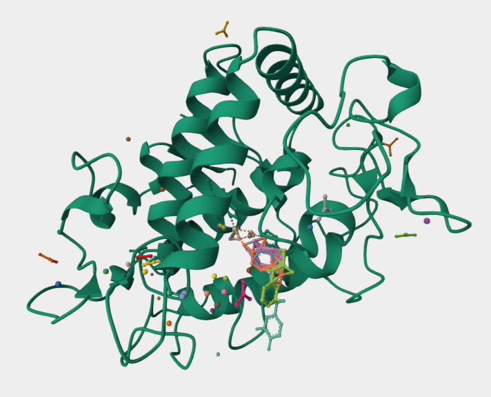
I selected MelC2 from Streptomyces antibioticus as the target enzyme for the first construct. I chose MelC2 because it is a soluble, cytosolic, oxygen-, copper-dependent and reasonably small enzyme of about 273 amino acids, and has a reviewed Swiss-Prot annotation, which made it a strong first candidate for cell-free TX-TL expression.
Its dependence on copper, oxygen, substrate availability, pH, and downstream polymerization also gives the project a clear set of tunable variables for validating melanin-like pigment formation.
Useful references for this step included the UniProt P07524 entry and WT sequence in Benchling.
And here is the predicted structural model used as part of my design context:
5.1.2.2 Codon optimization
I codon-optimized the MelC2 sequence for E. coli K-12 expression in Benchling**
Codon-optimization of P07524 for E. coli K-12, to avoid BsaI/BsmBI/BbsI and add a C-terminal His-tag to quantify enzyme expression cleanly -> Results in Benchling here.
I’ve selected the region of the AA sequence I wish to back translate and right clicked on the highlighted region. From the the codon optimization tab:
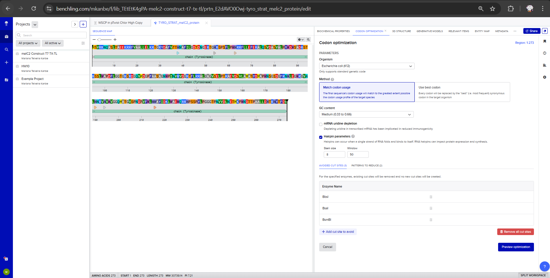
- Host: E. coli K-12
- Method: Match codon usage
- GC content: Medium (0.33 to 0.66) cause the extremes may be inconvenient. High GC can create strong secondary structures and low GC can cause instability/repeats and can make synthesis harder.
- Uridine depletion: off (not relevant for bacterial expression)
- Hairpin parameters: Stem size: 8 and Window 50
- Restriction sites: avoid BsaI, BsmBI, BbsI (Type IIS restriction enzymes, the workhorses of Golden Gate assembly)
- Patterns to reduce: AAAAAA and ATATATATA
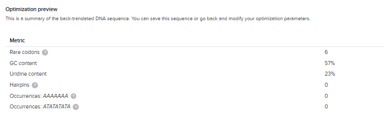
I clicked on “Preview Optimization” and got this result, which I’ve saved in the same Benchling folder here:
BLASTP verification of codon-optimized sequence:
I translated the codon-optimized DNA and ran BLASTP against nr/ClusteredNR. The top hits were MelC2 tyrosinases from Streptomyces spp., with 100% query coverage, E-value 0.0, 92% identity (251/273), 95% positives (261/273), and 0 gaps. Conserved domain analysis identified the Tyrosinase domain across the full sequence length. This confirms the optimized DNA still encodes a canonical tyrosinase.
melC2 tyrosinase (Streptomyces antibioticus, P07524, codon-optimized for E. coli K-12) DNA sequence Benckling link here.
5.1.2.3 Protein-detection design
I added a C-terminal 6xHis tag (CACCACCACCACCACCAC) before the stop codon to support future protein-level detection / quantification.
5.1.2.4 Expression cassette assembly
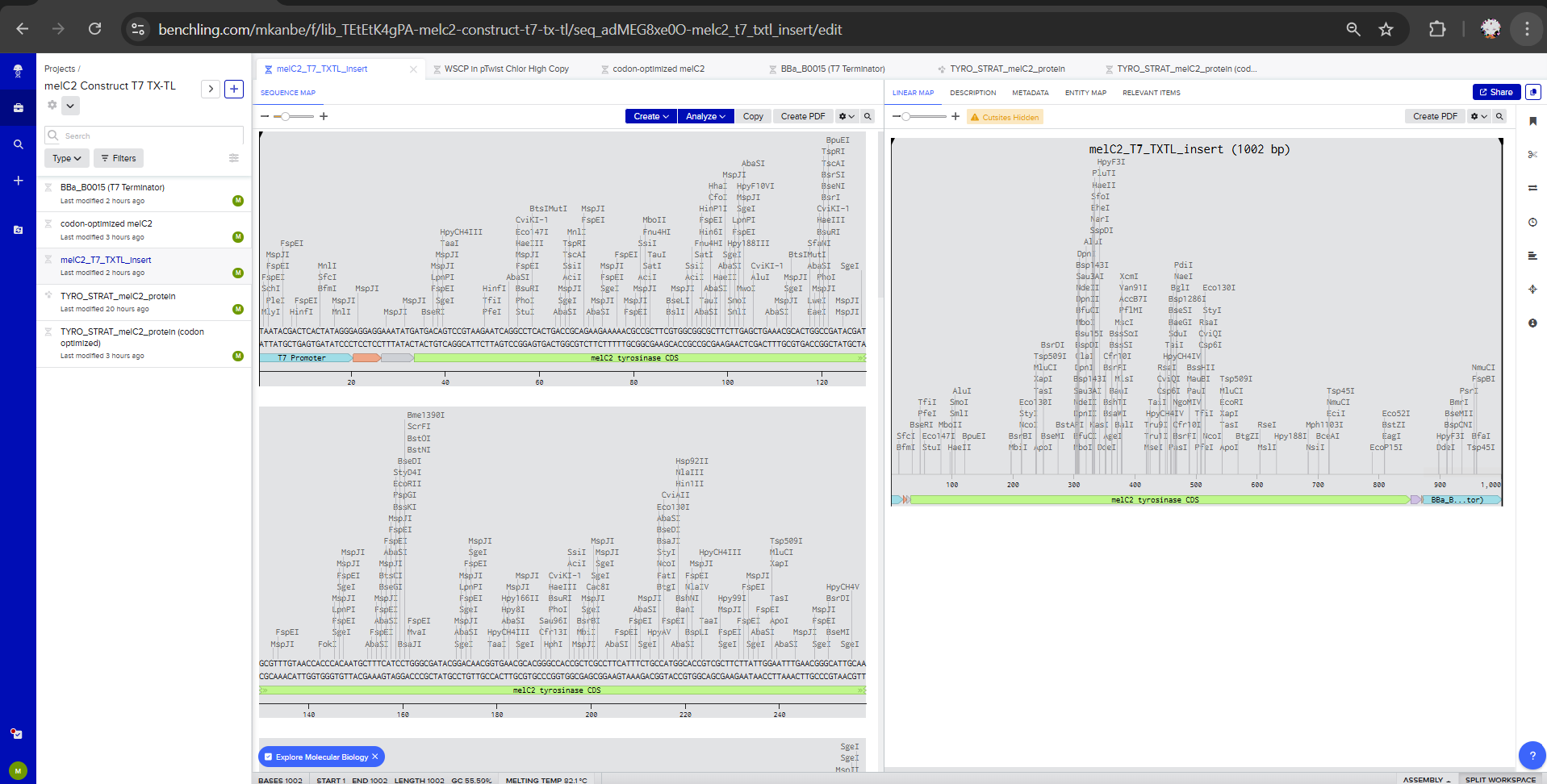
I assembled the TX-TL expression cassette using a T7 Promoter, RBS (Shine Delgarno) / AAATAT Spacer, codon-optimized melC2 CDS, C-terminal 6xHis tag, TAA stop codon, and T7 terminator BBa_B0015 Benchling link here.
To be considered: T7 can maximize protein yield but also overwhelm folding capacity, causing inactive protein accumulation (increase the likelihood of tyrosinases misfolds, aggregation, or fail to incorporate copper correctly). I’d replace it by a moderated construct and compare the results in reference to the BBa_K2481108 (control).
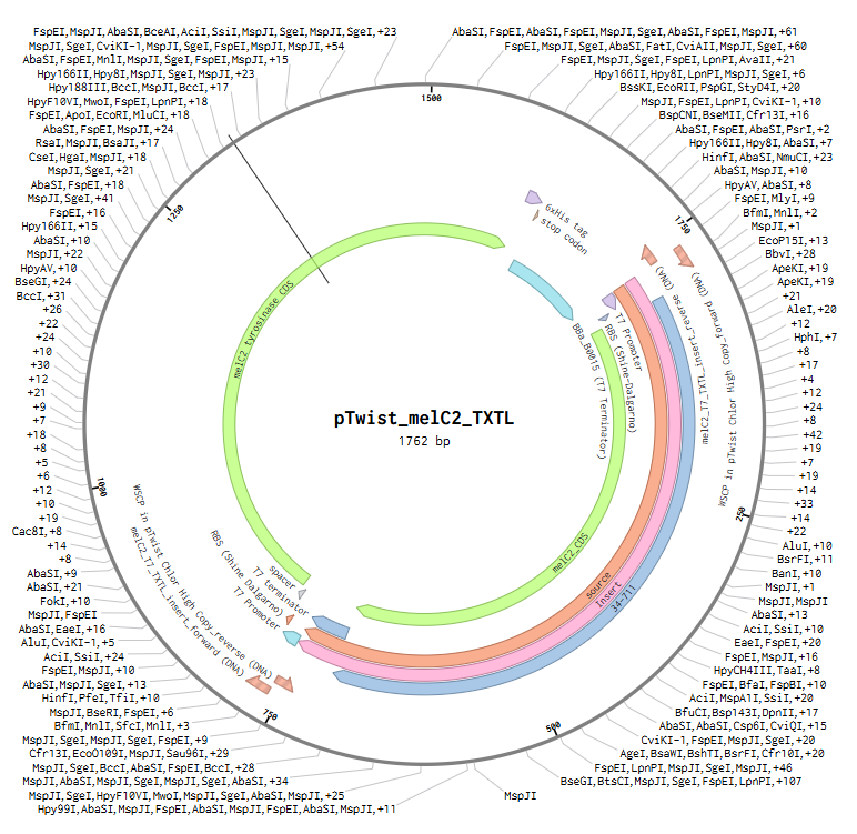
So here’s my final melC2 tyrosinase CDS with annotations.
5.1.2.5 Vector assembly and construct inspection
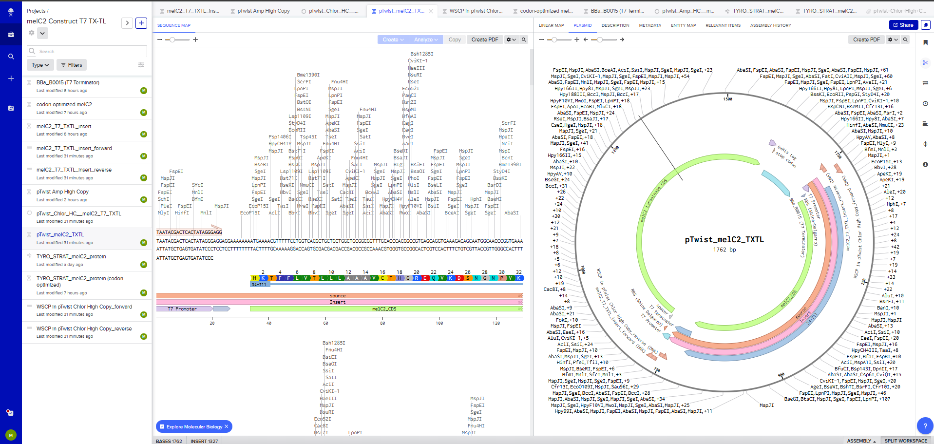
I placed the full expression cassette into a pTwist Amp High Copy vector. Why: high-copy propagation in E. coli for easy plasmid prep; selection marker is standard.**
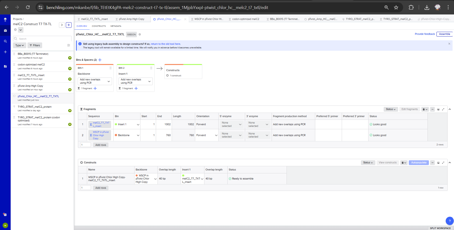
I inspected the final construct map in Benchling to confirm the organization of the insert, vector, promoter, terminator, and annotated CDS. Assemblings on Benchling here.
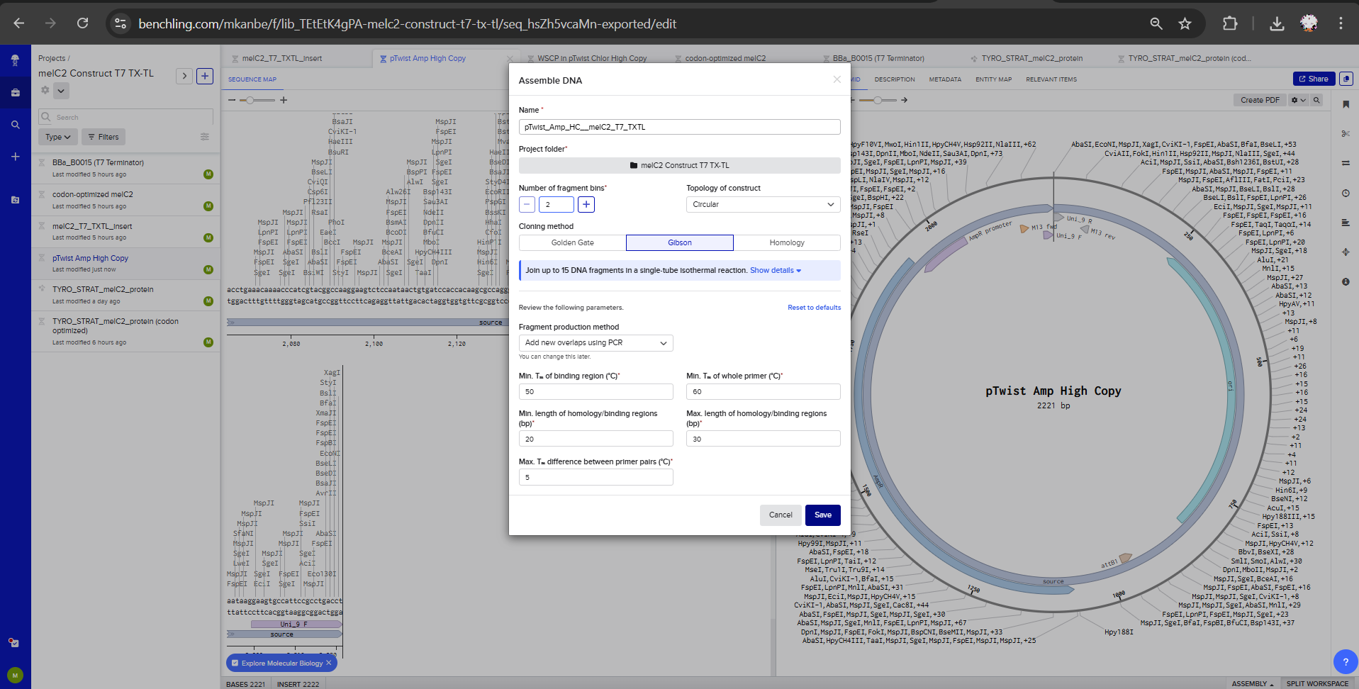
Final Construct: My melC2 construct submitted assembled into pTwist Amp High Copy on Benchling interface.
5.1.2.6 Twist submission
I submitted the final construct for synthesis through Twist. My Twist order for final Construct here
This completed the validated DNA-design layer of the project.
Planned next validation
After synthesis, the construct will be tested in a staged cell-free workflow: fluorescent protein controls for TX-TL capacity, visible darkening and OD 400-500 nm for pigment formation, SDS-PAGE / His-tag detection for MelC2 expression, and future LC-MS for tyrosine / L-DOPA-related intermediates.
I also began planning Ginkgo RAC-style cell-free reaction conditions to test key bottlenecks such as copper availability, tyrosine concentration, pH buffering, magnesium, incubation time, and reporter choice.
Prepare reagents and workflow (Ginkgo & Open AI)
I’ll first test my brainstormed HW 11C Cell-Free Master Mix experiment and then iterate aiming for a debugged and optimized workflow.
Here are some variables I had in mind when formulating this first 8 master mix compostion
Melanin production in E. coli or in a cell-free system is influenced by several parameters that act at the level of melC2 expression and enzyme activity / downstream reactions:
L-tyrosine concentration (substrate, limited solubility)
CuSO4 concentration: since this tyrosinase is a type 3 copper-containing enzyme, Cu2+ is a cofactor of the enzyme. Too much copper can also stress cells or inhibit cell-free reactions.
Magnesium
Energy mix
Molecular oxigen avaliability for tyrosinase reactions
pH: tyrosinase activity and melanin polymerization are pH-dependent. If the reaction acidifies over time, enzyme activity or pigment formation may decrease.
My first 8 experiments at Ginkgo - aim is to successfully produce fluorescent protein and generate an initial dataset for analysis.
sfGFP → system calibration (TX-TL health) Melanin has a broad absorbance spectrum, but it absorbs much more strongly at shorter wavelengths (blue/green) than at longer wavelengths (red). Melanin interferes with optical readout since we will be trying to measure fluorescence in a reaction that is simultaneously getting darker, which creates optical interference broadening the wavelength spectrum of signal.
mScarlet-I → expression readout for melC2 tyrosinase specifically fluorescence is less sensitive to melanin, so it better tracks expression alone (sfGFP → Ex ~488 nm / Em ~510 nm → high overlap with melanin absorbance; mTurquoise2 → even worse (blue region); mScarlet-I → Ex ~569 nm / Em ~594 nm → less overlap).
For optimizing the Master Mix design for mScarlet-I in my melC2 tyrosinase cell-free system, I’d supplement CuSO4 since my analyte is a copper-dependent enzyme, HEPES-KOH pH 7.5 to have an additional buffer against acidification and magnesium glutamate to improve translation capacity.
At first I thought about adding glucose since it could extend energy regeneration, but then I wondered that it may also increase acidification. Since you’re worried about fluorescence readout in a pigment-producing system, I’d prioritize pH stability over extra glucose.
I’d actually supplement L-tyrosine that serves as a functional validation that my protein of interest MelC2 tyrosinase is being expressed and active.
Master Mix designs to be tested using mScarlet-I and sfGFP, the 8 reactions outlined are available here in Week 11 HW Documentation.
5.1.3 Synthetic biology techniques used
The main synthetic biology technique used was DNA construct design. I designed a codon-optimized MelC2 tyrosinase cassette for TX-TL / E. coli expression, added a C-terminal 6xHis tag for future protein detection, assembled the cassette in Benchling, and prepared it for synthesis through Twist.
I also used database-based sequence selection and verification. UniProt and Benchling were used to select and inspect the MelC2 sequence, while BLASTP and conserved-domain analysis were used to confirm that the codon-optimized DNA still encoded a canonical tyrosinase.
A third relevant technique was cell-free system planning. The construct was designed specifically for TX-TL / E. coli use, and the next validation workflow was planned around fluorescent protein controls, visible darkening, OD 400-500 nm absorbance, SDS-PAGE / His-tag detection, and future LC-MS analysis.
Finally, I used lab automation planning by preparing a Ginkgo RAC-style reaction matrix to test variables expected to affect MelC2 pigment formation, including copper availability, L-tyrosine concentration, pH buffering, magnesium, incubation time, and reporter choice.
5.1.4 Data and analysis
The validation data for this stage are design-level and sequence-level results generated during construct preparation. These data show that the MelC2 construct is synthesis-ready and still encodes the intended tyrosinase target.
| Validation item | Result / quantitative expectation | Interpretation |
|---|---|---|
| Target enzyme | MelC2 tyrosinase from Streptomyces antibioticus | Selected as the first melanin-forming enzyme candidate |
| Protein length | ~273 amino acids | Small enough for practical TX-TL expression testing |
| Codon optimization host | E. coli K-12 | Matches the intended TX-TL / E. coli expression context |
| GC content after optimization | 57% | Within a workable synthesis and expression range |
| Rare codons | 6 | Low enough to support expression feasibility |
| Hairpins detected | 0 | Reduces risk of problematic RNA secondary structure |
| AAAAAA occurrences | 0 | Removes a problematic repetitive A-rich pattern |
| ATATATATA occurrences | 0 | Removes a problematic repetitive AT-rich pattern |
| Avoided restriction sites | BsaI, BsmBI, BbsI | Improves compatibility with future Type IIS cloning workflows |
| Detection feature | C-terminal 6xHis tag | Enables future protein-level validation |
| BLASTP query coverage | 100% | Optimized sequence still aligns across the full tyrosinase sequence |
| BLASTP E-value | 0.0 | Strong sequence-level match |
| BLASTP identity / positives | 92% identity / 95% positives | Confirms the optimized construct still encodes a MelC2-like tyrosinase |
| Gaps | 0 | No major sequence disruption introduced by optimization |
| Conserved domain | Tyrosinase domain across full sequence | Confirms the intended enzyme family was preserved |
These data validate the first build layer of the project: the DNA module is codon-optimized, annotated, compatible with the intended TX-TL / E. coli context, and submitted for synthesis. The results do not prove melanin production, but they confirm that the construct is coherent enough to justify downstream expression testing. The next quantitative expectation is that successful cell-free expression should produce detectable MelC2 by SDS-PAGE / His-tag detection and measurable pigment accumulation by OD 400-500 nm if the enzyme is active under the tested conditions.
5.2 Unexpected challenges, limitations, and alternatives
The main limitation is that a correct DNA construct does not automatically prove protein activity or melanin-like pigment formation. Tyrosinase expression can be detected while pigment remains absent if folding, copper incorporation, substrate availability, oxygen, pH, or downstream polymerization chemistry is limiting.
Another challenge is that melanin is a chemically heterogeneous output, so visible darkening alone is not enough to validate the system. To address this, the next validation stage separates TX-TL expression capacity, MelC2 protein production, pigment accumulation, and pathway-level chemistry using fluorescence controls, OD 400-500 nm, SDS-PAGE / His-tag detection, and future LC-MS. If the T7 design produces inactive or misfolded protein, an alternative strategy would be to test a moderated promoter, adjust copper and substrate concentrations, or compare purified enzyme / synthetic melanin-like polymer approaches before moving into more complex biomaterial systems.
SECTION 6: ADDITIONAL INFORMATION
6.1 References cited in this assignment
- Menichetti, L. et al. “Melanin as a photoprotective material,” 2025. https://www.mdpi.com/3235558
- Dadachova, E. and Casadevall, A. “Ionizing radiation: how fungi cope, adapt, and exploit with the help of melanin,” 2009. https://pmc.ncbi.nlm.nih.gov/articles/PMC2677413/
- Cordero, R. J. B. et al. “Fungal melanin-polymer biocomposites exposed to low Earth orbit conditions,” 2025. https://www.pnas.org/doi/10.1073/pnas.2427118122
- Yue, X. and Zhao, L. “Melanin-like materials for photothermal and bioelectronic applications,” 2021. https://www.mdpi.com/947490
- UniProt. Streptomyces antibioticus tyrosinase MelC2, P07524. https://www.uniprot.org/uniprotkb/P07524/entry
- iGEM Registry. BBa_I14032, P(lac)IQ promoter. https://parts.igem.org/Part:BBa_I14032
- iGEM Registry. BBa_K193600, melA tyrosinase. https://parts.igem.org/Part:BBa_K193600
- iGEM Registry. BBa_K193602, pLacIQ-RBS-melA composite construct. https://parts.igem.org/Part:BBa_K193602
- iGEM Registry. BBa_K2481108, MelA expression construct for E. coli BL21(DE3). https://parts.igem.org/Part:BBa_K2481108
6.2 Supply list and budget
This budget estimates the next practical stage of the project: validating the MelC2 construct in a cell-free TX-TL system before moving into material integration.
The cost ranges below were estimated with the assistance of ChatGPT and should be treated as approximate planning values. The estimation method was to break the project into major experimental cost categories - DNA synthesis, TX-TL reactions, controls, reagents, consumables, protein validation, chemical validation, and material integration - and assign conservative low/high ranges for each category based on typical small-scale synthetic biology workflows.
The lower end of each range assumes access to shared lab equipment, existing stocks of common reagents, and limited reaction numbers. The higher end assumes new reagent purchases, larger reaction matrices, external analytical services, or the need to purchase or arrange access to readout equipment. Exact costs would need to be confirmed through vendor quotes, institutional core facility pricing, or cloud-lab pricing.
| Category | Supplies / services | Estimated cost | Notes |
|---|---|---|---|
| DNA synthesis | MelC2 TX-TL expression cassette in pTwist Amp High Copy vector | $150-300 | One synthesis-ready expression cassette |
| Cell-free TX-TL reaction system | E. coli TX-TL master mix or freeze-dried cell-free reaction kit | $300-800 | Enough material for expression controls and an initial MelC2 reaction matrix |
| DNA / expression controls | sfGFP control plasmid or template; mScarlet-I control plasmid or template | $100-300 | Used to confirm that the TX-TL system supports protein expression |
| Substrates and cofactors | L-tyrosine; CuSO4; magnesium glutamate; nuclease-free water | $100-250 | Core reaction components for testing tyrosinase activity |
| Buffering and reaction-condition reagents | HEPES-KOH pH 7.5; additional salts or energy-mix supplements if needed | $100-250 | Used to adjust pH, magnesium, and reaction stability |
| Consumables | PCR tubes or reaction tubes; pipette tips; microcentrifuge tubes; plate or strip-tube format for reaction imaging | $100-250 | Disposable materials for small-volume reactions |
| Optical readout equipment | Plate reader or spectrophotometer capable of OD 400-500 nm; fluorescence readout for sfGFP / mScarlet-I | $0 if shared; $5,000+ if purchased | The project requires access to the instrument, not necessarily purchase |
| Protein-expression validation | SDS-PAGE gel system access; protein ladder; gel stains; optional His-tag detection reagents | $150-500 | Confirms whether MelC2 protein is produced independently of pigment output |
| Chemical validation | LC-MS access for L-tyrosine, L-DOPA, and related intermediates; analytical standards | $300-1,500 | Cost depends on shared facility access, outsourcing, and sample number |
| Automation / cloud lab testing | Ginkgo RAC-style cell-free reaction matrix, if available | Variable | Not included in the main estimate because pricing depends on platform access |
| Future material-integration supplies | Bacterial cellulose sheets or Komagataeibacter culture materials; coating / embedding materials | $200-700 | Future stage after biochemical validation |
Estimated total for first validation stage: approximately $850-3,600, assuming access to shared lab equipment.
This total excludes major equipment purchases and uncertain cloud-lab pricing. It includes the core experimental costs needed to move from a designed MelC2 construct to initial TX-TL expression, pigment-production screening, and basic validation.
Estimated total including purchased equipment or external analytical services: could exceed $5,000-10,000, depending on instrument access, number of samples, and whether LC-MS or optical readout must be outsourced or purchased.
This documentation was developed with the assistance of ChatGPT, which was used to support drafting, editing, organization, and figure generation. All scientific decisions, final content, and interpretations were reviewed and approved by the author.