Week 9 HW: Cell-Free Systems
Homework Part A: General and Lecturer-Specific Questions
General homework questions
- Advantages of cell‑free protein synthesis
Direct access to the reaction environment and no constraints from cell viability. Cell‑free synthesis is preferable for producing toxic proteins and for rapid screening of many variants without maintaining live cultures.
- Main components of a cell‑free system and their roles
Cell extract (ribosomes, tRNAs, translation factors, polymerase), energy system (ATP plus regeneration), amino acids, DNA template, buffer and salts.
- Why energy regeneration is critical and how to ensure continuous ATP supply
Protein synthesis rapidly depletes ATP; regeneration is essential. Method: creatine phosphate / creatine kinase system (creatine phosphate donates phosphate to ADP, generating new ATP).
- Comparison of prokaryotic and eukaryotic cell‑free systems. Choosing a protein for each
Prokaryotic system (E. coli extract) is suitable for simple proteins, e.g. GFP. Eukaryotic system (wheat germ extract) is suitable for proteins with post‑translational modifications, e.g. human immunoglobulin IgG.
- Designing a cell‑free experiment to optimise membrane protein expression
Challenges: poor solubility, aggregation, misfolding. Solutions: add detergents (DDM) or liposomes, use lipid nanodiscs, optimise temperature and detergent concentration.
- Low protein yield: three possible reasons and troubleshooting
For DNA degradation, add nuclease inhibitors and use fresh template. For energy depletion, increase ATP regeneration components. For protein aggregation, lower temperature and add chaperones or detergents.
Homework question from Kate Adamala
- Pick a function and describe it.
a. What would your synthetic cell do? What is the input and what is the output? The synthetic cell will detect mechanical envelope stress caused by sound vibration. Input: acoustic vibration (mechanical force transmitted through agar). Output: red fluorescence (mScarlet‑I protein).
b. Could this function be realized by cell‑free Tx/Tl alone, without encapsulation? No. Without encapsulation there is no cell envelope, and envelope stress is required to activate the Rcs cascade. A cell‑free system in a test tube cannot sense mechanical stress.
c. Could this function be realized by a genetically modified natural cell? Yes. E. coli carrying the P_rprA‑B0034‑mScarlet‑I‑B0015 construct works as a vibration biosensor. This is a natural cell with modified DNA.
d. Describe the desired outcome of your synthetic cell operation. Upon acoustic stimulation, the cell displays red fluorescence. Fluorescence intensity is proportional to vibration strength up to a certain threshold.
- Design all components that would need to be part of your synthetic cell.
a. What would be the membrane made of? For a synthetic minimal cell, the membrane would consist of POPC and cholesterol lipids (for stability).
b. What would you encapsulate inside? Enzymes, small molecules. Inside the liposome, one would encapsulate a cell‑free transcription‑translation (Tx/Tl) system based on E. coli extract (containing all necessary enzymes, including Rcs cascade components), amino acids, ATP, NTPs, buffer, and the DNA template with the P_rprA‑B0034‑mScarlet‑I‑B0015 cassette.
c. Which organism your Tx/Tl system will come from? A bacterial system from E. coli is suitable because the Rcs signalling pathway is endogenous to E. coli.
d. How will your synthetic cell communicate with the environment? Mechanical vibration is transmitted through the agar and creates envelope stress, which activates the Rcs cascade. All necessary substrates are stored inside the capsule.
- Experimental details
a. List all lipids and genes. Lipids: POPC, cholesterol (for stability). Genes: P_rprA (RcsB‑dependent promoter), B0034 (strong RBS), mScarlet‑I (red fluorescent protein), B0015 (terminator).
b. How will you measure the function of your system? Place Petri dishes with bioprinted bacteria on a vibration shaker. Apply controlled acoustic stimulation that produces vibration. Take photographs and quantify the fluorescence intensity using ImageJ software. Compare with control dishes that were not exposed to vibration.
Homework Part B: Individual Final Project
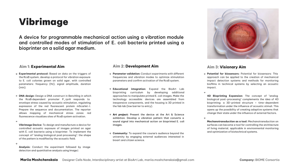
Vibrimage progect
DNA construct
The DNA construct includes the reporter cassette P_rprA — B0034 — mScarlet-I — B0015, where P_rprA is an RcsB‑dependent promoter and mScarlet-I encodes a red fluorescent protein.
Ссылка на Benchling: https://benchling.com/s/seq-aufGtzyeJfh49bTNUeT3?m=slm-vhiZGRT1DLF1aRg8DO0T
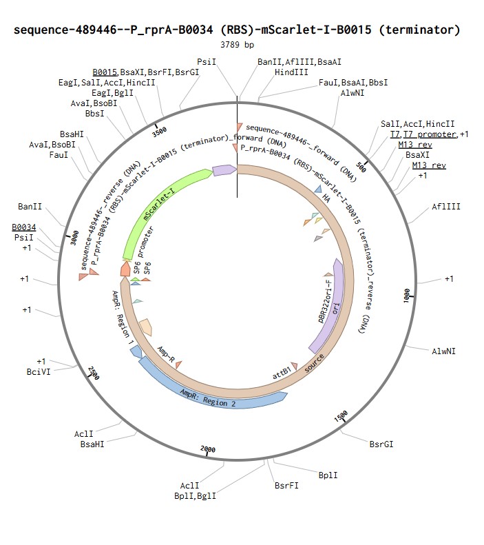
Plasmid for mScarlet-I expression in E. coli
Homework question from Peter Nguyen
Application Field: Architecture
The Concept: A living, breathable building brick made from fungal mycelium that acts as a continuous, low‑cost biosensor, visually detecting the presence of airborne heavy metal pollutants by changing colour.
- One‑Sentence Pitch
A living mycelium building brick that changes colour upon contact with toxic heavy metals in the air or water, serving as a built‑in, maintenance‑free pollution detector.
- How the Idea Works (in detail)
Fungal mycelium grown into a brick or tile is a living network. Certain fungi, such as Aspergillus niger, possess a natural ability for the biosorption of heavy metal ions (e.g., lead, cadmium, mercury) from the environment. When these ions bind to the fungal cell wall, biochemical reactions occur that produce a visible colour signal. For example, it has been demonstrated that fungal mycelium can turn blue upon contact with even very low concentrations of copper ions.
- Societal Challenge / Market Need Addressed
Monitoring urban air and water quality requires expensive sensors and electricity. Heavy metal pollution is often invisible and uneven. This brick creates a distributed, passive sensor network within the fabric of the building itself. This makes environmental monitoring cheap, accessible and visually understandable for residents, allowing them to identify local pollution “hotspots” in real time without complex instruments.
- Addressing Cell‑Free System Limitations
This proposal uses living fungi instead of freeze‑dried cell‑free systems, which directly addresses their limitations. First, activation by water is not required, because the hygroscopic mycelium naturally extracts moisture from the air. Second, stability is not a concern: as long as the fungus is alive and receives nutrients from the carrier material (e.g., agricultural waste), it remains active. Third, single‑use is also not an issue. The sensor is not “consumed” after a single event; it continues to react and accumulate the signal. The permanent colour change serves as a long‑term archival record of pollution, rather than a transient, disappearing signal.
Homework question from Ally Huang
Genes in Space Proposal: A Cell‑Free Biosensor for Early Biofilm Detection in Spacecraft Water Systems
- Background Information
Biofilms are a recognized threat in space habitats. They can clog water recovery systems, corrode metal surfaces, and harbour opportunistic pathogens (e.g., Pseudomonas aeruginosa). In microgravity, bacteria form more robust biofilms, making monitoring crucial. Current detection relies on time‑consuming culturing. A rapid, low‑resource cell‑free biosensor would allow astronauts to detect early contamination, safeguard equipment, and protect health on long‑duration missions.
Biofilms pose a serious risk to spacecraft water systems and crew health. In microgravity, they grow faster and are more resistant. Existing detection methods are slow and resource‑intensive. A quick, easy‑to‑use cell‑free biosensor would enable early warning and timely countermeasures, which is critical for long‑duration space exploration where resupply is impossible.
- Molecular or Genetic Target
A DNA construct encoding a fluorescent reporter protein (e.g., sfGFP) under the control of a promoter that is specifically activated by N‑acyl homoserine lactones (AHLs) – the quorum sensing molecules used by many biofilm‑forming bacteria, such as P. aeruginosa.
A genetic circuit consisting of a quorum‑sensing promoter (e.g., PluxR) fused to a superfolder GFP gene. This promoter is induced by AHL molecules that are key signals for biofilm formation.
- Relation to the Space Biology Challenge
AHLs diffuse into water as biofilms develop. Our cell‑free BioBits system will detect these molecules. Upon rehydration of the freeze‑dried reaction with a water sample, AHLs will bind to a constitutively expressed transcription factor (e.g., LuxR), which then activates the reporter promoter, driving GFP expression. Fluorescence, read with the P51 viewer, directly correlates with AHL concentration and thus biofilm biomass. This provides a rapid, semi‑quantitative readout of biofilm status in the water system.
As a biofilm grows, bacteria release AHL molecules into the surrounding water. Our BioBits sensor will detect these AHLs: they activate a transcriptional activator, which turns on GFP production. The resulting green fluorescence can be easily seen by an astronaut using the P51 viewer. No complex equipment or cell culture is needed.
- Hypothesis or Research Goal
Hypothesis: A cell‑free BioBits sensor can detect AHL molecules from developing biofilms in spacecraft water samples, producing a fluorescence signal proportional to the AHL concentration. Goal: To design, optimize, and validate a portable, shelf‑stable biosensor that provides an early, non‑invasive warning of biofilm contamination in ISS water systems. This will be tested both on Earth (with simulated microgravity controls) and in spaceflight conditions.
We hypothesize that our sensor will specifically and sensitively detect AHLs in water. The research goal is to produce a ready‑to‑use, freeze‑dried test that requires only adding a water sample, waiting 1‑2 hours, and looking for green fluorescence. This would enable routine, autonomous monitoring of water quality on the ISS and future deep‑space missions.
- Experimental Plan
Samples: Water samples from the ISS Water Processor Assembly (WPA) collected at different time points. Controls: Negative – sterile deionized water; Positive – water spiked with synthetic AHL (C4‑HSL or 3OC12‑HSL). Procedure: Each sample is mixed with freeze‑dried BioBits sensor pellets (contains all CFPS components). After 1‑2 h incubation at 22°C, fluorescence is measured using the P51 viewer. Tests will be run in triplicate. Data: Qualitative (visible glow) and semi‑quantitative (image analysis via smartphone photo). Comparison of Earth and space results will reveal microgravity effects on sensor performance.
We will test our sensor with real and simulated ISS water samples. Controls include pure water (no signal) and water with added AHLs (strong signal). The experiment is simple: rehydrate a freeze‑dried pellet, incubate, and observe fluorescence. Astronauts will document results with photos. The same procedure will be run on Earth to validate performance and then transferred to the ISS for space validation.