Individual Final Project
Vibrimage
SECTION 1: ABSTRACT
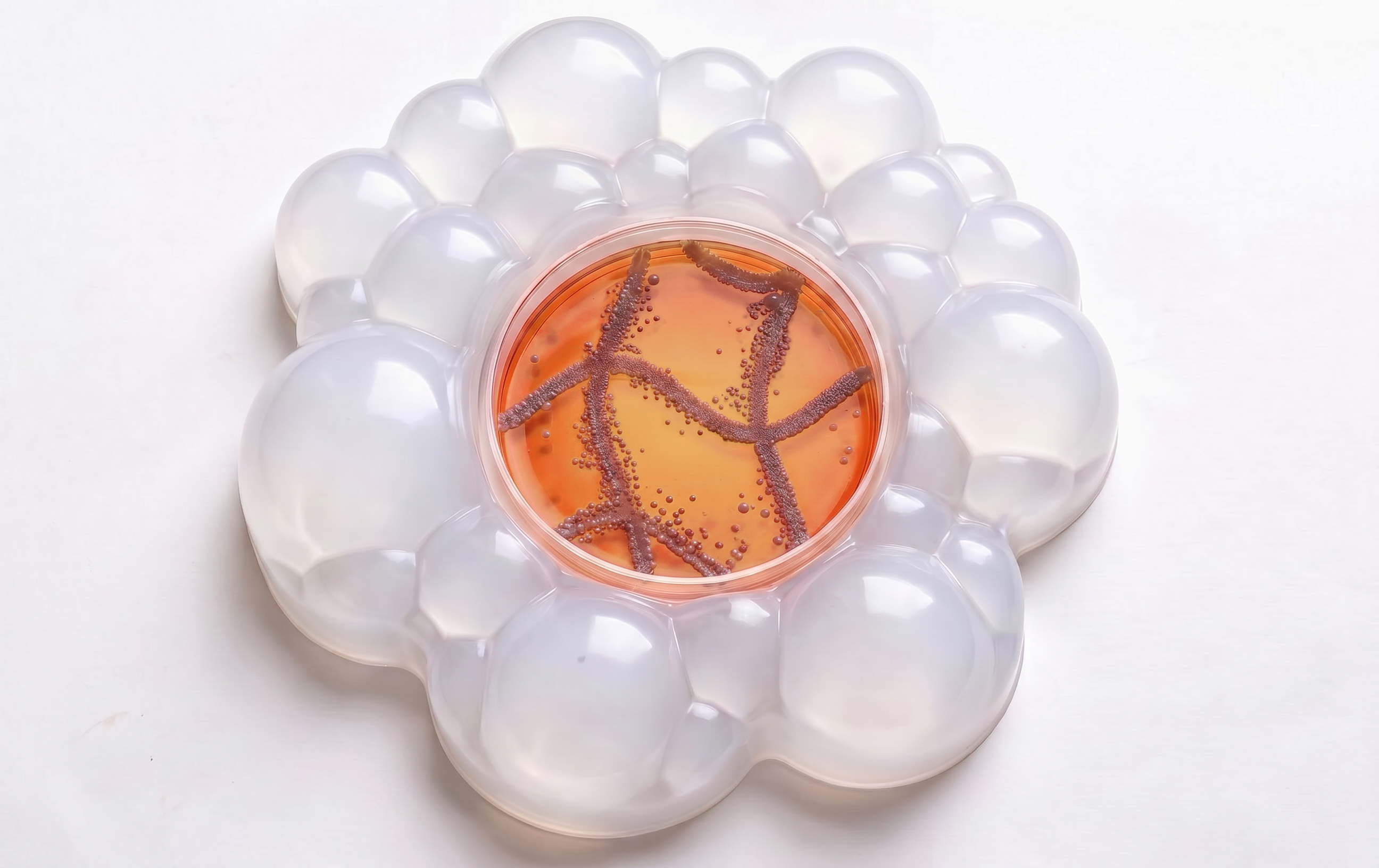
Vibrimage is a device for affecting the growth of bioprinted E. coli on solid agar medium using programmable acoustic exposure. The basic idea is to do post‑processing of a bacterial image using sound. In this case, audible frequencies act as a “bio‑Photoshop”. For DNA design, I will use the P_rprA promoter. It makes E. coli fluoresce red in areas of acoustic exposure. This makes ‘sound sculpting’ visible.
Most research focuses on removing bacteria using high frequencies in liquid media. The Vibrimage project studies the effect of sound frequencies on bacteria in a biosensor context. That is why it is important for me to also understand how to promote bacterial growth. Another important aspect of the project was to recreate conditions that could be found in real life. Bacteria grow on a solid medium. The exposure to audible sound frequencies is 500 hertz and 1000 hertz. The image will be printed by E. coli bacteria in the form of waves. It is 500 hertz and 1000 hertz waves, forming a kind of grid. This work is also presented as a way to share skills with the bioart community, so the focus is on building an easy‑to‑make device.
Acoustic exposure causes vibration and mechanical stress of the cell envelope, which activates the Rcs system. This triggers RcsB‑dependent signalling and activates the P_rprA promoter, ultimately leading to expression of mScarlet‑I and red fluorescence of E. coli.
In the future, the project is planned to be developed and presented as an Art & Science project with musical compositions, as well as to continue research into how it can be implemented in nutritional technologies.
 Device
SECTION 2: PROJECT AIMS
Aim 1: Experimental Aim
Experimental protocol: Based on data on the triggers of the Rcs system, develop a protocol for vibration exposure to E. coli colonies grown on solid agar, with controlled parameters: frequency (Hz), signal amplitude, duration (min).
DNA design: Design a DNA construct in Benchling in which the RcsB‑dependent promoter P_rprA responds to envelope stress caused by acoustic stimulation, regulating expression of the red fluorescent protein mScarlet-I. Prepare the sequence and documentation. The reporter allows mapping of mechanical stress zones: red fluorescence visualizes sites of RcsB system activation.
Vibrimage Device: To design and manufacture a device for controlled acoustic exposure of images printed on agar with E. coli bacteria using a bioprinter. To -implement the concept of “analog biological post‑processing”: the shape of the pattern is modified by the acoustic field.
Analysis: Conduct the experiment followed by image detection and quantitative analysis using ImageJ.
Aim 2: Development Aim
Parameter validation: Conduct experiments with different frequencies and vibration modes to optimize stimulation parameters and confirm activation of the Rcs system.
Educational integration: Expand the BioArt Lab bioprinting curriculum by developing additional approaches to manipulate printed E. coli images. Make the technology accessible: devices are assembled from inexpensive components, and the housing is 3D printed in the fab lab (low barrier to entry).
Art project: Present the device at the Art & Science exhibition. Develop a vibration pattern that converts a sound signal into mechanical action on bioprinted E. coli images.
Community: To expand the course’s audience beyond the university by engaging external audiences interested in bioart and citizen science.
Aim 3: Visionary Aim
Potential for biosensors: Potential for biosensors: This approach can be applied to the creation of mechanical impact detection systems and methods for monitoring biofilms in technical systems by selecting an acoustic impact.
4D Bioprinting Expansion: The concept of “analog biological post-processing” complements the idea of 4D bioprinting: a 3D-printed structure + time-dependent transformation under the influence of acoustic stimuli. This opens up the possibility of creating adaptive systems that change their state under the influence of external factors.
Mechanotransduction as a tool: Mechanotransduction on surfaces can become a tool for controlling the architecture of living material, applicable in environmental monitoring and optimization of biotechnical systems.
SECTION 3: BACKGROUND
- Murphy, M. F., Edwards, T., Hobbs, G., Shepherd, J., & Bezombes, F. (2016). Acoustic vibration can enhance bacterial biofilm formation. Journal of bioscience and bioengineering, 122(6), 765-770.
In this study, published in the Journal of Bioscience and Bioengineering, the scientists demonstrated that low-frequency acoustic vibration can significantly enhance biofilm formation in the bacteria Pseudomonas aeruginosa and Staphylococcus aureus. To do this, they developed a speaker-based system that transmitted vibration to the bottom of a Petri dish containing liquid culture. Exposure to vibration at frequencies of 100, 800, and 1600 Hz for 48 hours led to a significant increase in the number of cells and biofilm biomass, with each bacterial species having its own optimal frequency range. This research is of key importance because it was the first to show that vibration could serve as a new, potentially inexpensive tool for controlling biofilm growth in medical and industrial processes.
- Bazzoli, D. G., Mahmoodi, N., Verrill, T. A., Overton, T. W., & Mendes, P. M. (2024). Nanovibrational stimulation of Escherichia coli mitigates surface adhesion by altering cell membrane potential. ACS nano, 18(44), 30786-30797.
In this study, published in the journal ACS Nano, the scientists applied nanoscale surface vibrations to mechanically stimulate E. coli bacteria and study their effect on adhesion. They used a device based on the inverse piezoelectric effect, which generated vertical vibrations at frequencies of 0.5, 1, and 2 kHz with an amplitude of up to 40 nm. Using laser vibrometry, the authors confirmed that the vibration amplitude increased linearly with increasing voltage and was uniform across the entire sample surface. The experiments revealed that nanovibrational stimulation consistently reduced the ability of E. coli to adhere to surfaces: adhesion decreased on average by 19% at 1 kHz and by 21% at 2 kHz. A key discovery was that vibration alters the membrane potential of the bacterial cell, which directly affects its adhesive properties, and the effect depended primarily on frequency rather than on stimulus intensity. This research represents the cutting edge of science, as it establishes a direct link between physical stimulation, bacterial electrophysiology, and bacterial behavior, and points to a new mechanism for controlling bacterial adhesion.
- Ying, J. L., Dayou, J., & Phin, C. K. (2009). Experimental investigation on the effects of audible sound to the growth of Escherichia coli. Modern Applied Science, 3(3), 124.
In this work, published in the journal Modern Applied Science, the direct effect of audible sound on the growth of E. coli on solid nutrient medium (agar) was studied. The researchers exposed the bacteria to pure tone sound at frequencies of 1, 5, and 15 kHz. The results showed that all selected frequencies promoted the growth of E. coli, with the most significant increase in cell number (by 34%) observed at a frequency of 5 kHz. It is important to note that the stimulating effect was recorded both in liquid and on solid nutrient medium.
Novel and Innovative Aspects of the Project
The novelty of the project lies in the study of acoustic stimulation of bacteria on solid nutrient medium in the sub-kilohertz frequency range (500 Hz and 1000 Hz). Although a few studies have demonstrated the effect of sound on E. coli growth on agar, the vast majority of research has focused on liquid cultures. Existing articles typically investigate bacterial removal using ultrasound. The Vibrimage project, in contrast, proposes to explore how bacterial growth can be modulated using acoustic vibration. Moreover, the DNA construct design — P_rprA (promoter) — B0034 (RBS) — mScarlet-I (reporter) — B0015 (terminator) — can, through fluorescence, indicate zones of active acoustic impact, which has potential for the development of biosensor applications.
 The experiment I conducted on bioprinting E. coli bacteria onto agar medium
SECTION 4: EXPERIMENTAL DESIGN, TECHNIQUES, TOOLS, AND TECHNOLOGY
Experiment
- Print an image of E. coli bacteria on agar using a bioprinter. – Incubate for 2 hours to allow adaptation and entry into the active growth phase.
- Exposure to 500 Hz, amplitude 1–10 μm during the active growth phase.
- Exposure to 5 kHz, 0.25–5 μm during the active growth phase.
- Control: no frequency exposure.
- Using ImageJ, determine: areas of greatest fluorescence from acoustic vibration, area of bacterial growth.
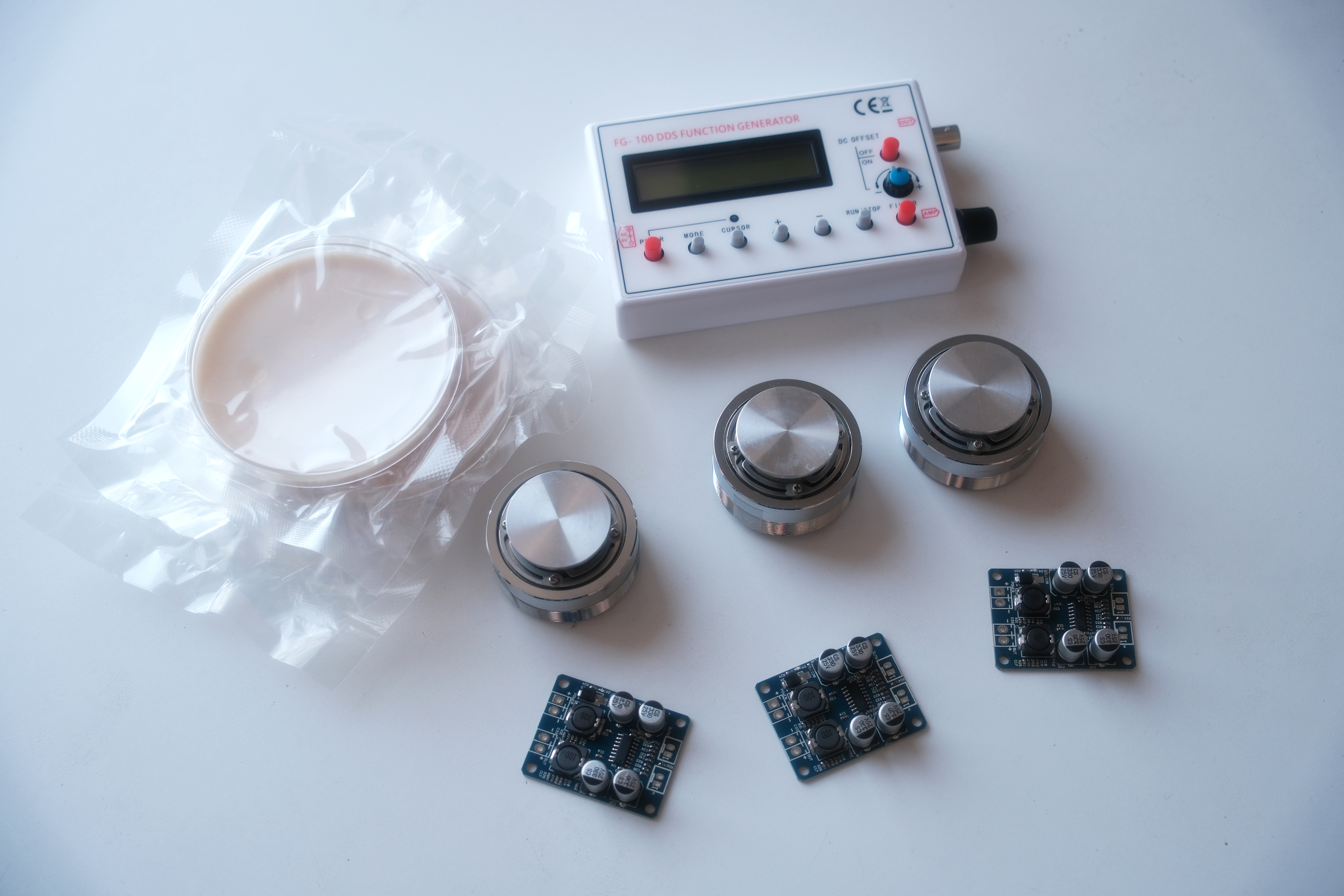
Device
- 3 vibrating speakers
- Amplifiers
- Signal generator
- Power supply
- Accelerometers for fine-tuning transmitted power
- App for monitoring audio parameters
- Endo agar (5.5 mm) – Biomorphic device casing made of photopolymer resin – Metal plates for Petri dishes – Radiators and fan
 The experiment I conducted on bioprinting E. coli bacteria onto agar medium
Techniques Used
- Bioethical Considerations
- Bioproduction
- Bacterial Culturing
- Quality Control/Analysis
- Bacterial Processing (e.g., Centrifugation, Lysis, DNA Purification)
- DNA Construct Design
- Databases (e.g., GenBank, NCBI, Ensembl, and UCSC Genome Browser)
- Gibson Assembly
- Use of Benchling
- Databases
Technique Expansion
Use of Benchling
“I would use Benchling to design, visualize, and annotate my DNA construct. This platform allows me to create detailed plasmid maps with color-coded features (promoter, RBS, CDS, terminator) and verify the correct assembly of genetic parts. Benchling also enables me to export sequences in various formats for future ordering or cloning. Additionally, I can share my design with collaborators and maintain detailed records of my construct design process.”
Databases “I would utilize databases such as NCBI and the iGEM Registry of Standard Biological Parts to obtain verified sequences for my genetic components. For example, I retrieved the B0034 RBS and B0015 terminator sequences from the iGEM Registry, and found the mScarlet-I sequence from NCBI. These databases ensure that I am using well-characterized parts with known functionality. This approach increases the reliability and success rate of my synthetic biology project.”
SECTION 5: Results & Quantitative Expectations
I validated the DNA construct design required for my project, as well as performed experimental testing of bioprinting and technical device assembly. In Benchling, I created the reporter cassette P_rprA — B0034 — mScarlet-I — B0015 and simulated its assembly into a plasmid vector. Additionally, real experiments were conducted to print E. coli bacteria using a bioprinter and to debug the technical setup for acoustic exposure. These steps confirmed the feasibility of the project.
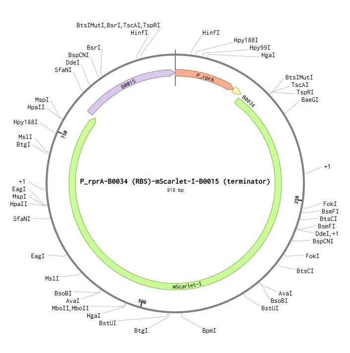
DNA construct design
SECTION 6: ADDITIONAL INFORMATION
Murphy, M. F., Edwards, T., Hobbs, G., Shepherd, J., & Bezombes, F. (2016). Acoustic vibration can enhance bacterial biofilm formation. Journal of bioscience and bioengineering, 122(6), 765-770.
Bazzoli, D. G., Mahmoodi, N., Verrill, T. A., Overton, T. W., & Mendes, P. M. (2024). Nanovibrational stimulation of Escherichia coli mitigates surface adhesion by altering cell membrane potential. ACS nano, 18(44), 30786-30797.
Ying, J. L., Dayou, J., & Phin, C. K. (2009). Experimental investigation on the effects of audible sound to the growth of Escherichia coli. Modern Applied Science, 3(3), 124.
Gu, S., Zhang, Y., & Wu, Y. (2016). Effects of sound exposure on the growth and intracellular macromolecular synthesis of E. coli k-12. PeerJ, 4, e1920.
Li, Z., Zhu, Y., Zhang, W., & Mu, W. (2024). Rcs signal transduction system in Escherichia coli: Composition, related functions, regulatory mechanism, and applications. Microbiological Research, 285, 127783.
Bindels, D. S., Haarbosch, L., Van Weeren, L., Postma, M., Wiese, K. E., Mastop, M., … & Gadella Jr, T. W. (2017). mScarlet: a bright monomeric red fluorescent protein for cellular imaging. Nature methods, 14(1), 53-56.