INDIVIDUAL FINAL PROJECT
A Cell-Free Biosensor Kit For Detecting Heavy Metal Mining Pollutants
Abstarct
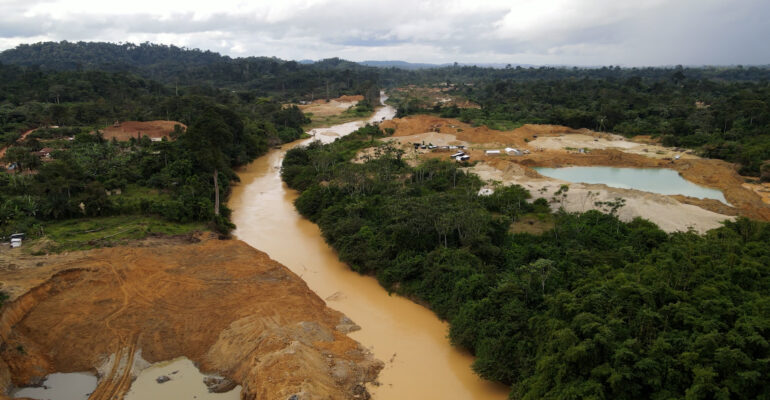
Illegal artisanal gold mining, locally known as galamsey, has devastated freshwater ecosystems across Ghana, particularly the Pra, Ankrobra, Ofiin, and Birim rivers. Contaminating them with toxic heavy metals such as mercury (Hg), lead (Pb), arsenic (As), and cadmium (Cd). This threatens the health of millions of Ghanaians in rural areas that rely on these rivers for domestic and agricultural purposes.
Access to safe drinking water is a global public health priority, yet current water quality monitoring infrastructure is sparse, expensive, inaccessible to rural communities most affected, and requires centralized laboratory infrastructure. My project is centered on the design, synthesis, and validation of a cell-free biosensor kit capable of detecting four heavy metals using engineered transcription factor circuits coupled to a colorimetric reporter producing results readable to the naked eye, requiring no laboratory equipment. It is built on four plasmid-based genetic circuits, one per metal, with each encoding a two-cassette transcriptional architecture: a constitutive J23119 promoter driving expression of a metal-responsive transcription factor (Cassette 1), and a cognate inducible promoter driving a visually distinct colorimetric reporter (Cassette 2). Metal-responsive transcription factors used are MerR (Hg²⁺), PbrR (Pb²⁺), ArsR (As³⁺), and CmtR (Cd²⁺). Reporters are eforRed (red), lacZ+CPRG (magenta), amilGFP (yellow-green), and tsPurple (purple) chromoproteins. All the circuits use sigma-70 compatible promoters and are expressed in E. coli BL21(DE3) cell-free protein synthesis (CFPS) extract, which is freeze-dried onto Whatman No.1 cellulose paper with 5% trehalose as cryoprotectant.
The central hypothesis of my project is that four orthogonal metal-responsive transcription factor systems(PbrR, ArsR, MerR, CadC), each coupled to a spectrally and chromaically distinct colorimetric reporter enzyme (LacZ, amilGFP, eforRed, tspurple) and integrated into freeze-dried cell-free extracts, can generate naked-eye detectable signals at or below Ghana Standards Authority regulatory limits (Hg ≤ 1 µg/L, Pb ≤ 10 µg/L, As ≤ 10 µg/L, Cd ≤ 3 µg/L).
Project Aims
Aim 1 - Experimental Aim
The first aim of my project is to design and computationally model in Asimov Kernel four plasmid-based genetic circuits. Each encodes a metal-responsive transcription factor, a cognate inducible promoter, and a visually distinct colorimetric reporter capable of detecting mercury, lead, arsenic, and cadmium at or above WHO and Ghana Standards Authority thresholds. By utilising Benchling sequence design and assembly, as well as Asimov Kernel for circuit layout and regulatory logic modelling.
Aim 2 - Developmental Aim
To integrate the four validated biosensor circuits into a portable paper-based cell-free diagnostic kit that produces a naked-eye colorimetric readout for all four heavy metals simultaneously.
Aim 3 - Developmental Aim
Develop an open-source blueprint for a community-operated heavy metal surveillance network across West Africa, enabling real-time regulatory-grade water testing to support future remediation efforts.
Background
Heavy metal contamination from illegal artisanal gold mining represents one of the most severe and underaddressed environmental health crises in Sub-Saharan Africa. Bempah and Ewusi (2016) documented alarming concentrations of mercury, arsenic, lead, and cadmium in surface water and sediments of the Ankobra River Basin in the Western Region of Ghana. Mercury levels exceed thresholds set by the World Health Organisation (WHO) for safe drinking water by an order of magnitude in communities downstream of active galamsey sites. Another study by Obiri et al.,(2016) documented significant correlations between proximity to mining sites and blood metal levels in Children, establishing a direct epidemiological link between galamsey activity and pediatric neurotoxicity. However, no systematic real-time monitoring infrastructure exists to alert affected populations, as existing heavy metal detection methods, such as inductively coupled plasma mass spectrometry (ICP-MS), atomic absorption spectroscopy, and electrochemical sensors, require expensive instrumentation and centralized laboratories inaccessible to rural communities at greatest risk.
Pardee et al.,(2016) demonstrated that cell-free protein synthesis reactions can be freeze-dried onto cellulose paper with trehalose as a cryoprotectant and remain functional for weeks of storage at room temperature, reactivating upon rehydration with a water sample. They apply this to detect Zika virus RNA using a toehold switch riboswitch reporter system, producing a visible output. Additionally, Wan et al.,(2019) demonstrated that a metal-responsive transcription factor biosensor could be engineered to detect heavy metals at concentrations relevant to environmental and clinical thresholds with sensitivity improvements achieved through cascaded signal amplification circuits. Together, both papers established the foundational platform technology on which my biosensor will be built. The core gap my project aims to fill is not necessarily scientific but technological in nature, as no published platform integrates multiple metal-responsive circuits into a single multiplexed colorimetric automation-compatible format suitable for the simultaneous field detection of lead, mercury, arsenic, and cadmium.
Inovation
In terms of innovation, my project combines four orthogonal transcription factor operator systems into a single multiplexed platform specifically designed against the Ghana Standards Authority and WHO regulatory thresholds, enabling simultaneous multi-metal detection from a single water sample. Additionally, the use of chromoproteins (eforRed, amilGFP, tsPurple) and the substitution of X-gal for Chlorophenol red-β-D-galactopyranoside (CPRG) in the lacZ lead sensor instead of enzyme-substrate reporters (xylE/catechol, phoA, bphC, lacZ/X-gal) eliminates toxic substrates, cold-chain substrate requirements, and colour ambiguities in a single design decision, creating a genuinely substrate-free platform for three of the four sensors.
Significance and Impact
The problem I seek to solve with my project is the absence of affordable, field-deployable, regulatory-grade heavy metal monitoring tools in communities most severely affected by heavy metal contamination due to illegal artisanal mining in West Africa. Galamsey operations across Ghana’s Ashanti, Western, Central, and Eastern regions have introduced mercury, lead, arsenic, and cadmium into river systems at concentrations documented at 10–50 times Ghana Standards Authority and EPA limits, affecting an estimated 4,500 km of rivers and the health of over 1.5 million people who rely on these waterways for drinking water, irrigation, and fishing. The importance of this problem cannot be overstated as chronic low-dose exposure to these metals during childhood causes irreversible neurological damage, lower IQ scores, behavioural disorders, and stunted development in children, while adult exposure is associated with kidney failure (cadmium), lung cancer (arsenic), cardiovascular disease (lead), and neurological degeneration (mercury). These are not merely public health statistics but ongoing, preventable harms being inflicted on communities that have neither the tools to detect contamination nor the data to demand remediation.
Ghana has tried to address some of the problems associated with illegal mining. In 2017, Citi FM launched a campaign against galamsey. In response, the government deployed military personnel to illegal mining areas under the operation Vanguard and the operation Halt initiatives to stop illegal mining. This failed, triggering unrest, which led to confrontations between officers and some residents. I believe the approach of my project will provide a sustainable solution with the biosensor kit, offering a scientific, preventive, and community-centered approach to monitoring mining pollution, rather than relying only on military enforcement. Additionally, it also reduces the cost of a four-metal heavy metal test from $100–500 (ICP-MS) to under $5, reducing the time for obtaining results from days to 60 minutes, and eliminating the requirement for laboratory infrastructure, enabling community water monitors with basic training to generate regulatory-grade screening data at the point of contamination.
When my project achieves its developmental aims, the open-source release of all plasmid sequences, protocols, and computational models will enable research groups at West African universities to manufacture the kit locally, creating a sustainable in-country supply chain that does not depend on imported reagents. At the field level, a surveillance network of 100 monitoring points across Ghana’s three most contaminated river basins could generate approximately 5,000 test results per week at a total reagent cost under $25,000 per year, compared to $500,000–$2.5 million for equivalent ICP-MS coverage, making continuous, spatially dense surveillance economically feasible for the first time. In the longer term, my project challenges the paradigm that regulatory-grade environmental monitoring requires centralised laboratory infrastructure, demonstrating that synthetic biology tools designed for field accessibility from the outset can transfer data power to affected communities and fundamentally change how environmental accountability is exercised in low-resource settings.
Ethics and Biosafety
At its core, my project is guided by the ethical principles of beneficence, non-maleficence, justice, autonomy, and responsible innovation. It emphasizes community co-ownership and responsible governance. Communities affected by illegal mining will not merely serve as research subjects but as active partners in the design, testing, interpretation, and management of the biosensor system. Because environmental data could potentially be exploited by mining companies, political actors, or external organizations, data ownership agreements will ensure that contamination data remains under community control and cannot be shared without local approval. In addition, the use of genetically engineered DNA components, even within non-living cell-free systems, may raise cultural or religious concerns regarding biotechnology. Therefore, field deployment will include open dialogue, public engagement, and transparent explanation of the technology, while SecureDNA screening of all constructs helps ensure biosafety and responsible synthetic biology practices.
Additionally, from a biosafety and implementation perspective, the cell-free design provides a significant safety advantage over whole-cell biosensors because it contains no living organisms capable of replication or environmental persistence. All reactions are freeze-dried, non-replicating, and safely degradable after use, minimizing ecological risk. Safe handling protocols, proper disposal procedures, and community training will be implemented alongside deployment, and regulatory organizations such as the Ghana Standards Authority and Environmental Protection Agency will help ensure compliance with national environmental and public health standards. Furthermore, partnerships with NGOs, public health agencies, and local authorities will ensure that positive detections lead to meaningful remediation and policy action rather than fear, stigma, or inaction.
Experimental Design
Plasmid and Circuit Design
To bring my project to life, I designed four plasmids in Benchling, with each plasmid detecting one of the target heavy metals. Each plasmid is made up of a constitutive promoter, a metal-sensing transcription factor, a terminator, a metal-responsive promoter, a reporter gene, and a terminator. All four plasmids used some common parts, which are:
- J23119 - Constitutive Promoter
- B0034 - RBS
- BBa_B0015 - Terminator
with the metal-sensing transcription factor, metal-responsive promoter, and reporter gene, each tailored exclusively to the target metal it detects.
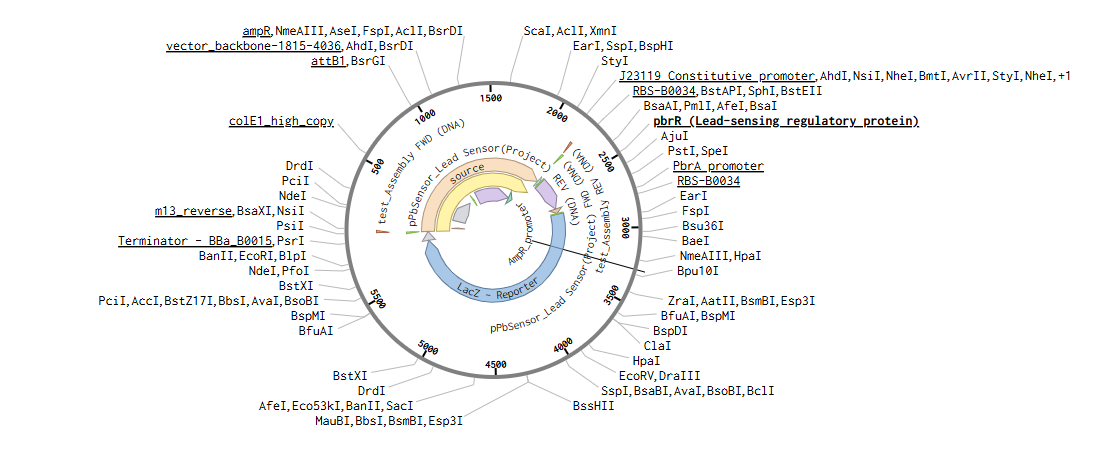
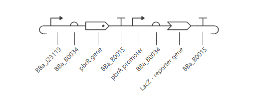
For the lead-sensing plasmid, I used the PbrR transcription factor with its cognate PpbrA promoter from Cuprivaridus metallidurans CH34 and the lacZ colorimetric reporter, which will be coupled with the Chlorophenol red-(beta)-D-galactopyranoside (CPRG) substrate to produce a magenta color when lead is present.
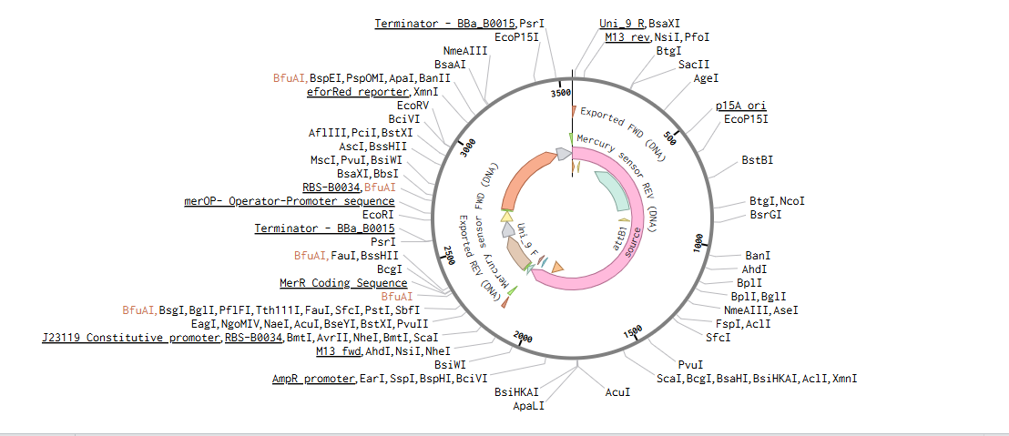
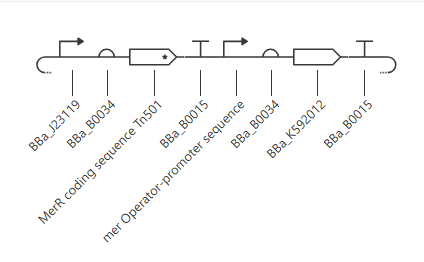
For the mercury-sensing plasmid, I used the MerR transcription factor with its cognate Pmert promoter from Escherichia coli, Tn21 transposon and the eforRed chromoprotein, which produces a red color as the reporter.
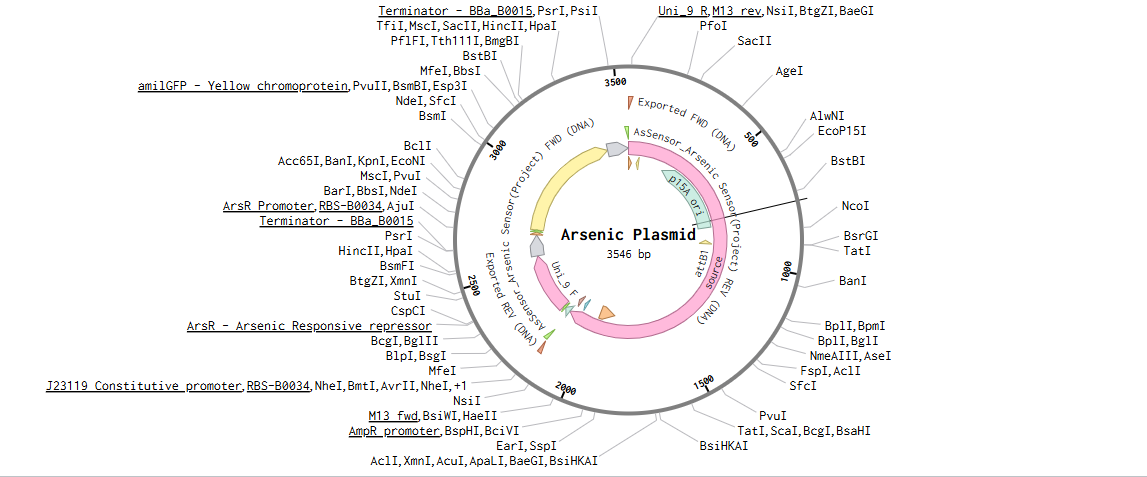
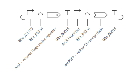
For the arsenic-sensing plasmid, I used the ArsR transcription factor with its cognate Pars promoter, which belongs to the SmtB/ArsR family from Escherichia coli K-12 and the amilGFP chromoprotein, which produces a yellow color as the reporter.
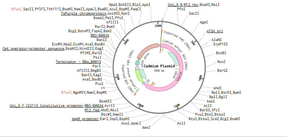
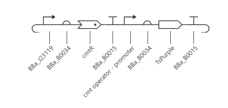
For the cadmium-sensing plasmid, I was initially going to use the SmtB transcription factor, but it is natively a zinc-responsive repressor, so I opted to use the CmtR transcription factor with its cognate Pcmt promoter from Mycobacterium tuberculosis, which has a higher cadmium specificity. I used the Tspurple chromoprotein, which produces purple color as the reporter.
This link contains my plasmid designs in Benchling https://benchling.com/nana_agyei/f_/TkfUeV9pDT-htgaa-2026-project/
Circuit Architecture
I modeled the circuit logic of each plasmid in Asimov Kenerl. Each plasmid has the same architecture and is made up of two cassettes: Cassette 1, responsible for the constitutive expression of the metal-sensing transcription factor, and Cassette 2, responsible for the expression of the reporter driven by the metal-responsive promoter.
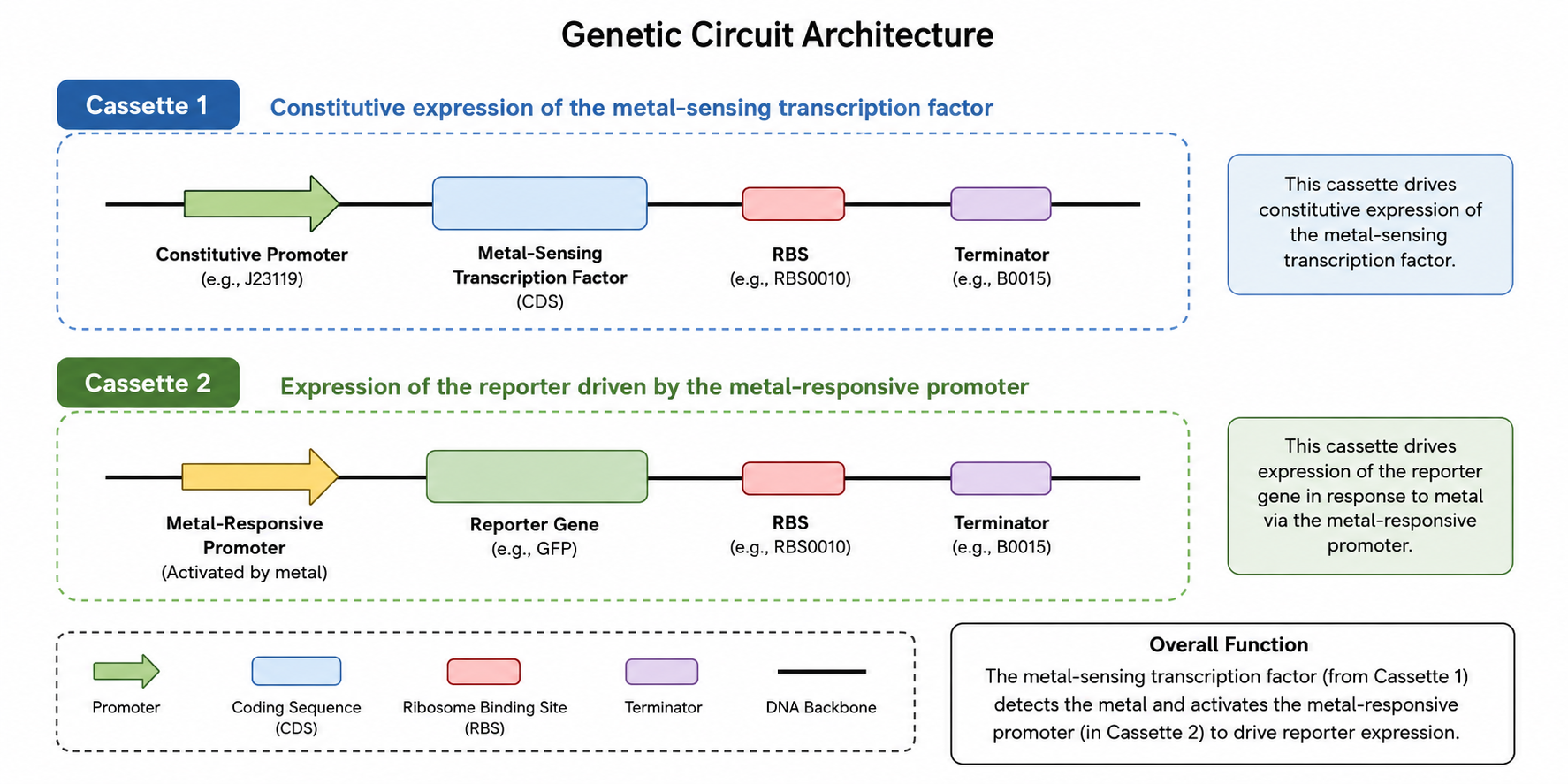
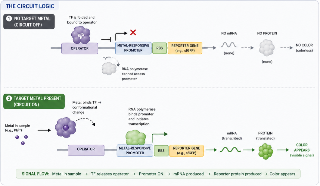
Together, both parts function using simple metal-triggered switch logic. In the absence of the target metal, the metal-sensing transcription factor is folded and binds to the operator sequence of the metal-responsive promoter, blocking RNA polymerase from transcribing the reporter gene, so no color is produced. However, when the water sample added contains the target metal, the metal ions bind to the transcription factor, inducing a conformational change that releases it from the operator. Allowing RNA polymerase to transcribe the reporter gene and the ribosomes in the cell-free reaction to translate it into the reporter protein, producing a visible color.
- Lead Plasmid Circuit
Simulation
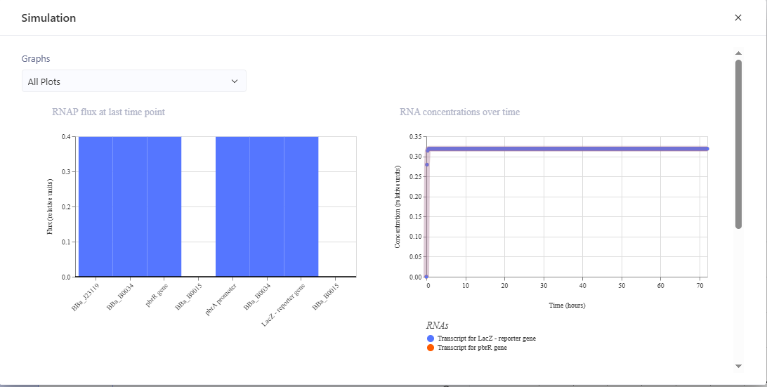
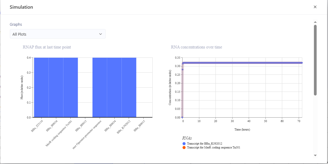
- Mercury Plasmid Circuit
Simulation
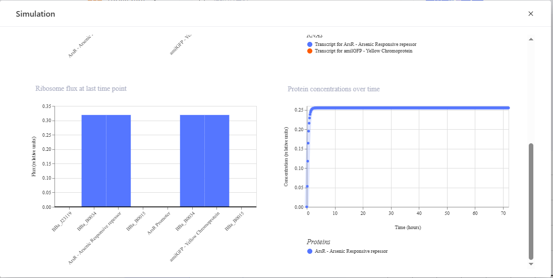
- Asernic Plasmid Circuit
Simulation

- Cadmium Plasmid Circuit
Simulation


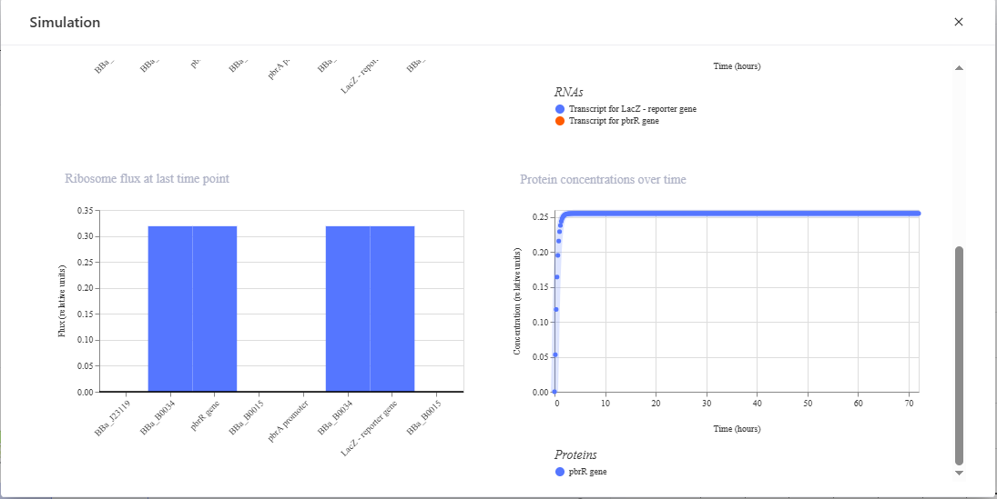
I simulated each circuit in the Asimov Kernel, with each simulation showing the geneic circuits are functioning properly. The RNA flux graph of each circuit demonstrates that RNA polymerase efficiently transcribes both cassette 1 and 2, indicating that the promoters are active and transcription proceeds without major bottlenecks. The RNA concentration graph shows a rapid increase in transcript levels followed by a plateau, indicating that transcription and RNA degradation eventually balance each other. This demonstrates stable mRNA production within the circuit, with the ribosome flux graph confirming that the ribosomes efficiently translate the produced mRNAs, meaning the ribosome binding sites and translation processes are functioning effectively. Finally, the protein concentration graph shows successful accumulation of the metal-sensing transcription factor protein before reaching steady-state equilibrium, where protein synthesis and degradation become balanced. Overall, the simulation suggests that the circuit is transcriptionally and translationally active, stable, and capable of sustained gene expression.
It was quite challenging to use the Asimov Kernel to model the circuit logic and get each part to work properly, as Kennel did not have some of the parts I needed to build the circuits, and I needed to add and define those parts, taking into account how they would interact with the other parts available. It took a lot of simulations and matching up different parts to ensure everything was working correctly.
While I focused on the dry-lab aspect of the project, I have outlined below how I envisioned the laboratory experimentation, validation, and development of the biosensor kit would be conducted.
Plasmid Verification
After each plasmid has been synthesized and delivered by Twist Bioscience, 100 ng will be transformed into chemically competent E. coli DH5α using a heat shock method at 42°C for 45 seconds. The bacteria will then be plated on LB media containing ampicillin (100 µg/mL), with 3 to 5 colonies picked per plasmid and grown overnight in 5 mL of LB with ampicillin. Next, a miniprep will be performed using the Qiagen QIAprep kit to extract the successfully transformed plasmids. Following this, NanoDrop spectrophotometry will be conducted to measure the concentration and purity of the DNA. Samples with an A260/A280 ratio of 1.8 or higher and a concentration of at least 50 ng/µL will be selected for further analysis. A restriction enzyme digest will then be performed for each plasmid, and the observed band sizes will be compared to the predicted digest fragment sizes in Benchling, allowing for an acceptable tolerance of ±5% from the predicted size. Additionally, Sanger sequencing will be conducted to ensure there are no non-synonymous mutations in the transcription factor coding sequences, no mutations at essential metal-binding residues, and no mutations within the promoters. This will confirm that the sequences match the original designs.
Cell-Free Protein Synthesis (CFPS) Extract Preparation
E. coli BL21(DE3) will be grown in 1 L of 2×YT medium in a 2.5 L baffled Erlenmeyer flask at 37°C while shaking at 250 rpm. The optical density (OD600) will be monitored every 20 minutes. The cells will be harvested when the OD600 reaches 0.6–0.8 and then centrifuged at 4,000 × g for 12 minutes at 4°C. After centrifugation, the cells will be washed three times with ice-cold S30A buffer (10 mM Tris-acetate, pH 8.2, 14 mM magnesium acetate, 60 mM potassium acetate, 1 mM DTT). Next, the cells will be lysed using bead milling with 0.1 mm glass beads, processing them three times for 30 seconds at maximum speed, with 1 minute of ice resting between cycles. The lysate will be clarified by centrifugation at 30,000 × g for 30 minutes at 4°C. It will then be run off at 37°C for 80 minutes without added DNA. A second clarification step will be performed by centrifuging at 15,000 × g for 15 minutes. Aliquots of 25 µL will be snap-frozen in liquid nitrogen and stored at −80°C. Before conducting any biosensor experiments, I will validate the quality of the extract using a GFP control plasmid. This involves setting up a 10 µL CFPS reaction containing 3.3 µL of the extract, reaction buffer, and 10 nM pET28-GFP or an equivalent plasmid, incubating at 37°C for 60 minutes. Fluorescence will be measured using a plate reader with excitation at 488 nm and emission at 509 nm. Acceptable extracts should show detectable fluorescence above background levels. If an extract fails the GFP test, I will troubleshoot the lysis efficiency by checking for inhibitor carryover from the washing steps or preparing a fresh batch.
In Vitro Dose-Response Validation
An automated Dose-Response assay will be set up to measure the colorimetric response of each cell-free reaction across a metal concentration series: 0 (blank control), 0.1, 0.5, 1, 3, 5, 10, 30, 100, and 500 µg/L. The Echo 525 will be used to dispense metal solutions into a 384-well Greiner black-well clear-bottom plate. The Bravo-96 liquid handling system will then add the TX-TL reaction mix (plasmid + extract) to each well. 1 mM of CPRG will be added to the cell-free reactions for the lead sensor only. The plate will be incubated at 37°C and 300 rpm, with absorbance readings taken every 10 minutes for a duration of 90 minutes. The specific wavelengths for reading are as follows: eforRed/tsPurple at 572 nm, lacZ + CPRG at 574 nm, and amilGFP at 507 nm. We will fit four-parameter logistic (4PL) dose-response curves to the data. The limit of detection (LOD) will be calculated as the mean of the blanks plus three times the standard deviation of the blanks. It is expected that all four sensors will demonstrate empirical LODs at or below the regulatory thresholds established by the Ghana Standards Authority: Hg ≤1 µg/L, Pb ≤10 µg/L, As ≤10 µg/L, and Cd ≤3 µg/L. Additionally, we aim for a signal-to-blank ratio of ≥3 at the LOD concentration.
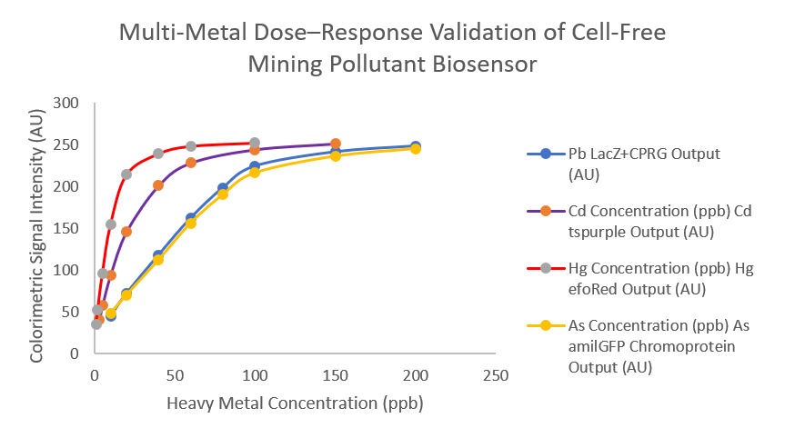
The figure above is a graph of expected results from the multi-metal dose-response validation of the cell-free biosensor, with colorimetric output (Absorbance Units, AU) plotted against heavy metal concentration (ppb/µg·L⁻¹).
Each curve represents a different metal-specific sensor:
- Blue — Lead (Pb²⁺): lacZ + CPRG reporter
- Purple — Cadmium (Cd²⁺): tsPurple chromoprotein
- Red — Mercury (Hg²⁺): eforRed chromoprotein
- Yellow — Arsenic (As³⁺): amilGFP chromoprotein
All four biosensors display a sigmoidal dose-response curve, indicating the increase of colourimetric signal at high concentrations of the target metals.
At low target metal concentrations (0–5 ppb), there is a small baseline signal due to normal background activity in the cell-free system. In the mid-range (about 5–60 ppb), the sensors are most sensitive and show a strong increase in output, meaning they can clearly detect changes in the metal concentration. At higher concentrations (above ~60 ppb), the response reaches a maximum plateau, where further increases in metal concentrations do not significantly increase the signal because the system is fully activated.
If the LODs do not meet the regulatory threshold, the constitutive promoter J23119 could be replaced with a weaker promoter to lower the transcription factor concentration, resulting in less repressor or activator occupying the metal-responsive promoter at baseline, reducing the metal concentration needed to shift the equilibrium. Alternatively, the ribosome binding site (RBS) upstream of the reporter could be replaced with a stronger one to increase reporter protein output per mRNA molecule without affecting the concentration of the transcription factors or the LOD thresholds directly, but improve the signal-to-noise ratio, making the colors more vivid at low metal concentrations. Additionally, the concentration of the plasmids could also be adjusted, as higher plasmid concentrations increase the number of transcription events per unit time, boosting the total signal. Finally, the reaction time could be extended as chromoproteins accumulate over time. Extending incubation from 60 to 90 minutes may allow the signal at the target LOD concentrations to cross the visible threshold without any other changes.
Selectivity Testing
Single-element standards for the target elements Hg²⁺, Pb²⁺, As³⁺, and Cd²⁺ will be prepared, along with potential interferents such as Zn²⁺, Cu²⁺, Fe³⁺, and Mn²⁺ at ten times their typical concentrations found in rivers. The cell-free reaction systems designed to sense the target metals will be challenged with each of the metals using a 96-well plate format, with three replicates (n=3) for each condition. Cross-reactivity will be calculated using the formula: Cross-reactivity (%) = (Signal from non-target metal / Signal from target metal at the same concentration) × 100. The pass criteria will be a cross-reactivity of less than 20% for non-target metals. Transcription factor - operator DNA complexes can be modeled using RoseTTAFold or HADDOCK to identify metal-coordinating residues and operator-contact residues that may enhance target metal affinity or reduce cross-reactivity.
Fabrication of Sensing Kit and Lyophilisation
Whatman No. 1 paper will be used to create the sensing kit, with circles of 6 mm in diameter pre-cut using a biopsy punch. These circles will be arranged on a printed template card to ensure they are properly positioned. The optimized CFPS (Cell-Free Protein Synthesis) reaction mixture will be combined with 5% w/v trehalose. To pre-load the substrate in the lead sensor zone, 1 mM of CPRG will be added, eliminating the need for a separate addition in the field. Next, 3 µL of the appropriate CFPS mixture will be pipetted onto each paper circle. The droplet will be absorbed within 30–60 seconds through capillary action. The spots will absorb the mixtures at room temperature for 5 minutes and should not be allowed to completely air dry before being freeze-dried. The card with the spotted paper circles will then be transferred onto the lyophilizer trays. The lyophilizer must be pre-frozen at −80°C for 2 hours before loading to ensure that all water present is fully crystallized prior to applying a vacuum. If liquid water is present when the vacuum starts, it will boil rather than sublimate, which could disrupt the structure of the paper spots.
Once the trays are pre-frozen, they will be loaded into the lyophilizer chamber, and the following cycle will be run:
- Condenser temperature: −50°C
- Chamber pressure: <100 mTorr (13 Pa)
- Primary drying phase: 12 hours — removes bulk free water by sublimation
- Secondary drying phase: 4 hours at −10°C shelf temperature — removes bound water from the trehalose glass
- Total cycle duration: 16 hours
At the end of the drying cycle, the chamber will be vented with dry nitrogen gas instead of ambient air to prevent moisture reabsorption during the removal of the card. The freeze-dried cards will be immediately transferred into pre-prepared aluminized Mylar foil pouches that contain a silica gel desiccant sachet, and will then be heat-sealed immediately.
Rehydration and Shelf-life stability testing
The card should be removed from its sealed pouch and placed flat on a clean surface. Next, pipette 10 µL of the metal standard onto the designated zone. Immediately seal the card with a pre-cut piece of transparent adhesive film to prevent evaporation during the incubation process. Incubate the card face-up in a closed humid chamber at 37°C for 60 minutes. To create a simple humid chamber, place the card on a wet paper towel inside a sealed container. After incubation, read the results by comparing them to the reference color card. Photograph the results under standardized white LED lighting for documentation. A visible color change at the limit of detection (LOD) concentration within 60 minutes, along with the negative control zone (buffer only) remaining colorless, indicates that the kit is functioning correctly.
To determine the shelf life of freeze-dried cards, set aside a batch of cards on the day of manufacture. You will need a minimum of 3 cards per storage condition for each time point. For 6 time points and 3 different conditions, this means you need at least 54 cards total. Be sure to label each card with the manufacturer’s date and the intended test date.
Immediately after sealing, distribute the cards into three different storage conditions:
- Condition 1: 4°C refrigerator (positive control — best-case stability)
- Condition 2: 25°C incubator or room temperature (with temperature monitoring)
- Condition 3: 37°C incubator (worst-case scenario representing typical ambient conditions in West Africa)
At each designated time point (weeks 0, 1, 2, 4, 8, and 12), remove 3 cards from each storage condition. Rehydrate the cards using 10 µL of a metal standard at the Ghana EPA limit of detection (LOD) concentration. Incubate the rehydrated cards at 37°C for 60 minutes, then measure the absorbance using a plate reader. To calculate signal retention at each time point and for each condition, use the formula: Signal retention (%) = (Signal at week N / Signal at week 0) × 100. Fit a first-order exponential decay model to the signal retention data for each storage condition using the following equation: Retention(t) = 100 × e^(−k_deg × t). Solve for k_deg by linear regression of ln(Retention) against time. Calculate the shelf life by determining the time at which the predicted retention falls below 80%. If the signal retention is greater than 80% at week 4 at 37°C, this indicates the minimum acceptable performance for a kit stored in a field office without refrigeration during a typical deployment cycle, and it will be considered to have passed the stability test.
Field Validation
The process will involve collecting water samples from locations affected by galamsey on the Pra, Ankobra, and Birim rivers. Using pre-cleaned HDPE sample bottles that have been acid-washed with 10% nitric acid (HNO₃) and rinsed with deionized water three times to collect samples from a depth of 30 cm below the water surface to avoid contamination from the surface film. At each site, two split samples will be collected: one for ICP-MS (Inductively Coupled Plasma Mass Spectrometry) reference analysis, which must be sent to an accredited laboratory within 48 hours on ice, and the other for immediate testing in the field using a kit. Before testing, pre-treat each sample by filtering it through a 0.2 µm syringe filter to remove any particulates and turbidity that may interfere with colorimetric readings. For arsenic detection, add one drop of a 10% ascorbic acid solution for every 1 mL of sample and wait for 5 minutes. Then, apply 10–15 µL of the pre-treated sample to each zone on the biosensor card and seal it with an adhesive overlay. Incubate the card at ambient temperature (25–37°C) for 60 minutes. After incubation, read the results visually against the reference color card. Take a photograph of each card with the reference card visible in the same frame for documentation. Record the GPS coordinates, site name, date, time, water source type, and results for each zone on the KoboToolbox reporting form. Finally, calculate the agreement between the kit and ICP-MS results for each metal at each site using the following formula:
Agreement (%) = (Concordant results / Total results) × 100
“Concordant” means that both the kit and ICP-MS indicate whether the sample is above or below the regulatory threshold, with the pass criteria being greater than 80% across all samples.
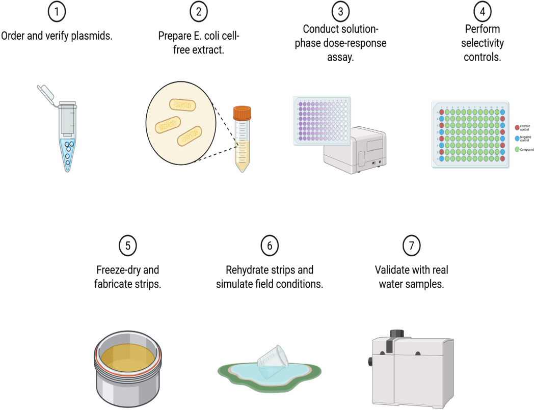
The figure below shows the entire experimental procedure and validation steps.
Implementation and Deployment
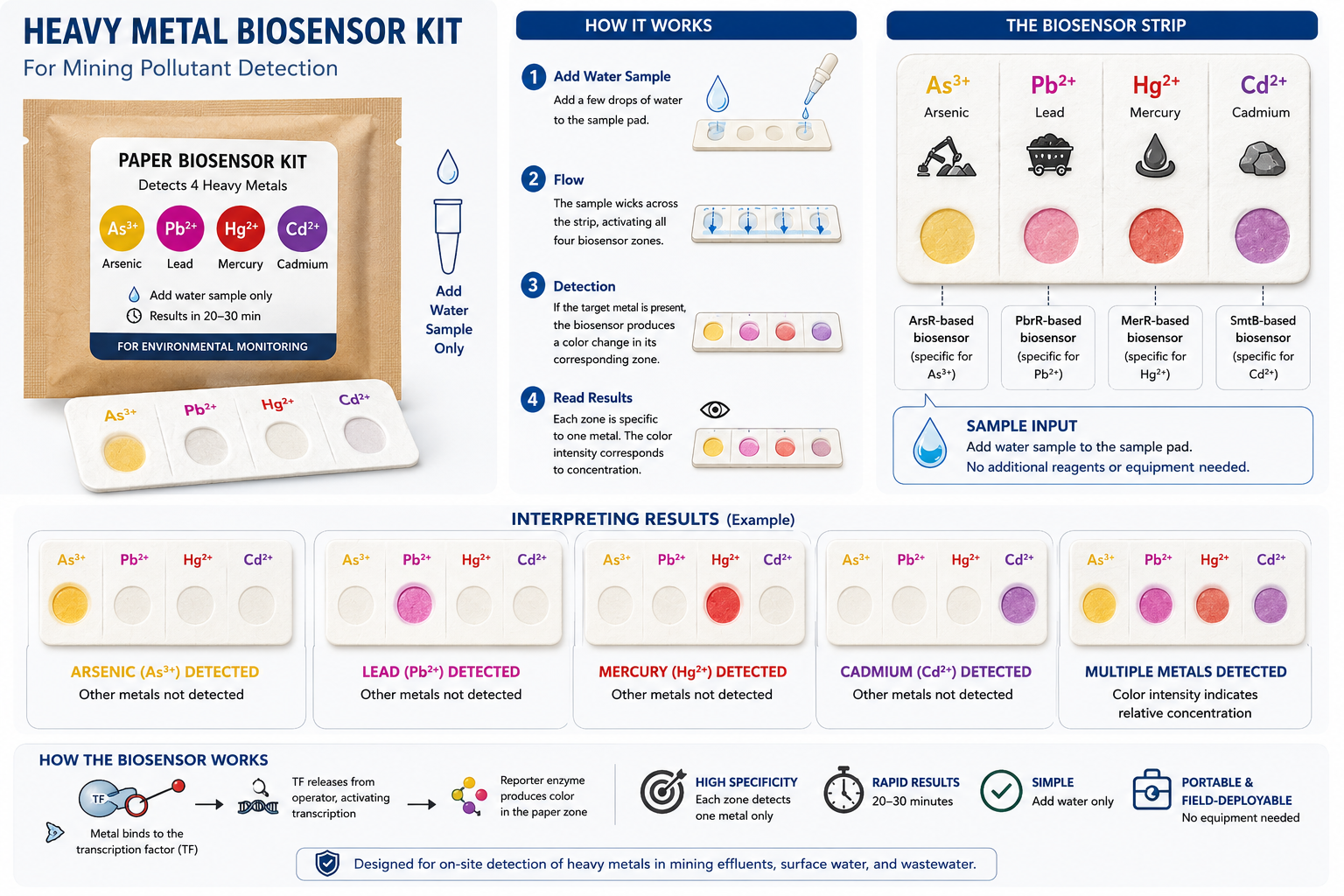
The figure below demonstrates how I envision the kit functioning, with a water sample added to the four sensing zones to detect the target heavy metals. You wait 15 to 60 minutes, and a colour change will be observed in each zone if the target metals are present in the water sample.
This is how I envision the deployment and use of the biosensor kit in the field.
A trained community water monitor collects their monthly supply of kits from a local community center or health post and travels to a nearby river, borehole, or town well. Using a collection tube, they obtain a 1mL water sample and dispense 15 µL onto each of the four detection zones on the biosensor kit. The card is then sealed and left to incubate for about 60 minutes. If any of the target heavy metals are present, the corresponding zone develops a visible color change specific to the detected heavy metal. The monitor then photographs the completed test card alongside the reference card and uploads the image, metadata, and GPS coordinates through a monitoring platform such as KoboToolBox. The results are immediately transmitted to a district-level Tier 2 technician who reviews the submission and flags a potential contamination event. The Environmental Protection Agency dispatches a field officer within 24 hours to collect confirmatory samples for ICP-MS analysis. If laboratory testing confirms elevated heavy metal levels, emergency interventions are initiated within the affected community, including the provision of safe drinking water and the subsequent implementation of remediation measures. The generated data can also be used to support regulatory enforcement and aid in identifying and holding polluters accountable.
Reference
Bansah, P. K. (2022, October 13). Ghana’s artisanal miners are a law unto themselves: Involving communities can help fix the problem. https://theconversation.com/ghanas-artisanal-miners-are-a-law-unto-themselves-involving-communities-can-help-fix-the-problem-192256?utm_source=chatgpt.com
Bempah, C. K., & Ewusi, A. (2016). Heavy metals contamination and human health risk assessment around Obuasi gold mine in Ghana. Environmental monitoring and assessment, 188(5), 261.
Gräwe, A., Dreyer, A., Vornholt, T., Barteczko, U., Buchholz, L., Drews, G., Ho, U. L., Jackowski, M. E., Kracht, M., Lüders, J., Bleckwehl, T., Rositzka, L., Ruwe, M., Wittchen, M., Lutter, P., Müller, K., & Kalinowski, J. (2019). A paper-based, cell-free biosensor system for the detection of heavy metals and date rape drugs. PloS one, 14(3), e0210940. https://doi.org/10.1371/journal.pone.0210940
Obiri, S., Yeboah, P. O., Osae, S., Adu-Kumi, S., Cobbina, S. J., Armah, F. A., … & Quansah, R. (2016). Human health risk assessment of artisanal miners exposed to toxic chemicals in water and sediments in the Prestea Huni Valley District of Ghana. International journal of environmental research and public health, 13(1), 139.
Pardee, K., Green, A. A., Takahashi, M. K., Braff, D., Lambert, G., Lee, J. W., … & Collins, J. J. (2016). Rapid, low-cost detection of Zika virus using programmable biomolecular components. Cell, 165(5), 1255-1266.
Wan, X., Volpetti, F., Petrova, E., French, C., Maerkl, S. J., & Wang, B. (2019). Cascaded amplifying circuits enable ultrasensitive cellular sensors for toxic metals. Nature Chemical Biology, 15(5), 540-548.
WHO (2022). Guidelines for Drinking-Water Quality, 4th ed., incorporating the 1st and 2nd addenda. https://www.who.int/publications/i/item/9789240045064
Additional Information
Igem parts used:
- https://registry.igem.org/parts/bba-j23119
- https://registry.igem.org/parts/bba-b0034
- https://registry.igem.org/parts/bba-b0015
- https://registry.igem.org/parts/bba-k592012
- https://registry.igem.org/parts/bba-k592010
- https://registry.igem.org/parts/bba-k1033906
Techniques Utilised
Pipetting & Lab Safety
- Pipetting
- Lab Safety
- Bioethical Considerations
DNA Design & Sequencing
- DNA Sequencing
- DNA Construct Design
- Restriction Enzyme Digestion
- Gel Electrophoresis
- Designing a Twist Order
Databases
- GenBank / NCBI
- iGEM Parts Registry
- Addgene
Protein Design Tools
- Use of Asimov Kernel
- Use of Benchling
- Models & Notebooks
Cell-Free Systems
- Cell-Free Reactions
- Freeze-Dried Cell-Free Systems
Bioproduction
- Chassis Selection (BL21(DE3))
- Registry of Standard Biological Parts
- Plasmid Preparation
- Bacterial Culturing
- Bacterial Processing (Centrifugation, Lysis)
- Quality Control / Analysis
Industry Council Companies
- Twist Biosciences — whole-plasmid synthesis of all four biosensor constructs
- Asimov Kernel — circuit design and regulatory logic modelling for all four genetic circuits
- Addgene — plasmid deposition and open-source distribution upon project completion
- New England Biolabs — EcoRI-HF, HindIII-HF restriction enzymes; CutSmart buffer for digest validation
- Opentrons — automated liquid handling for consistent CFPS reaction setup in 384-well plate format
- Thermo Fisher Scientific — cell culture reagents, NanoDrop spectrophotometry, reaction buffer components
- Waters Corporation** — ICP-MS or LC-MS validation of metal concentrations in field water samples for ground-truth comparison against biosensor readouts.
- Millipore Sigma — CPRG substrate, heavy metal standards, and molecular biology reagents.
Supply List and Estimated Budget
| Item | Supplier | Catalog # | Estimated Cost | Link |
|---|---|---|---|---|
| Whole plasmid synthesis ×5 (4 circuits + 1 control) | Twist Bioscience | Custom order | ~$500 | https://www.twistbioscience.com/products/genes |
| myTXTL Cell-Free Expression Kit | Arbor Biosciences | 507024 | ~$400 | https://arborbiosci.com/cell-free-biology/txtl-kits/mytxtl-sigma-70-master-mix/ |
| CPRG (Chlorophenol Red β-D-galactopyranoside) | Millipore Sigma | 10884308001 | ~$120 | https://www.sigmaaldrich.com/US/en/product/roche/10884308001 |
| 384 Greiner black-well clear-bottom plates ×10 | Greiner Bio-One | 781096 | ~$180 | https://www.sigmaaldrich.com/US/en/product/sigma/m0062 |
| 384-well Echo PP source plates ×5 | Labcyte/Beckman | LP-0200 | ~$100 | https://www.beckman.com |
| NaAsO₂, Pb(NO₃)₂, HgCl₂, CdCl₂ standards | Millipore Sigma | Various | ~$200 | https://www.sigmaaldrich.com |
| DH5α competent cells | NEB | C2987I | ~$80 | https://www.neb.com/en-us/products/c2987-neb-5-alpha-competent-e-coli-high-efficiency |
| LB Broth + Ampicillin | Thermo Fisher Scientific | BP1426-500 | ~$60 | https://www.thermofisher.com/order/catalog/product/BP1426-500 |
| Miniprep Kit | Qiagen | 27104 | ~$90 | https://www.qiagen.com/us/products/discovery-and-translational-research/dna-rna-purification/dna-purification/plasmid-dna/qiaprep-spin-miniprep-kit/ |
| SDS-PAGE gel + Coomassie stain | Bio-Rad | 4561096 | ~$80 | https://www.bio-rad.com/en-us/product/mini-protean-tgx-precast-protein-gels |
| Sanger sequencing (20 reactions) | Genewiz/Azenta | Per reaction | ~$60 | https://www.azenta.com/products/sanger-sequencing |
| Total Estimated Cost | ~$1,870 |