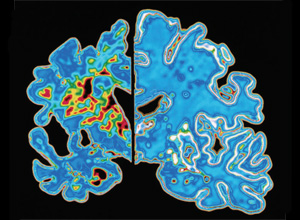
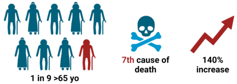
Section 1: Abstract Alzheimer’s disease (AD) is a neurodegenerative disorder characterized by amyloid plaques and neurofibrillary tangles. As the most common cause of dementia, AD affects 1 in 9 Americans aged 65 and older and was the seventh leading cause of death in the United States in 2021. While deaths from stroke and heart disease decreased from 2000 to 2021, deaths from AD increased more than 140% during this period, and AD is projected to affect 13.8 million Americans by 2060 (figure 1). Given that AD begins 20 years or more before symptom onset, there is a substantial window in which medical interventions may alter the disease progression. By 2050, the number of individuals affected by AD is projected to surpass 150 million, yet effective therapeutic interventions to halt or reverse the disease remain elusive. [1,2]
Alzheimer’s disease (AD) is a neurodegenerative disorder characterized by amyloid plaques and neurofibrillary tangles. As the most common cause of dementia, AD affects 1 in 9 Americans aged 65 and older and was the seventh leading cause of death in the United States in 2021. While deaths from stroke and heart disease decreased from 2000 to 2021, deaths from AD increased more than 140% during this period, and AD is projected to affect 13.8 million Americans by 2060 (figure 1). Given that AD begins 20 years or more before symptom onset, there is a substantial window in which medical interventions may alter the disease progression. By 2050, the number of individuals affected by AD is projected to surpass 150 million, yet effective therapeutic interventions to halt or reverse the disease remain elusive. [1,2]
Traditional diagnostic approaches, including cerebrospinal fluid (CSF) analysis and positron emission tomography (PET) imaging, are limited by their invasive nature, high costs, and limited availability, hindering their widespread use in clinical practice. [2] PCR-based methods are currently considered the gold standard of detection; however, PCR-based methods usually require expensive instruments, complete laboratory settings, and professionals, thereby limiting their deployment in low-resource regions, such as Africa. Antigen–antibody reaction exists disadvantages of cross-reactivity and low sensitivity. Genome sequencing is time-consuming and highly expensive. Compared to these traditional diagnostic tools, CRISPR/Cas13a based detection methods are able to simultaneously achieve high sensitivity, high specificity, low cost and low time-consuming, by combining Cas13a system with isothermal amplification technique, visual readout, extraction-free method, etc [3]
Recent breakthroughs in blood based biomarkers offer a promising, minimally invasive, and cost-effective alternative for early AD diagnosis and disease monitoring. [2] Reliable biomarkers are needed for early detection of preclinical AD to delay or prevent the development of clinical AD. [1]
MicroRNA (miRNA) expression in peripheral blood has been examined as a biomarker to evaluate AD progression. It has been reported that more than 10 miRNAs were deregulated in AD pathogenesis, and the concentration of miRNA34a in peripheral blood could be used to distinguish AD patients from healthy people.[2]
MicroRNAs (miRNAs) are a class of small non-coding RNAs, typically 18–24 nucleotides in length, which exert negative regulation on gene expression. They emerged as potential biomarkers for various diseases [5] MicroRNAs play a crucial role in gene regulation through two main mechanisms: mRNA cleavage and translational repression. MicroRNAs have gained attention as biomarkers in various scientific fields owing to their numerous desirable properties. The ideal biomarker must be particular, sensitive, and predictive. In the complex network of AD pathology, miRNAs function as regulatory elements within the post-translational modification (PTM), orchestrating molecular events that contribute to the onset and progression of the disease. These PTMs encompass a diverse array of processes, including acetylation, carbamylation, glycation, methylation, nitration, sumoylation, truncation, ubiquitination, and phosphorylation. Through the regulation of these pathways, miRNAs assume crucial positions in molding the molecular processes driving AD development and etiology. The miRNAs can influence tau protein expression in AD through multiple mechanisms like phosphorylation, splicing and acetylation. [6]
The CRISPR system is a natural adaptive immune system of prokaryotes. Among the CRISPR-Cas type VI systems, the CRISPR/Cas13a system has been the most widely characterized for its application in molecular diagnostics, gene therapy, gene editing, and RNA imaging. Moreover, because of the trans-cleavage activity of Cas13a and the high specificity of its CRISPR RNA, the CRISPR/ Cas13a system has enormous potential in the field of molecular diagnostics. [3]
Among CRISPR/Cas-nucleases, Cas13a nuclease forms a complex with the RNA molecule of a specific sequence (gRNA). The sequence of gRNA is composed of a part responsible for interaction with Cas13a (‘direct repeat’) and a part (28 nt long ”spacer”) complementary to a target sequence (“protospacer”). Cas13a activates only if an almost perfect heteroduplex between spacer and protospacer is formed, resulting in a nicking of a target sequence. Upon activation, Cas13a additionally acquires the so called “collateral” or trans-activity, which is an ability of the nuclease to unselectively cleave, alongside with the target RNA, all RNA molecules present around. [4]
The visionary aim of the project is to develop a CRISPR-Cas13a based detection system that it capable of detecting multiple disease biomarkers simultaneously in a fast and accurate manner. For the first aim, it will detect three microRNA biomarkers associated with AD and upon their binding with the Cas13a system, a fluorescence will be produced.
Section 2: Project Aims
Aim 1: Experimental Aim
The first aim of my project is to design the DNA parts necessary for the successful biomarkers detection using DNA design tools such as Benchling and Twist platforms.
The DNA designs involve the following:
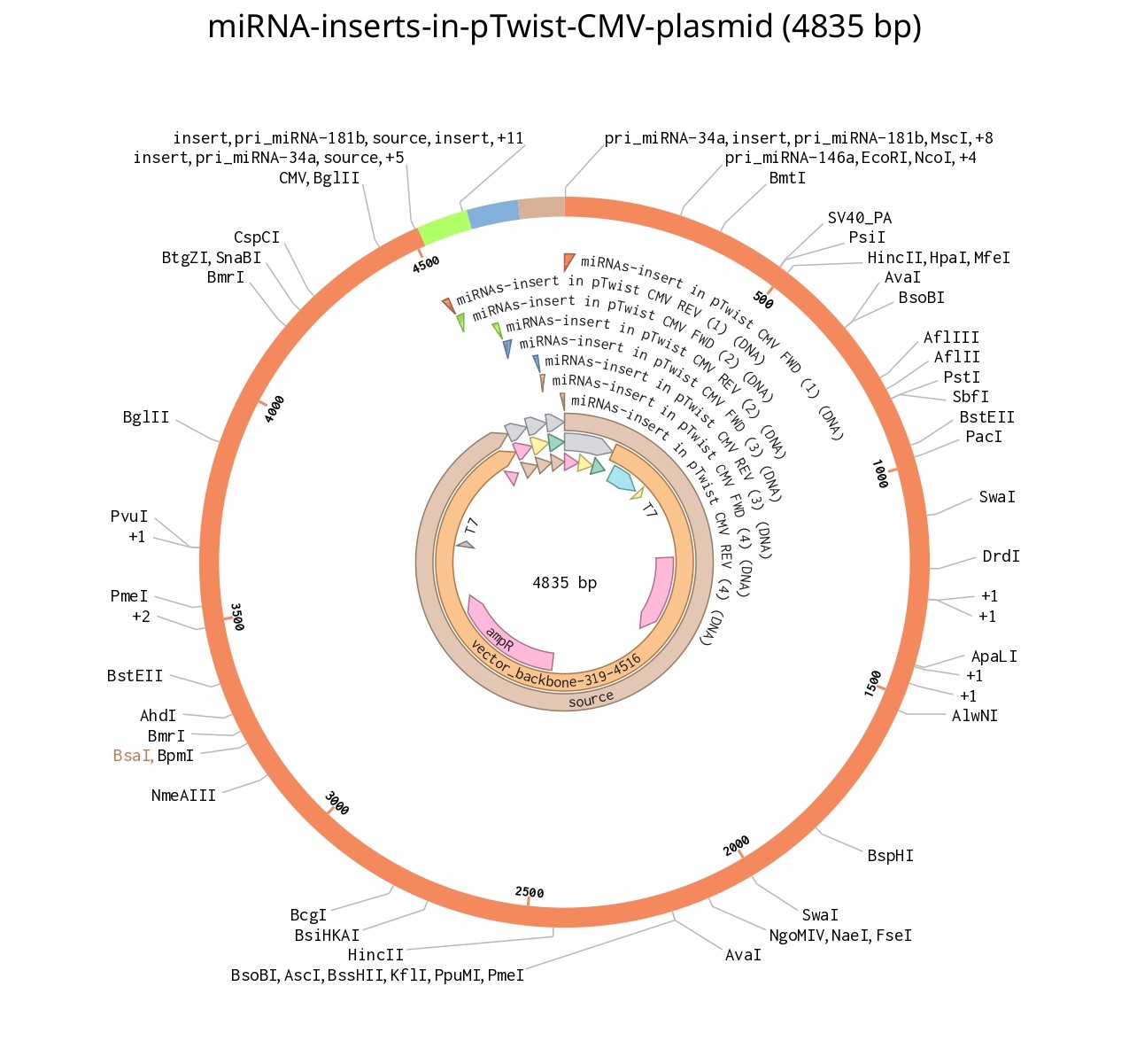
Plasmid design of three miRNA biomarkers (to be used as a positive control)
gRNA Plasmid design of the CRISPR-Cas13a system
Aim 2: Developmental Aim
The next aims are:
to test these design through miRNA expression in eukaryotic cells and the mature miRNA detection through fluorescence generation.
to develop the CRISPR system to detect proteins.
develop any technical limitations
Aim 3: Visionary Aim
A visionary aim is for the project to be developed into a paper-based detection system that is a customizable platform for different diseases detection. The paper-based detection should be easily accessible, accurate and low cost.
Section 3: Background
1. Briefly summarize two peer-reviewed research citations relevant to your research
CRISPR-AD: Combinational Detection of Blood Protein and miRNA with Digital CRISPR-Based Assay Enable to Improve the Diagnostic Performance of Alzheimer’s Disease [7]
The paper used a noninvasive approach for detecting and assessing Alzheimer’s disease (AD) pathophysiology using blood-based biomarkers. They called their system CRISPR-AD, a CRISPR/Cas-based digital assay designed for the combined detection of protein and microRNA in blood. This method achieves a limit of detection (LOD) as low as 60 fg/mL for phosphorylated tau217 (p-tau217) and 0.5 fM for microRNA-34a-5p (miRNA34a), enabling successful detection in both AD patients and healthy individuals. They found that the combined use of these biomarkers improves the ability to distinguish between AD patients and healthy participants, particularly in individuals with mild cognitive impairment (MCI). Additionally, they developed a portable device that integrates a smartphone as an imaging system for point-of-care testing (POCT), offering the potential for early stage AD screening. The study represents the first effort to evaluate the combined detection of blood protein and microRNA biomarkers for AD, underscoring the potential of multiple biomarker combinations for more accurate AD diagnosis.
Direct Detection of MiRNAs miR-34a, -145, and -218 with CRISPR/Cas13a-nuclease [8]
The paper demostrated the direct detection of three miRNAs, miR-34a, -145, and -218 using CRISPR/Cas13a-nuclease. The detection was based on registration of a cleavage of molecular reporters bearing a fluorophore and a quencher by the complex of CRISPR/Cas13a-nuclease and guide RNA (gRNA) with a spacer
of 21-23 nucleotides long. The detection sensitivity varied among miRNAs tested by 10-fold, presumably due to the unwanted intramolecular
partial base paring of gRNA. The miRNA detection with Cas13a nuclease strongly depended on the presence of background RNA thus potentially
compromising its direct application to complex media in a general case. The authors suggested the further optimization of measurement conditions to directly detect miR-34a, -145, and -218 in biological samples.
2. Explain how your project is novel or innovative.
The novality of the project lies in the simulataneous detection of the different biomarkers types such as microRNA, proteins and genes and the visionary aim of incorporating the system to be paper-based to be easily accessible.
3. Explain why your project matters and what impact it could have.
Early diagnosis can prevent disease deterioration, especially the ones that we can’t fully treat yet. The project aims to produce a detection system that is easily accessible and provide reliable results. Therefore, we can easily detect diseases earlier and prevent its deterioration.
4. Describe the ethical implications associated with your project and identify relevant ethical principles
The ethical implications associated with my project include:
Beneficence: Early detection enabling prevention
Non-maleficence: Protecting from psychological harm of early diagnosis
Justice: Equitable access to all populations (especially low-resource regions)
Responsibility: Ethical oversight, not profit-driven
The measures that should be taken to ensure the ethical implementation of my project are:
What actions do you propose?
Rigorous validation in diverse populations
Informed consent/counseling protocols
Equitable access partnerships
Independent ethics oversight
Data privacy safeguards
What are potential unintended consequences?
Increased development costs/timeline
Deterrence from testing due to consent protocols
Need for clinical proof that early detection actually helps
What could you be wrong about?
Assumption that early biomarker detection = better outcomes
Need longitudinal trials to prove intervention effectiveness
What are alternatives?
Focus on symptomatic individuals first (lower harm, less prevention)
Public health/government-funded model (more access, autonomy questions)
Addresses critical gap in preclinical detection (15-20 year window)
Transformative potential for prevention paradigm
Particularly benefits Low- and Middle-Income countries with limited diagnostics access
Section 4: Experimental design, techniques, tools and technology
Whole Project Experimental Workflow
The whole project experimental workflow covers three phases across seven weeks: DNA construct design and synthesis (Weeks 1–4), reagent preparation (Week 5), and automated detection assay execution and analysis (Weeks 6–7). All automation is performed at Ginkgo Bioworks. Three whole plasmids are ordered from Twist Bioscience.
Literature review; retrieve miR-34a, miR-146a, miR-181b sequences (week 1)
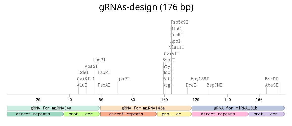
Design all three plasmids in Benchling: pCDNA3.1-3xmiRNA (CMV + Golden Gate), pUC57-3xgRNA (T7 + 22nt spacers), pUC57-LwaCas13a (codon-optimized, His6-tag) (week 1-2)
Order all three constructs as whole plasmids from Twist Bioscience (week 2 -4)
Sanger sequence verification of received plasmids (≥99% identity) (week 4)
Cell-free expression of LwaCas13a: add pUC57-LwaCas13a (10 nM) to Ginkgo CFPS lysate; incubate 37°C / 4h; verify by western blot (week 5)
IVT of gRNAs from pUC57-3xgRNA (HiScribe T7, NEB); purify by LiCl precipitation; verify size by PAGE (week 5)
Reconstitute fluorescent reporters: FAM-UUUUU-BHQ1, HEX-UUUUU-BHQ2, Cy5-UUUUU-BHQ3 at 10 µM; store at −80°C (week 5)
Prepare 10-point serial dilution of synthetic miRNA controls (100 nM → 0.1 fM) for standard curves (week 5)
RT-qPCR confirmation of gRNA transcript size and yield (week 6)
Fit 4-PL standard curves; calculate LOD (target: < 1 pM); assess cross-channel bleed-through (target: < 10%) (week 7)
First Aim Experimental Workflow
The first experimental aim was to design the DNA parts (plasmid design with the 3 microRNA, gRNA design of crispr-cas13a) necessary for the successful biomarkers detection using DNA design tools such as Benchling and Twist platforms.
The first step is the design of the plasmid incorporating the microRNA. The produced plasmid will be transfected in eukaryotic cells to express and process them to mature RNA that will be used as a positive control for the system.
Conduct a literature review identifying the most common microRNA for detecting Alzheimer’s disease
Get their sequences
Choose a suitable eukaryotic plasmid in Twist that will be used to express the microRNA to mature RNA similar to the ones present in AD patients
Insert the three microRNA sequences in benchling
insert that design in the plasmid in Twist
make golden gate assembly of the produced plasmid in Benchling for the expression of three microRNA in one plasmid
The second step is designing the 3 gRNA for the 3 miRNA. Knowing that the gRNA consist of the direct repear (DR) sequence that will bind to the cas protein and the spacer sequence binding to the miRNA
Get the direct repeat (DR) sequence for LwCas13a Cas protein
Get the mature sequences of the three microRNA
Get the reverse complementary sequences of the three microRNA
Insert them in Benchling
Add them to a plasmid at Twist Bioscience
Techniques relevant to my project.
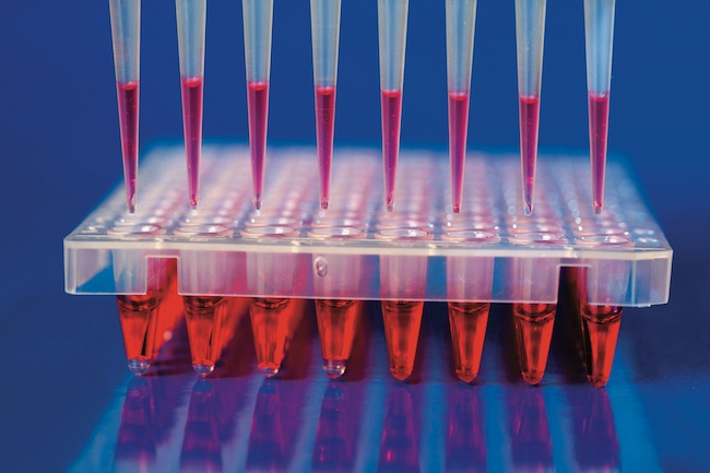
Pipetting
Bioethical Considerations
DNA Gel Art
DNA Sequencing
DNA Construct Design
Databases (e.g., GenBank, NCBI, Ensembl, and UCSC Genome Browser)
Lab Automation
Designing a Twist Order
Protein Design
Use of Benchling
Cell-Free Systems
Cell Free Reactions
Gibson Assembly
Other Cloning Methods (e.g., Restriction Enzyme Digestion or Gateway Cloning)
CRISPR
CRISPR/Cas13a
I would utilize Cell-free protein synthesis (CFPS) using the Ginkgo Bioworks BL21 DE3 lysate kit is used to express functional LwaCas13a directly from the pUC57-LwaCas13a plasmid — no cell culture or purification required. The plasmid is added to the pre-prepared lysate, incubated at 37°C for 4 hours, and the resulting Cas13a-containing lysate is dispensed directly into 384-well detection plates alongside the three gRNAs and FAM/HEX/Cy5 reporters via the Echo525. A positive fluorescence signal on the PHERAstar FSX when synthetic miRNA controls are present confirms that the cell-free expressed Cas13a is functional.
Benchling will be used as the primary DNA design platform for all three constructs before ordering from Twist. Mapping out the Golden Gate assembly scheme for the miRNA expression plasmid, designing and checking the three gRNA spacer sequences for secondary structure issues using mFold, and annotating the codon-optimized LwaCas13a coding sequence.
The companies associated with my project:
Twist Biosciences
Ginkgo Bioworks
Opentrons
Addgene
Thermo Fisher Scientific
Millipore Sigma
Section 5: Results and quantitative expectations
For the first step, after literature search, the microRNA chosen were: miR-34a, miR-181b, miRNA-146a with the following sequences [9,10,11]: