Week 6: GENETIC CIRCUITS PART I
Week # 6 Genetic Circuits Part I
GENETIC CIRCUITS PART I
To learn core molecular biology tools and techniques for processing and assembling DNA, including PCR and Gibson Assembly.
Assignment: DNA Assembly
Answer these questions about the protocol in this week’s lab:
- What are some components in the Phusion High-Fidelity PCR Master Mix and what is their purpose?
Phusion High-Fidelity PCR Master Mix is a comprehensive formulation that supplies all the essential components required for precise and efficient DNA amplification through the polymerase chain reaction (PCR). The mixture contains Phusion polymerase, an enzyme renowned for its exceptional accuracy in synthesizing new DNA strands during the amplification process. It also includes deoxynucleotide triphosphates (dNTPs), which serve as the molecular building blocks that polymerase incorporates into the growing DNA chains. Additionally, magnesium chloride (MgCl₂) is present as a critical cofactor—an enabling molecule that the polymerase enzyme requires to function optimally and catalyze the formation of new DNA bonds. Finally, the formulation includes a reaction buffer solution that maintains the proper chemical environment throughout the PCR process. This buffer preserves stable pH levels and regulates salt concentration, ensuring that all enzymatic reactions proceed smoothly and that the overall amplification process achieves maximum efficiency. In essence, Phusion High-Fidelity PCR Master Mix eliminates the need to manually combine individual components—it is a ready-to-use formulation where all necessary ingredients are already optimized and proportioned for reliable, high-fidelity DNA amplification.
- What are some factors that determine primer annealing temperature during PCR?
The temperature at which primers attach to target DNA during PCR is determined by several interconnected factors that collectively influence how effectively and specifically the primers bind to their complementary DNA sequences. The melting temperature (Tm) of the primers is a central parameter that determines this annealing temperature. Melting temperature represents the specific thermal point at which approximately half of all DNA-primer complexes denature and separate from each other. This value is not fixed—it varies depending on the chemical composition of the primer sequence itself. A major factor affecting Tm is the GC content of the primer. Since guanine-cytosine base pairs form stronger hydrogen bonds compared to adenine-thymine base pairs, primers with a higher percentage of G and C nucleotides exhibit higher melting temperatures. Conversely, primers rich in adenine and thymine have lower Tm values. This difference in bond strength directly correlates with thermal stability. Beyond the primer sequence itself, the salt concentration within the PCR reaction buffer also significantly influences binding stability. Salts present in the solution help reinforce and stabilize the interaction between primers and their target DNA by shielding the negative charges on the DNA backbone, reducing electrostatic repulsion and promoting tighter primer-template binding. In essence, optimizing the primer annealing temperature requires balancing the intrinsic properties of the primer sequence (particularly its GC content and resulting Tm) with the chemical conditions of the reaction environment (salt concentration).
- There are two methods from this class that create linear fragments of DNA: PCR, and restriction enzyme digests. Compare and contrast these two methods, both in terms of protocol as well as when one may be preferable to use over the other.
Two Complementary Approaches to DNA Fragmentation Two distinct strategies can be employed to generate linear DNA fragments: polymerase chain reaction (PCR) amplification and restriction enzyme digestion. Each method operates on different principles and offers distinct advantages depending on the experimental objective.
PCR Method PCR is a DNA amplification technique that simultaneously increases the quantity of DNA while allowing for deliberate sequence modifications. During amplification, this method can introduce desired changes into the DNA—such as specific mutations or sequence overlaps that facilitate subsequent cloning steps. A key characteristic of PCR is that it relies on primer binding to guide amplification rather than targeting specific nucleotide sequences for cutting. This means the method is not constrained by the presence or absence of predefined restriction sites.
Restriction Enzyme Digestion In contrast, restriction enzyme digestion employs specialized proteins that function as molecular scissors. These enzymes recognize and cut DNA exclusively at specific sequence motifs that serve as their recognition sites. However, this method has a critical limitation: it can only be used successfully if the target DNA contains those specific recognition sequences at the desired locations. Choosing the Appropriate Method PCR is the preferred approach when your goal is to alter or engineer the DNA sequence or when the precise locations where you want to separate the DNA are unknown or unavailable as restriction sites. Restriction digestion becomes the better choice when you have already identified the exact locations where cuts should be made and the DNA contains appropriate restriction sites at those positions.
- How can you ensure that the DNA sequences that you have digested and PCR-ed will be appropriate for Gibson cloning?
Preparing DNA Fragments for Gibson Assembly To successfully prepare DNA fragments for Gibson cloning, several critical requirements must be met to ensure seamless assembly of the final construct.
Overlapping Sequence Regions Each DNA fragment must contain complementary overlapping sequences at its ends, typically ranging from 20 to 40 base pairs in length. These overlapping regions are essential for the assembly process—they allow the individual DNA pieces to recognize and bind to each other, providing the molecular “glue” that holds the fragments together during Gibson cloning.
Absence of Interfering Restriction Sites It is crucial to verify that the DNA fragments do not contain internal restriction enzyme recognition sites that could be problematic. If such sites are present within the fragments themselves, they could lead to unwanted cutting or interference with the joining process, compromising the success of the cloning procedure.
Proper Fragment Orientation The fragments must be arranged and oriented in the correct order and direction relative to one another. This proper orientation ensures that when the pieces assemble, they form a functional gene or plasmid with the correct genetic sequence and regulatory elements. Incorrect orientation would result in non-functional or defective constructs. In summary, successful Gibson cloning depends on careful design of overlapping sequences, verification of fragment integrity, and precise positioning of all component fragments.
- How does the plasmid DNA enter the E. coli cells during transformation?
Getting Plasmid DNA into Bacterial Cells During the transformation process, plasmid DNA successfully enters E. coli cells by temporarily disrupting the normally impermeable bacterial cell membrane, allowing DNA passage through the otherwise sealed barrier.
Two Primary Transformation Techniques Two main strategies accomplish this membrane permeabilization:
Heat shock: This method applies a sudden, dramatic shift in temperature, destabilizing the membrane structure and opening transient channels through which DNA can enter.
Electroporation: This approach uses a brief, high-voltage electrical pulse that creates temporary microscopic pores across the cell membrane, providing pathways for the plasmid DNA to cross into the cytoplasm. In both cases, the temporary openings allow DNA molecules to pass through the membrane barrier before the cell membrane reseals itself. Post-Transformation Recovery Following successful DNA uptake, the transformed bacterial cells require placement in a nutrient-rich growth medium where they can recover and restore normal membrane integrity. During this recovery period, the cells repair the membrane damage and activate gene expression, including the production of antibiotic resistance proteins encoded by genes on the plasmid. This recovery step is essential for ensuring that successfully transformed cells can survive when exposed to selective conditions. Only cells that have successfully integrated the plasmid will express the antibiotic resistance gene, allowing them to survive and grow on culture plates containing antibiotics, while untransformed cells die.
Describe another assembly method in detail (such as Golden Gate Assembly)
- Explain the other method in 5 - 7 sentences plus diagrams (either handmade or online).
- Model this assembly method with Benchling or Asimov Kernel!
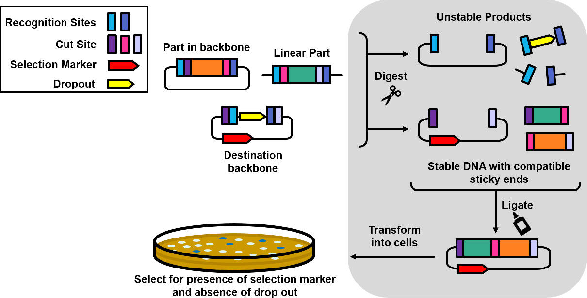
When Golden Gate Assembly Is Advantageous Golden Gate assembly is the preferred cloning method when your DNA sequences already contain Type IIS restriction sites and you need to assemble multiple fragments simultaneously at high efficiency. Unlike Gibson assembly, which offers greater flexibility in fragment design, Golden Gate is specifically optimized for situations where you have many DNA pieces—up to approximately 35 fragments—that need to be joined into a single construct all at once. This method operates as a “one-pot” process, meaning all components react together in a single tube, streamlining the workflow compared to multi-step procedures.
Essential Components and Mechanism Golden Gate assembly requires several key ingredients: (1) DNA fragments obtained from PCR or previously cloned sources, each flanked by Type IIS restriction enzyme recognition sites (such as BsaI or BsmBl); (2) a destination vector—a linearized plasmid backbone containing inward-facing Type IIS recognition sites; (3) Type IIS restriction enzymes (for example, BsaI-HFv2 or BsmBl-v2) that cut DNA at specific recognition sequences; (4) T4 DNA ligase, an enzyme that catalyzes strand joining; (5) reaction buffer to maintain proper chemical conditions; and (6) nuclease-free water as the reaction medium.
The Assembly Process All components are combined in a single reaction tube and then placed in a thermocycler, where the temperature alternates between 37°C (for restriction enzyme cutting) and 16°C (for DNA ligation). This thermal cycling allows cutting and joining to occur sequentially within the same reaction, maximizing efficiency. The key to Golden Gate’s elegance is the Type IIS restriction enzymes, which cut DNA slightly offset from their recognition sequence, generating sticky end overhangs that are perfectly complementary to adjacent DNA fragments. Once cutting is complete, the ligase enzyme seamlessly joins the sticky ends together, assembling all fragments in the correct order and orientation. This approach is particularly powerful for combining multiple genetic elements such as promoters, coding sequences, and regulatory regions. After the thermocycler reaction finishes, purification and bacterial transformation procedures are essentially identical to Gibson assembly.
References
- The use of LLM to help with finding information and reporting