Week 9: CELL FREE SYSTEMS
Week # 9 Cell Free Systems
CELL FREE SYSTEMS
To learn synthesis of proteins using cellular machinery outside of a cell.
General homework questions
- Explain the main advantages of cell-free protein synthesis over traditional in vivo methods, specifically in terms of flexibility and control
- over experimental variables. Name at least two cases where cell-free expression is more beneficial than cell production.
Cell-Free Protein Synthesis Advantages Cell-free protein synthesis (CFPS) provides substantial benefits compared to conventional cell-based protein production methods, particularly in terms of experimental flexibility and precise control over reaction parameters. In contrast to traditional in vivo approaches that require cell transformation, growth in culture media, and cell disruption, CFPS enables rapid protein production without these intermediate steps, significantly accelerating the research timeline.
Greater Experimental Control and Customization CFPS operates in an open, accessible reaction environment where researchers enjoy extensive freedom to manipulate conditions on the fly. This accessibility allows for straightforward modification of reaction parameters, supplementation with molecular chaperones and folding assistants, incorporation of non-standard amino acids not found in natural proteins, and inclusion of tagged or labeled molecular markers. These capabilities make CFPS exceptionally well-suited for investigating translational control mechanisms, conducting ribosome display experiments for directed evolution, or analyzing intricate protein-protein interaction networks.
Two Primary Applications Where CFPS Excels Producing toxic or cytotoxic proteins: Cell-free systems enable the synthesis of proteins that would be harmful or lethal to living cells if produced inside them. Since there is no intact cellular machinery to be damaged, toxic proteins can be safely manufactured in vitro. Engineering protein sequences with labeled components: CFPS greatly simplifies the incorporation of specialized building blocks, such as amino acids labeled with stable isotopes. This type of customization is vastly more straightforward to accomplish in a cell-free environment compared to achieving the same modifications within living organisms, where metabolic pathways create complications and inefficiencies. In essence, CFPS provides researchers with an unparalleled degree of control and experimental versatility that traditional cell-based systems simply cannot match.
- Describe the main components of a cell-free expression system and explain the role of each component.
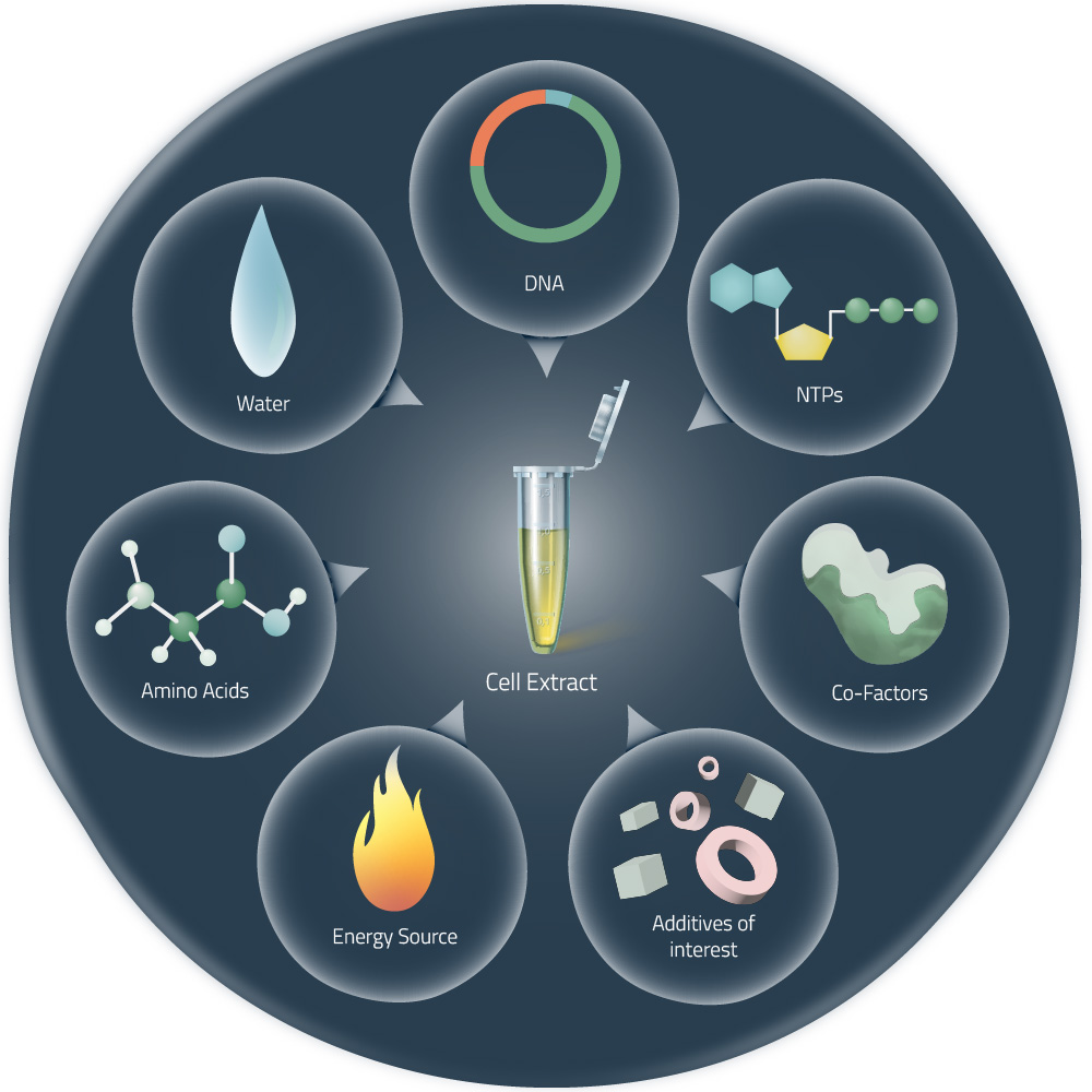
Cell-Free Extract The cell-free extract represents the core biochemical engine of the CFPS system. This liquid extract is derived from disrupted cells and contains the fundamental molecular machinery required for synthesizing proteins—specifically ribosomes (which build proteins), transfer RNAs (which deliver amino acids), enzymes (which catalyze chemical reactions), and numerous regulatory factors. The source organism selected to prepare this extract depends on the characteristics of the target protein: E. coli extracts are commonly used for straightforward proteins, rabbit reticulocyte extracts are chosen when higher eukaryotic protein qualities are needed, and wheat germ extracts are employed for particularly complex or post-translationally modified proteins.
DNA Template The DNA template serves as the genetic blueprint that contains all instructions for creating the desired protein. This template comprises two essential regions: a promoter sequence that signals where transcription should begin and initiate RNA synthesis, and a coding sequence that encodes the precise linear arrangement of amino acids that will form the target protein’s primary structure.
Energy Sources and Essential Cofactors Protein synthesis is an energy-intensive process that demands substantial quantities of adenosine triphosphate (ATP) and guanosine triphosphate (GTP). The CFPS system must continuously regenerate these energy molecules to sustain the reaction and prevent depletion. Beyond these energy sources, metal ion cofactors—particularly magnesium and potassium—are critical for maintaining the chemical environment necessary for both transcription and translation to proceed efficiently and accurately.
- Why is energy provision regeneration critical in cell-free systems? Describe a method you could use to ensure continuous ATP supply in your cell-free experiment.
The Critical Role of Energy Regeneration Maintaining adequate ATP levels is essential for successful cell-free protein synthesis because adenosine triphosphate serves as the primary energy currency driving both transcription (the conversion of DNA to RNA) and translation (the conversion of RNA to protein). During the CFPS reaction, ATP is consumed rapidly and continuously, as nearly every step of protein synthesis requires energy input. If ATP levels are not actively replenished and maintained, the biochemical machinery will exhaust its energy supply, the reaction will halt, and protein production will plummet significantly.
ATP Regeneration Strategy in E. coli Systems To overcome this energy limitation, E. coli-based cell-free systems employ a coupling strategy using two key components: phosphoenolpyruvate (PEP) and the enzyme pyruvate kinase. This enzymatic pair works together to continuously regenerate ATP from adenosine diphosphate (ADP), ensuring a steady supply of fresh energy molecules throughout the reaction. By maintaining consistent ATP availability through this regeneration mechanism, the system can sustain extended and robust protein synthesis, preventing the energy depletion that would otherwise terminate the reaction prematurely and severely reduce overall protein yield. In essence, energy regeneration transforms CFPS from a short-lived, low-yield process into a sustainable, high-productivity system capable of extended protein manufacturing.
- Compare prokaryotic versus eukaryotic cell-free expression systems. Choose a protein to produce in each system and explain why.
Prokaryotic cell-free systems (e.g., E. coli extracts) are typically faster to produce protein because transcription and translation are closely coupled, while eukaryotic systems separate these processes and are generally slower. Prokaryotic extracts often yield higher total protein amounts and are less expensive to run than eukaryotic extracts. Eukaryotic cell-free systems (e.g., rabbit reticulocyte, wheat germ, insect, or mammalian extracts) better support complex post-translational modifications such as glycosylation and disulfide bond formation. Prokaryotic systems lack most native eukaryotic chaperones and modification enzymes, which can limit correct folding and activity of many eukaryotic proteins. Eukaryotic systems can more accurately translate mRNAs with complex regulatory elements (e.g., internal ribosome entry sites, Kozak sequences) and handle eukaryotic codon usage more naturally. Prokaryotic systems are more amenable to genetic and biochemical optimization (e.g., energy regeneration, component supplementation) for high-throughput screening and rapid prototyping. Eukaryotic extracts are preferred for membrane proteins and multi-subunit complexes when proper folding, assembly, or membrane insertion requires eukaryotic-specific factors or microsomes. Prokaryotic systems often require codon optimization and may produce inclusion bodies or inactive protein for eukaryotic targets without additional folding aids. Eukaryotic systems are generally costlier, have lower batch-to-batch yields, and can be more variable, but they increase the likelihood of obtaining functionally active eukaryotic proteins. Choice depends on the goal: use prokaryotic systems for speed, yield, and cost-effective screening; use eukaryotic systems when native folding, modifications, or activity are required.
Recommended Proteins and Rationales
Prokaryotic System: Green Fluorescent Protein (GFP) Why GFP is ideal for E. coli CFPS: GFP is a small, robust protein (~27 kDa) that folds autonomously without requiring post-translational modifications or extensive chaperone assistance. The chromophore (fluorescent group) forms spontaneously through auto-oxidation of its own amino acid sequence, making it self-contained and independent of cellular machinery. The rapid synthesis capability of prokaryotic systems would allow researchers to quickly produce labeled protein variants for high-throughput screening, fluorescence assays, or incorporation of non-canonical amino acids at specific positions to tune optical properties. The low cost and ease of E. coli extract preparation make this system economically practical for producing large quantities needed for research or diagnostic applications. Additionally, GFP doesn’t require disulfide bonds or glycosylation, which are unnecessary complications in a prokaryotic environment.
Eukaryotic System: Antibody (Immunoglobulin) Why antibodies are ideal for eukaryotic CFPS: Antibodies are large, multimeric proteins (~150 kDa per molecule, often functioning as dimers or larger complexes) that require sophisticated post-translational processing. They contain multiple disulfide bonds that stabilize the structure and are critical for antigen binding and immune function—a feature that eukaryotic systems handle naturally through their oxidizing endoplasmic reticulum environment. Antibodies also undergo glycosylation at specific sites, which is essential for their effector functions (complement activation, antibody-dependent cellular cytotoxicity, and Fc receptor binding). The rich folding environment provided by eukaryotic extracts, with resident molecular chaperones like BiP and protein disulfide isomerases, ensures proper tertiary and quaternary structure formation. While eukaryotic CFPS is more expensive and time-consuming, the production of functional, correctly modified antibodies justifies this investment for therapeutic development, diagnostic reagents, or research applications where authentic biological activity is non-negotiable.
Summary Choose prokaryotic systems for rapid, simple, cost-effective production of robust proteins that self-fold and don’t require post-translational modifications. Choose eukaryotic systems when investing in complex proteins that demand sophisticated folding assistance, disulfide bond formation, or chemical modifications essential for biological function. The choice ultimately depends on balancing speed and cost against biological complexity and functional requirements of your target protein.
Prokaryotic cell-free expression systems, such as those based on E. coli, are fast, cost-effective, and capable of producing high protein yields. However, they lack the ability to perform post-translational modifications and often struggle with proper folding of complex or membrane-bound proteins. In contrast, eukaryotic systems like wheat germ extract are slower and more expensive but offer better support for folding and modifications, making them suitable for expressing complex eukaryotic proteins. For a prokaryotic system, I would choose to express
How would you design a cell-free experiment to optimize the expression of a membrane protein? Discuss the challenges and how you would address them in your setup.
Imagine you observe a low yield of your target protein in a cell-free system. Describe three possible reasons for this and suggest a troubleshooting strategy for each.
Homework question from Kate Adamala
Design an example of a useful synthetic minimal cell as follows:
- Pick a function and describe it. a. What would your synthetic cell do? What is the input and what is the output? b. Could this function be realized by cell-free Tx/Tl alone, without encapsulation? c. Could this function be realized by genetically modified natural cell? d. Describe the desired outcome of your synthetic cell operation.
- Design all components that would need to be part of your synthetic cell. a. What would be the membrane made of? b. What would you encapsulate inside? Enzymes, small molecules. c. Which organism your Tx/Tl system will come from? Is bacterial OK, or do you need a mammalian system for some reason? (hint: for example, if you want to use small molecule modulated promotors, like Tet-ON, you need mammalian) d. How will your synthetic cell communicate with the environment? (hint: are substrates permeable? or do you need to express the membrane channel?)
- Experimental details a. List all lipids and genes. (bonus: find the specific genes; for example, instead of just saying “small molecule membrane channel” pick the actual gene.) b. How will you measure the function of your system?
AHL-Quenching Synthetic Cell for Biofilm Disruption What the Cell Does This engineered synthetic minimal cell detects the quorum-sensing molecule 3-oxo-C12-HSL, which is released by the pathogenic bacterium Pseudomonas aeruginosa. When exposed to this signal, the cell produces the enzyme AiiA (a lactonase), which breaks down the AHL molecules within the enclosed vesicle. As new AHL continuously enters from the surrounding environment, the trapped enzyme works to destroy it, establishing a depletion zone that gradually reduces the available AHL in the nearby area. This reduction in quorum-sensing signaling dampens the pathogen’s ability to express disease-causing proteins and build biofilm structures.
Input and Output Input: The AHL signal (3-oxo-C12-HSL) from the surrounding liquid. Output: Reduced AHL concentration in the environment; the lactonase enzyme stays trapped inside the vesicle and does not escape. The primary measurable indicator is a decline in AHL levels over time.
Feasibility of Alternative Realizations Using only cell-free transcription-translation in solution: This approach is technically possible—mixing DNA and AHL in a test tube would produce AiiA and allow degradation. However, without encapsulation, the system lacks physical boundaries, the enzyme becomes diluted and exposed, and it cannot be precisely delivered to an infection site. A vesicle creates a controlled, protected microreactor that can be positioned exactly where treatment is needed. Using a genetically engineered living bacterium: An engineered E. coli strain programmed to express the LasR receptor and AiiA enzyme would theoretically accomplish the same sensing and degradation. However, living microorganisms present significant concerns: they may multiply uncontrollably, share modified genes with other bacteria, and face regulatory approval obstacles. A synthetic cell avoids these risks because it is non-living, incapable of reproduction, and inherently safer for medical use.
Desired Outcome Introducing a collection of these synthetic vesicles into a P. aeruginosa biofilm would lower AHL concentrations in that region to below the level needed for quorum-sensing activation. This would suppress the production of harmful proteins (such as elastase and pyocyanin) and destabilize the biofilm structure, making the bacteria vulnerable to destruction by the immune system or antimicrobial drugs.
Design of the Synthetic Cell Membrane Composition The synthetic cell consists of a single-layer lipid sphere containing: • POPC (70% of lipid composition) – promotes fluid membrane behavior and is compatible with living tissue • Cholesterol (30% of lipid composition) – strengthens the membrane structure and controls how easily molecules pass through The AHL molecule is sufficiently lipid-soluble (logP ≈ 3.5) to naturally seep through the lipid barrier without requiring specialized transport proteins. Encapsulated Components Biological machinery: An E. coli cell-free system that includes the natural RNA polymerase (with σ⁷⁰ factor), ribosomes, and all components needed for protein synthesis. This bacterial system is ideal because it recognizes standard bacterial promoters without needing more complex mammalian machinery. Fuel and building blocks: Energy sources (PEP, ATP, GTP, UTP, CTP), amino acids, transfer RNA, nucleotides, creatine phosphate, and necessary salts—all standard components for keeping bacterial lysate functional. Genetic instructions: Two circular DNA plasmids:
- pLuxR – continuously produces the LasR receptor protein using a powerful σ⁷⁰ promoter (J23119), with the lasR gene from P. aeruginosa PAO1 (GenBank reference: NP_250121.1)
- pAiiA produces the AiiA lactonase enzyme in response to AHL binding, controlled by the lasI promoter region (which contains the DNA binding site for the LasR-AHL complex) and containing the aiiA gene from Bacillus sp. 240B1 (GenBank reference: AAF62398.1) (These can be placed on separate plasmids for flexible tuning or combined on a single plasmid.)
Interaction with Surroundings AHL freely passes through the membrane via passive diffusion; no special channels are required. By contrast, the lactonase protein cannot escape because it carries no export signal and the membrane pores are too small (~0.5 nanometers) for proteins to exit. This design confines the synthetic cell to act purely as an AHL sink, eliminating any release of the enzyme into the environment.
Experimental Details Key Genetic and Chemical Components Component Specification POPC 1-palmitoyl-2-oleoyl-sn-glycero-3-phosphocholine Cholesterol Cholest-5-en-3β-ol Promoter (constitutive) J23119 (Anderson collection) lasR gene Pseudomonas aeruginosa PAO1, locus PA1430, protein NP_250121.1 PlasI promoter ~200 base pairs upstream of lasI (PA1432), includes las box binding site aiiA gene Bacillus sp. 240B1 lactonase, GenBank reference AAF62398.1
Measuring Performance Testing AHL removal from the environment: Expose a known quantity of synthetic vesicles to 1–5 µM of 3-oxo-C12-HSL in a buffered solution. At regular time points, collect samples of the surrounding liquid and measure remaining AHL using either liquid chromatography–mass spectrometry (LC-MS/MS) or a bacterial biosensor (E. coli carrying lasR and a fluorescent reporter gene under the lasI promoter). A decline in fluorescence demonstrates the quenching effect. Confirming enzyme activity inside the vesicle: Break open vesicles using freeze-thaw cycles and assess how efficiently AiiA degrades AHL by employing a color-changing dye that reacts to the acid released when lactones hydrolyze, or by using a synthetic lactone compound linked to a fluorescent tag (e.g., carboxyfluorescein) and monitoring the fluorescence change. Evaluating impact on the pathogen: Combine the synthetic cells with live P. aeruginosa PAO1 and assess whether virulence markers (elastase activity, pyocyanin pigment levels) decrease or whether the biofilm becomes thinner or less dense (using crystal violet staining). This approach creates a clearly defined, non-replicating, safe therapeutic system that uses a minimal lipid compartment and standard bacterial protein-making machinery to neutralize a pathogenic communication pathway.
Homework question from Peter Nguyen
Freeze-dried cell-free systems can be incorporated into all kinds of materials as biological sensors or as inducible enzymes to modify the material itself or the surrounding environment. Choose one application field — Architecture, Textiles/Fashion, or Robotics — and propose an application using cell-free systems that are functionally integrated into the material. Answer each of these key questions for your proposal pitch: • Write a one-sentence summary pitch sentence describing your concept. • How will the idea work, in more detail? Write 3-4 sentences or more. • What societal challenge or market need will this address? • How do you envision addressing the limitation of cell-free reactions (e.g., activation with water, stability, one-time use)?
BioChroma—Biosensor in Enhanced Textiles for Athlete and Worker Health Monitoring Product Concept BioChroma is an innovative textile technology that incorporates freeze-dried, cell-free biosensing components directly into fabric fibers. The system undergoes a lasting color shift when it encounters specific sweat-based health indicators, enabling athletes and industrial workers to monitor hydration status and electrolyte balance in real time without needing electronic devices or specialized equipment.
Operational Mechanism The system uses freeze-dried protein-synthesis machinery equipped with regulatory proteins that respond to specific molecular triggers and genes that encode color-producing proteins. These components are packaged into tiny droplets suspended within a gel and then applied as a coating to sections of fabric designed to absorb moisture. When sweat contacts the fabric, it rehydrates the dormant system, which then activates a series of reactions that identify target molecules (such as lactate or sodium) and rapidly manufacture a colored pigment—all without needing living microorganisms or electrical input. The chemical cascade is engineered to be one-directional and irreversible, ensuring that the color change becomes a permanent marker of the wearer’s peak stress levels rather than a temporary indicator. By placing several different sensor zones at various locations on a single garment, the design allows simultaneous tracking of multiple different biomarkers from different areas of the body.
The Problem and Market Opportunity This technology tackles an important unmet need: many outdoor workers, military personnel, and endurance athletes are vulnerable to heat-related medical emergencies but lack practical access to real-time body monitoring systems. Traditional electronic smart textiles are expensive, fragile, depend on batteries, and may violate workplace safety rules. Additionally, these devices deteriorate when laundered and generate electronic waste. The biological approach presented here offers a compelling alternative—significantly cheaper, single-use, and completely compostable—making physiological monitoring accessible to broad consumer populations seeking practical health tracking options.
Overcoming Technical Challenges with Cell-Free Systems To prevent unwanted activation and maintain long-term viability, the freeze-dried biosensing material is enclosed in a water-repellent silica coating that dissolves only when exposed to sweat conditions (particular pH and salt concentration), guarding against false activation from rain or everyday moisture and allowing the product to remain stable for more than two years at normal temperatures. The one-time-use nature of the system is reframed as an intentional advantage, functioning similarly to radiation exposure badges that accumulate and record total stress over time. A modular patch architecture means users can replace only the sensors that have been activated rather than discarding the entire garment. Additionally, the dried biological material is treated with protective compounds (trehalose sugar and stabilizing proteins) that preserve enzyme function even after the fabric experiences physical stress and repeated washing before the sensors are triggered.
Homework question from Ally Huang
Freeze-dried cell-free reactions have great potential in space, where resources are constrained. As described in my talk, the Genes in Space competition challenges students to consider how biotechnology, including cell-free reactions, can be used to solve biological problems encountered in space. While the competition is limited to only high school students, your assignment will be to develop your own mock Genes in Space proposal to practice thinking about biotech applications in space! For this particular assignment, your proposal is required to incorporate the BioBits® cell-free protein expression system, but you may also use the other tools in the Genes in Space toolkit (the miniPCR® thermal cycler and the P51 Molecular Fluorescence Viewer). For more inspiration, check out https://www.genesinspace.org/ .
- Provide background information that describes the space biology question or challenge you propose to address. Explain why this topic is s ignificant for humanity, relevant for space exploration, and scientifically interesting. (Maximum 100 words)
- Name the molecular or genetic target that you propose to study. Examples of molecular targets include individual genes and proteins, DNA and RNA sequences, or broader -omics approaches. (Maximum 30 words)
- Describe how your molecular or genetic target relates to the space biology question or challenge your proposal addresses. (Maximum 100 words)
- Clearly state your hypothesis or research goal and explain the reasoning behind it. (Maximum 150 words)
- Outline your experimental plan - identify the sample(s) you will test in your experiment, including any necessary controls, the type of data or measurements that will be collected, etc. (Maximum 100 words)
Cell-Free Biosensing for Radiation Damage Assessment in Space Background Astronauts traveling beyond Earth’s protective magnetic field are exposed to high-energy cosmic radiation that causes harmful changes to DNA, elevating their lifetime risk of malignancy and cellular deterioration. Directly evaluating the biological impact of this damage in living cells is challenging, particularly during space missions where resources are limited. Cell-free protein-making systems such as BioBits® offer a practical solution: they enable rapid measurement of how radiation-damaged DNA impairs the production of mRNA and proteins, creating a simple, lightweight detection method for monitoring genetic damage during extended journeys through deep space.
Molecular or Genetic Target The experimental system uses a superfolder GFP (sfGFP) gene carried on a plasmid, which functions as a fluorescent signal indicating how efficiently genes are transcribed and translated after the DNA has been damaged by radiation.
Connection to Space Biology Challenge Radiation damage creates breaks and chemical alterations in the DNA backbone that obstruct the movement of RNA polymerase enzymes, thereby suppressing mRNA production. When researchers introduce radiation-damaged sfGFP DNA into a BioBits reaction mixture, the reduced fluorescence signal directly correlates with the proportion of DNA templates that have been rendered non-functional. This approach translates the amount of DNA injury into a measurable decrease in gene expression, resembling what occurs when critical genes are damaged in the cells of actual space travelers.
Research Hypothesis and Objectives The research team predicts that plasmids exposed to radiation mimicking galactic cosmic ray exposure will generate diminished amounts of sfGFP protein in a cell-free reaction system, with the reduction increasing proportionally to radiation dose. Since BioBits reactions contain all necessary enzymes for transcription and translation but lack the DNA repair machinery found in live cells, any reduction in fluorescence must come directly from physical damage to the DNA template itself. By plotting the relationship between radiation dose and fluorescence loss, this assay will establish a reliable, space-suitable method for quantifying how radiation affects gene expression capability, potentially enabling continuous health assessment during long-term space missions.
Experimental Approach Identical samples of isolated pSFGFP plasmid will be exposed to iron ion (⁵⁶Fe) radiation at four levels: no radiation (as a control), 5 gray, 10 gray, and 20 gray. Each radiation-treated DNA sample (100 nanograms per reaction) will then be mixed into reconstituted BioBits components and allowed to incubate for two hours at 30 degrees Celsius. Fluorescence intensity will be measured at the end of the reaction using a P51 Molecular Fluorescence Viewer. The experiment will include three identical reactions per radiation dose plus a blank reaction containing no DNA to serve as a background reference. The resulting fluorescence measurements will be plotted against radiation dose to establish a curve showing the progressive decline in gene expression as DNA damage increases.
References
- The use of LLM to help with finding information and reporting