Ermine Moth Caterpillar Silk Matrices as Biofilters for Airborne PFAS Particles
SECTION 1: ABSTRACT
This project is centred around the remediation of PFAS pollution. PFAS, per- and polyfluoroalkyl substances, commonly known as “forever chemicals” are a group of chemical compounds that have a significant negative effect on human and ecological health. They are synthetic chemicals characterised by chains of carbon atoms bound to fluorine atoms, which are the reason why they do not break down in nature as carbon-fluorine bonds are incredibly strong.
However, a paper published in early 2025 entitled “PFAS biodegradation by Labrys portucalensis F11: Evidence of chain shortening and identification of metabolites of PFOS” describes promising evidence that a bacterial species isolated from PFAS contaminated soil in Portugal managed to enzymatically biodegrade PFOS, 6:2 FTS, and 5:3 FTCA into shorter-chain compounds.
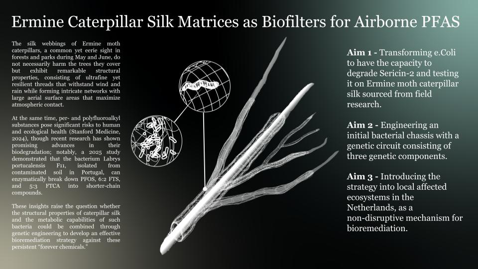
On the other hand, the silk webbings of the Ermine moth caterpillar are a recurring, eerie sight in forests and parks around May and June. Luckily, infestations by this caterpillar do not necessarily harm the trees affected. However, these silk networks boast remarkable structural qualities: they are composed of ultrafine yet resilient threads that endure wind and rain and their intricate matrix yields a vast aerially exposed surface area, maximizing atmospheric contact.
This project hypothesises that the webbing’s properties could be utilised to create naturally occurring filtration systems through genetic engineering, by transforming bacteria to have the capacity to proliferate on the silk while also having the capacity to degrade various PFAS compounds.
The development of the project is formulated in three key objectives. The primary objective is to transform the chassis e.coli K12 to have the capacity to survive on silk by degrading sericin-2 with the enzyme Cocoonase. If successful, the second objective is to develop a three-component genetic circuit that leverages this sericin-2 degrading mechanism and combine it with a PFAS degrading enzyme and a signaling system. A final, big picture objective would be to implement the fully developed mechanism in real world scenarios, in compliance with regulatory systems in regards to genetically modified organisms
Within the context of How To Grow Almost Anything as a course, this project primarily becomes a tool to learn about the fundamentals of synthetic biology framed within themes like ecology, unconventional bioremediation strategies, bacterial metabolic mechanisms and biomaterials. The technical and experimental aspects of the project include but are not limited to DNA design, plasmid design, protein design, experiment design, Twist orders, data validation, AlphaFold modeling
This project combines several technical and experimental approaches, including DNA and plasmid design, protein engineering, codon-aware construct building, and in silico validation using AlphaFold. It also involves experimental planning for E. coli chassis engineering, cocoonase expression, silk degradation assays, CFU measurements, and protein purification and quantification. In addition, the project draws on bioinformatics, literature review, and biosensor design to develop a modular PFAS-responsive genetic circuit. Overall, the work integrates synthetic biology, biomaterials, and environmental bioremediation into one interdisciplinary framework.
Presentation Slides
SECTION 2: PROJECT AIMS
Define three aims for your final project (minimum one sentence per aim).
Aim 1 - Transforming e.Coli to have the capacity to degrade Sericin-2 and testing it on Ermine moth caterpillar silk sourced from field research.
A key component of the envisioned genetic circuit in aim 2 is the capacity to degrade the silk protein sericin-2 as a carbon source in conditions when PFAS particles are not readily available.
Cocoonase and Sericin-2
Cocoonase is a proteolytic enzyme produced by silk moths during adult development and used for softening the end of the cocoon to permit exit of the adult moth. In recent development, cocoonase is being studied as an ecofriendly alternative for silk-degumming. Silk primarily consists of fibroin and sericin, where fibroin is the core structural component of the fibers and sericin acts as a gum coating that allows for the fibres to stick together. The degumming process targets the latter.
Ermine moth caterpillar silk is similar in structural and chemical composition, consisting of a fibroin core, combined with four types of sericin protein and a mucin protein. This gave me the idea to look into the degradation of sericin for the development of this aim.
Leveraging suboptimal conditions
Cocoonase functions optimally at a pH between 8.0 and 9.8, and while exact data on the pH of ermine moth silk is not readily available, it is likely that the silk has the same pH as the silk gland. This would be between 5 and 7 pH; clearly this is outside of the optimal range. However, these suboptimal conditions could prove to be advantageous in these conditions. The goal is not to completely degum the silk, compromising its structural integrity, but rather to degrade enough silk for the bacteria to proliferate.
This supported the hypothesis that a cocoonase-based sericin degradation mechanism could be an effective strategy in sustaining transformed bacterial chassis.
This finding made me strategise to focus on the expression of cocoonase for my aim 1 experimental goal. For which I developed a Twist order and an experimental planning, the latter can be found in section 4.
DNA sequence of BmCoc:
atgattgtgggcggcgaagaaattagcattaacaaagtgccgtatcaggcgtatctgctg
ctgcagaaagataacgaatattttcagtgcggcggcagcattattagcaaacgccatatt
ctgaccgcggcgcattgcattgaaggcattagcaaagtgaccgtgcgcattggcagcagc
aacagcaacaaaggcggcaccgtgtataccgcgaaaagcaaagtggcgcatccgaaatat
aacagcaaaaccaaaaacaacgattttgcgattgtgaccgtgaacaaagatatggcgatt
gatggcaaaaccaccaaaattattaccctggcgaaagaaggcagcagcgtgccggataaa
accaaactgctggtgagcggctggggcgcgaccagcgaaggcggcagcagcagcaccacc
ctgcgcgcggtgcatgtgcaggcgcatagcgatgatgaatgcaaaaaatattttcgcagc
ctgaccagcaacatgttttgcgcgggcccgccggaaggcggcaaagatagctgccagggc
gatagcggcggcccggcggtgaaaggcaacgtgcagctgggcgtggtgagctttggcgtg
ggctgcgcgcgcaaaaacaacccgggcatttatgcgaaagtgagcgcggcggcgaaatgg
attaaaagcaccgcgggcctg
AA sequence:
MIVGGEEISINKVPYQAYLLLQKDNEYFQCGGSIISKRHILTAA HCIEGISKVTVRIGSSNSNKGGTVYTAKSKVAHPKYNSKTKNNDFAIVTVNKDMAIDG KTTKIITLAKEGSSVPDKTKLLVSGWGATSEGGSSSTTLRAVHVQAHSDDECKKYFRS LTSNMFCAGPPEGGKDSCQGDSGGPAVKGNVQLGVVSFGVGCARKNNPGIYAKVSAAAKWIKSTAGL
Plasmid Design
In Benchling, I designed a gene fragment consisting of the sequence for BmCoc, tagged with a His-tag for protein purification later on down the line. I put this fragment in a Twist order, in vector pTwist Amp High Copy.
Originally I designed a full plasmid in Benchling using a found backbone on Snapgene. However, after lots of confusion and frustration I realised that in order to fill out my order from Twist, I would have to use one of their vectors.
As stated prior, the experiment design for my project involves exposing sterilised silk fibres to engineered BmCoc-expressing cells, alongside uninduced and no-cell controls. Over a 48-hour incubation, I will assess degradation and system performance using three readouts:
1. Weight loss of the silk fibres to quantify sericin degradation,
2. CFU assays to determine whether E. coli can proliferate using silk-derived nutrients.
3. Protein quantification (Bradford assay following His-tag purification) to confirm expression and secretion of functional cocoonase.
Together, this should demonstrate both material breakdown and biological utilisation within the engineered system. A full protocol can be found in section 4.
Aim 2 - Engineering an initial bacterial chassis with a genetic circuit consisting of three genetic components:
Under normal conditions, the chassis is able to survive on the ermine moth silk through degradation of sericin-2. However, when exposed to PFAS-particles, the cell’s sericin-2 degradation is repressed, incentivising it to degrade PFAS. Once the particles are degraded, the sensor is no longer engaged and the cell continues producing cocoonase (BmCoc) to degrade sericin-2.
A gene that encodes for an enzyme that breaks down Sericin 2
See Aim 1 for further information on this aspect.
A gene that encodes for enzymatic degradation of PFOS, 6:2 FTS, and 5:3 FTCA into shorter-chain compounds
Due to the hazardous nature of PFAS particles, I knew early on that further development working with these particles was not possible. Moreover, I found very little genetic data related to the specific enzymes described in the key source paper.
The mechanism that combines the three components of the circuit is inspired by the classic repressilator first introduced during the biobootcamp lecture day 2 as well as genetic cascades .
The repressilator consists of three transcriptional repression system where a lacI represses the transcription of tetR, tetR represses the transcription of cl and cl represses the transcription of lacI.
To be honest, at first I didn’t get it, because I thought the protein would bind to the sequence it was described to repress. Only later it clicked that the repressor proteins bind to a corresponding operon upstream in the sequence.
A PFAS detecting biosensor – hlFABP/split-tetR sensor
A paper published in 2023, describes a novel biosensor that utilises hlFABP ligated to a GFP. When PFOA molecules bind to the hlFABP, its structure slightly shifts, shifting the GFP’s fluorescent read-out slightly. This made me wonder whether this mechanism can be adapted where the sensor directly triggers a repression mechanism. This led me to a split tetracycline repressor (TetR).
In the final stretch of this course I investigated the feasibility of a hlFABP/split-tetR sensor where the structural shift of hlFABP could dictate tetR component recombination. I
While the exemplary sensor was tested on PFOA and the enzymatic degradation by L. Portucalensis F11 was shown to degrade PFOS, 6:2 FTS, and 5:3 FTCA, the application of a similar sensor mechanism for PFOS could be possible considering PFOS’ binding affinity to hlFABP due to its structural similarity to lipids.
Modular cascade
In further development, the mechanism could be modulated to fit remediation strategies of various air pollutants.
The puzzle pieces of the hlFABP/split-Tetr and this aspect of the circuit system came together to inform the circuit design depicted below.
hlFABP/split-tetR sensor validation
I investigated the feasibility of a hlFABP/split-tetR sensor where the structural shift of hlFABP could dictate tetR component recombination. The paper “A split transcriptional repressor that links protein solubility to an orthogonal genetic circuit” by Zeng et al., details the potential use of a split TetR repressor.
The paper describes the construction of a TetR fission library by randomly fragmenting the TetR coding sequence and screening the resulting variants for those that still functioned as repressors when the two fragments were brought together by interacting peptide pairs. Using this approach, the authors identified three successful split positions—after residues 180, 184, and 193—which were the only unique fission sites recovered from the screened clones and were located in the regulatory core of TetR.
In my project, I am using these experimentally validated fission sites as candidate fusion points to build hlFABP–split TetR constructs and test them in silico. By comparing the predicted structures of the fusion proteins to native hlFABP, I aim to assess whether these split sites preserve the hlFABP fold and therefore are unlikely to disrupt PFAS binding.
hlFABP
DNA:
atgagctttagcggcaaatatcagctgcagagccaggaaaactttgaagcgtttatgaaagcgattggcctgccggaagaactgattcagaaaggcaaagatattaaaggcgtgagcgaaattgtgcagaacggcaaacattttaaatttaccattaccgcgggcagcaaagtgattcagaacgaatttaccgtgggcgaagaatgcgaactggaaaccatgaccggcgaaaaagtgaaaaccgtggtgcagctggaaggcgataacaaactggtgaccacctttaaaaacattaaaagcgtgaccgaactgaacggcgatattattaccaacaccatgaccctgggcgatattgtgtttaaacgcattagcaaacgcatt
Amino Acid sequence:
MSFSGKYQLQSQENFEAFMKAIGLPEELIQKGKDIKGVSEIVQNGKHFKFTITAGSKVIQNEFTVGEECELETMTGEKVKTVVQLEGDNKLVTTFKNIKSVTELNGDIITNTMTLGDIVFKRISKRI
AlphaFold Image
pTM = 0.9
TetR
DNA sequence:
atgagccgcctggataaaagcaaagtgattaacagcgcgctggaactgctgaacgaagtg
ggcattgaaggcctgaccacccgcaaactggcgcagaaactgggcgtggaacagccgacc
ctgtattggcatgtgaaaaacaaacgcgcgctgctggatgcgctggcgattgaaatgctg
gatcgccatcatacccatttttgcccgctggaaggcgaaagctggcaggattttctgcgc
aacaacgcgaaaagctttcgctgcgcgctgctgagccatcgcgatggcgcgaaagtgcat
ctgggcacccgcccgaccgaaaaacagtatgaaaccctggaaaaccagctggcgtttctg
tgccagcagggctttagcctggaaaacgcgctgtatgcgctgagcgcggtgggccatttt
accctgggctgcgtgctggaagatcaggaacatcaggtggcgaaagaagaacgcgaaacc
ccgaccaccgatagcatgccgccgctgctgcgccaggcgattgaactgtttgatcatcag
ggcgcggaaccggcgtttctgtttggcctggaactgattatttgcggcctggaaaaacag
ctgaaatgcgaaagcggcagc
Amino acid sequence:
MSRLDKSKVINSALELLNEVGIEGLTTRKLAQKLGVEQPTLYWHVKNKRALLDALAIEMLDRHHTHFCPLEGESWQDFLRNNAKSFRCALLSHRDGAKVHLGTRPTEKQYETLENQLAFLCQQGFSLENALYALSAVGHFTLGCVLEDQEHQVAKEERETPTTDSMPPLLRQAIELFDHQGAEPAFLFGLELIICGLEKQLKCESGS
180TetR
atgagccgcctggataaaagcaaagtgattaacagcgcgctggaactgctgaacgaagtgggcattgaaggcctgaccacccgcaaactggcgcagaaactgggcgtggaacagccgaccctgtattggcatgtgaaaaacaaacgcgcgctgctggatgcgctggcgattgaaatgctggatcgccatcatacccatttttgcccgctggaaggcgaaagctggcaggattttctgcgcaacaacgcgaaaagctttcgctgcgcgctgctgagccatcgcgatggcgcgaaagtgcatctgggcacccgcccgaccgaaaaacagtatgaaaccctggaaaaccagctggcgtttctgtgccagcagggctttagcctggaaaacgcgctgtatgcgctgagcgcggtgggccattttaccctgggctgcgtgctggaagatcaggaacatcaggtggcgaaagaagaacgcgaaaccccgaccaccgatagcatgccgccgctgctgcgccaggcgattgaactgtttgatcatcag
184TetR
atgagccgcctggataaaagcaaagtgattaacagcgcgctggaactgctgaacgaagtgggcattgaaggcctgaccacccgcaaactggcgcagaaactgggcgtggaacagccgaccctgtattggcatgtgaaaaacaaacgcgcgctgctggatgcgctggcgattgaaatgctggatcgccatcatacccatttttgcccgctggaaggcgaaagctggcaggattttctgcgcaacaacgcgaaaagctttcgctgcgcgctgctgagccatcgcgatggcgcgaaagtgcatctgggcacccgcccgaccgaaaaacagtatgaaaccctggaaaaccagctggcgtttctgtgccagcagggctttagcctggaaaacgcgctgtatgcgctgagcgcggtgggccattttaccctgggctgcgtgctggaagatcaggaacatcaggtggcgaaagaagaacgcgaaaccccgaccaccgatagcatgccgccgctgctgcgccaggcgattgaactgtttgatcatcagggcgcggaaccg
193TetR
atgagccgcctggataaaagcaaagtgattaacagcgcgctggaactgctgaacgaagtgggcattgaaggcctgaccacccgcaaactggcgcagaaactgggcgtggaacagccgaccctgtattggcatgtgaaaaacaaacgcgcgctgctggatgcgctggcgattgaaatgctggatcgccatcatacccatttttgcccgctggaaggcgaaagctggcaggattttctgcgcaacaacgcgaaaagctttcgctgcgcgctgctgagccatcgcgatggcgcgaaagtgcatctgggcacccgcccgaccgaaaaacagtatgaaaccctggaaaaccagctggcgtttctgtgccagcagggctttagcctggaaaacgcgctgtatgcgctgagcgcggtgggccattttaccctgggctgcgtgctggaagatcaggaacatcaggtggcgaaagaagaacgcgaaaccccgaccaccgatagcatgccgccgctgctgcgccaggcgattgaactgtttgatcatcagggcgcggaaccggcgtttctgtttggcctggaactgatt
hlFABP/180TetR
DNA
atgagctttagcggcaaatatcagctgcagagccaggaaaactttgaagcgtttatgaaagcgattggcctgccggaagaactgattcagaaaggcaaagatattaaaggcgtgagcgaaattgtgcagaacggcaaacattttaaatttaccattaccgcgggcagcaaagtgattcagaacgaatttaccgtgggcgaagaatgcgaactggaaaccatgaccggcgaaaaagtgaaaaccgtggtgcagctggaaggcgataacaaactggtgaccacctttaaaaacattaaaagcgtgaccgaactgaacggcgatattattaccaacaccatgaccctgggcgatattgtgtttaaacgcattagcaaacgcattagccgcctggataaaagcaaagtgattaacagcgcgctggaactgctgaacgaagtgggcattgaaggcctgaccacccgcaaactggcgcagaaactgggcgtggaacagccgaccctgtattggcatgtgaaaaacaaacgcgcgctgctggatgcgctggcgattgaaatgctggatcgccatcatacccatttttgcccgctggaaggcgaaagctggcaggattttctgcgcaacaacgcgaaaagctttcgctgcgcgctgctgagccatcgcgatggcgcgaaagtgcatctgggcacccgcccgaccgaaaaacagtatgaaaccctggaaaaccagctggcgtttctgtgccagcagggctttagcctggaaaacgcgctgtatgcgctgagcgcggtgggccattttaccctgggctgcgtgctggaagatcaggaacatcaggtggcgaaagaagaacgcgaaaccccgaccaccgatagcatgccgccgctgctgcgccaggcgattgaactgtttgatcatcag
Amino Acid Sequence
MSFSGKYQLQSQENFEAFMKAIGLPEELIQKGKDIKGVSEIVQNGKHFKFTITAGSKVIQNEFTVGEECELETMTGEKVKTVVQLEGDNKLVTTFKNIKSVTELNGDIITNTMTLGDIVFKRISKRISRLDKSKVINSALELLNEVGIEGLTTRKLAQKLGVEQPTLYWHVKNKRALLDALAIEMLDRHHTHFCPLEGESWQDFLRNNAKSFRCALLSHRDGAKVHLGTRPTEKQYETLENQLAFLCQQGFSLENALYALSAVGHFTLGCVLEDQEHQVAKEERETPTTDSMPPLLRQAIELFDHQ
pTM = 0.66
hlFABP/184TetR
DNA
atgagctttagcggcaaatatcagctgcagagccaggaaaactttgaagcgtttatgaaagcgattggcctgccggaagaactgattcagaaaggcaaagatattaaaggcgtgagcgaaattgtgcagaacggcaaacattttaaatttaccattaccgcgggcagcaaagtgattcagaacgaatttaccgtgggcgaagaatgcgaactggaaaccatgaccggcgaaaaagtgaaaaccgtggtgcagctggaaggcgataacaaactggtgaccacctttaaaaacattaaaagcgtgaccgaactgaacggcgatattattaccaacaccatgaccctgggcgatattgtgtttaaacgcattagcaaacgcattagccgcctggataaaagcaaagtgattaacagcgcgctggaactgctgaacgaagtgggcattgaaggcctgaccacccgcaaactggcgcagaaactgggcgtggaacagccgaccctgtattggcatgtgaaaaacaaacgcgcgctgctggatgcgctggcgattgaaatgctggatcgccatcatacccatttttgcccgctggaaggcgaaagctggcaggattttctgcgcaacaacgcgaaaagctttcgctgcgcgctgctgagccatcgcgatggcgcgaaagtgcatctgggcacccgcccgaccgaaaaacagtatgaaaccctggaaaaccagctggcgtttctgtgccagcagggctttagcctggaaaacgcgctgtatgcgctgagcgcggtgggccattttaccctgggctgcgtgctggaagatcaggaacatcaggtggcgaaagaagaacgcgaaaccccgaccaccgatagcatgccgccgctgctgcgccaggcgattgaactgtttgatcatcagggcgcggaaccg
Amino acid
MSFSGKYQLQSQENFEAFMKAIGLPEELIQKGKDIKGVSEIVQNGKHFKFTITAGSKVIQNEFTVGEECELETMTGEKVKTVVQLEGDNKLVTTFKNIKSVTELNGDIITNTMTLGDIVFKRISKRISRLDKSKVINSALELLNEVGIEGLTTRKLAQKLGVEQPTLYWHVKNKRALLDALAIEMLDRHHTHFCPLEGESWQDFLRNNAKSFRCALLSHRDGAKVHLGTRPTEKQYETLENQLAFLCQQGFSLENALYALSAVGHFTLGCVLEDQEHQVAKEERETPTTDSMPPLLRQAIELFDHQGAEP
pTM = 0.66
hlFABP/193TetR
DNA
atgagctttagcggcaaatatcagctgcagagccaggaaaactttgaagcgtttatgaaagcgattggcctgccggaagaactgattcagaaaggcaaagatattaaaggcgtgagcgaaattgtgcagaacggcaaacattttaaatttaccattaccgcgggcagcaaagtgattcagaacgaatttaccgtgggcgaagaatgcgaactggaaaccatgaccggcgaaaaagtgaaaaccgtggtgcagctggaaggcgataacaaactggtgaccacctttaaaaacattaaaagcgtgaccgaactgaacggcgatattattaccaacaccatgaccctgggcgatattgtgtttaaacgcattagcaaacgcattagccgcctggataaaagcaaagtgattaacagcgcgctggaactgctgaacgaagtgggcattgaaggcctgaccacccgcaaactggcgcagaaactgggcgtggaacagccgaccctgtattggcatgtgaaaaacaaacgcgcgctgctggatgcgctggcgattgaaatgctggatcgccatcatacccatttttgcccgctggaaggcgaaagctggcaggattttctgcgcaacaacgcgaaaagctttcgctgcgcgctgctgagccatcgcgatggcgcgaaagtgcatctgggcacccgcccgaccgaaaaacagtatgaaaccctggaaaaccagctggcgtttctgtgccagcagggctttagcctggaaaacgcgctgtatgcgctgagcgcggtgggccattttaccctgggctgcgtgctggaagatcaggaacatcaggtggcgaaagaagaacgcgaaaccccgaccaccgatagcatgccgccgctgctgcgccaggcgattgaactgtttgatcatcagggcgcggaaccggcgtttctgtttggcctggaactgatt
Amino Acid Sequence
MSFSGKYQLQSQENFEAFMKAIGLPEELIQKGKDIKGVSEIVQNGKHFKFTITAGSKVIQNEFTVGEECELETMTGEKVKTVVQLEGDNKLVTTFKNIKSVTELNGDIITNTMTLGDIVFKRISKRISRLDKSKVINSALELLNEVGIEGLTTRKLAQKLGVEQPTLYWHVKNKRALLDALAIEMLDRHHTHFCPLEGESWQDFLRNNAKSFRCALLSHRDGAKVHLGTRPTEKQYETLENQLAFLCQQGFSLENALYALSAVGHFTLGCVLEDQEHQVAKEERETPTTDSMPPLLRQAIELFDHQGAEPAFLFGLELI
pTM = 0.7
Through visual comparison, the structural integrity of the hlFABP component of the sensor remains intact. The ptm-score of the predicted structures are all above 0.5, which indicates a high confidence in structural accuracy.
Combining the data on split-TetR sensor design and the predicted protein structure obtained through Alphafold, all of these DNA designs have potential to be functional as a biosensor. However, further validation would need to be done in the lab.
Lyophilized alternative
During the homework development, I also reviewed the possibility of translating this mechanism into a cell-free system as opposed to a system that is founded on silk degradation:
“I propose a freeze-dried, cell-free enzymatic system designed to degrade PFAS contaminants in water, building on recent evidence of bacterial PFAS biodegradation pathways. By incorporating these enzymes into a lyophilized platform, the system can be easily stored, transported, and deployed globally, with activation triggered simply by contact with contaminated water. This approach directly addresses the growing environmental and public health challenge posed by persistent PFAS pollution, particularly in aquatic systems where conventional remediation methods are limited or costly. While cell-free systems typically face constraints such as water dependence and single-use functionality, these characteristics are reframed here as advantages: water acts as a natural activation mechanism in polluted environments, and the temporary activity reduces the risk of long-term ecological accumulation. Overall, the concept offers a scalable, controllable, and potentially safer alternative for targeted PFAS remediation.”
Aim 3 - Introducing the strategy into local affected ecosystems in the Netherlands, as a non-disruptive mechanism for bioremediation, after rigorous testing to meet EU ecological risk assessment regulations (ERAs) and conducting EU mandated post-market environmental monitoring (PMEM). (European Commission)
I first explored the implication of my aim 3 early on in the course, focussing my governance research on this project proposal. The research was developed with “de Alblasserwaard", a region within the province of Zuid-Holland, the Netherlands, in mind. I grew up in this area. In recent years, it has become known that the entire area is heavily contaminated with PFAS originating from industrial emissions by a local DuPont factory.
I envisioned the following policy/governance goals in relation to this project:
Biosecurity
Keeping a rigorous overview over the whole process
Designing inherently safe genetic modifications
Ecological Preservation
By protecting the ecological balance of target ecosystems
Equity
By making it accessible to those that need it
Justice
For humans
For non-humans
As well as the following potential governance actions:
R&D phase researchers:
P - Ensuring the core mechanism of PFAS degradation by genetically engineered bacteria within a fine meshed network-substrate works efficiently and does not leave residual harmful chemicals.
D - Researching to what capacity the principle can be scaled up for real world deployment.
A - Ensuring that no harmful organisms are produced and released into the wild throughout laboratorial development.
R - Ensuring that potentially implemented engineered microorganisms are rigorously tested to meet EU ecological risk assessment regulations (ERAs) for genetically engineered microorganisms.
Deployment researchers:
P - Localising areas affected by airborne PFAS pollution.
D - Finding suitable host sites for the caterpillar-biofilter within affected areas throughout the development process, allowing for efficient application.
A - Assess potential environmental factors that could undermine the efficacy of implementation.
R - Conducting post-market environmental monitoring (PMEM)* as mandated by the EU.
Environmental Rights litigators
P - Use legal tools to safeguard responsible application of the biofilter.
D - Draft funding mandates for known local major polluters to fund local bioremediation pilots like this biofilter.
A - Advocating for the legal recognition of the trees targeted for the application under the Right of Nature, protecting them from overexploitation through caterpillar infestation. building towards legally protecting the entire local ecosystems they reside in.
R - Compose long term Risk and Success assessment based on legal precedents in relation to ‘Right of Nature’ and funding mandate rulings.
Local Governmental Authorities
P - Spreading informative campaigns to local communities that transparently communicates the project and informs people not to destroy the webs
D - Forming a recognised mediative contact point between the executive parties and impacted communities, allowing communities to voice potential concerns.
A - Assist interested members of the public to find ways to get involved, incentivising people to (re)connect with their local ecosystems and giving back a sense of agency in their own wellbeing.
R - Formulate contingency plans to manage misinformation and potential community protest, as well as rapid response protocols to pause activities in case community concerns escalate.
*Post-market environmental monitoring (PMEM) is a mandatory, legally required process in the EU for GMOs and authorized products, designed to detect direct, indirect, or unforeseen adverse effects on human health or the environment after market release. It consists of Case-Specific Monitoring (CSM) to verify assumptions in the Environmental Risk Assessment (ERA) and General Surveillance (GS) to identify unexpected effects.
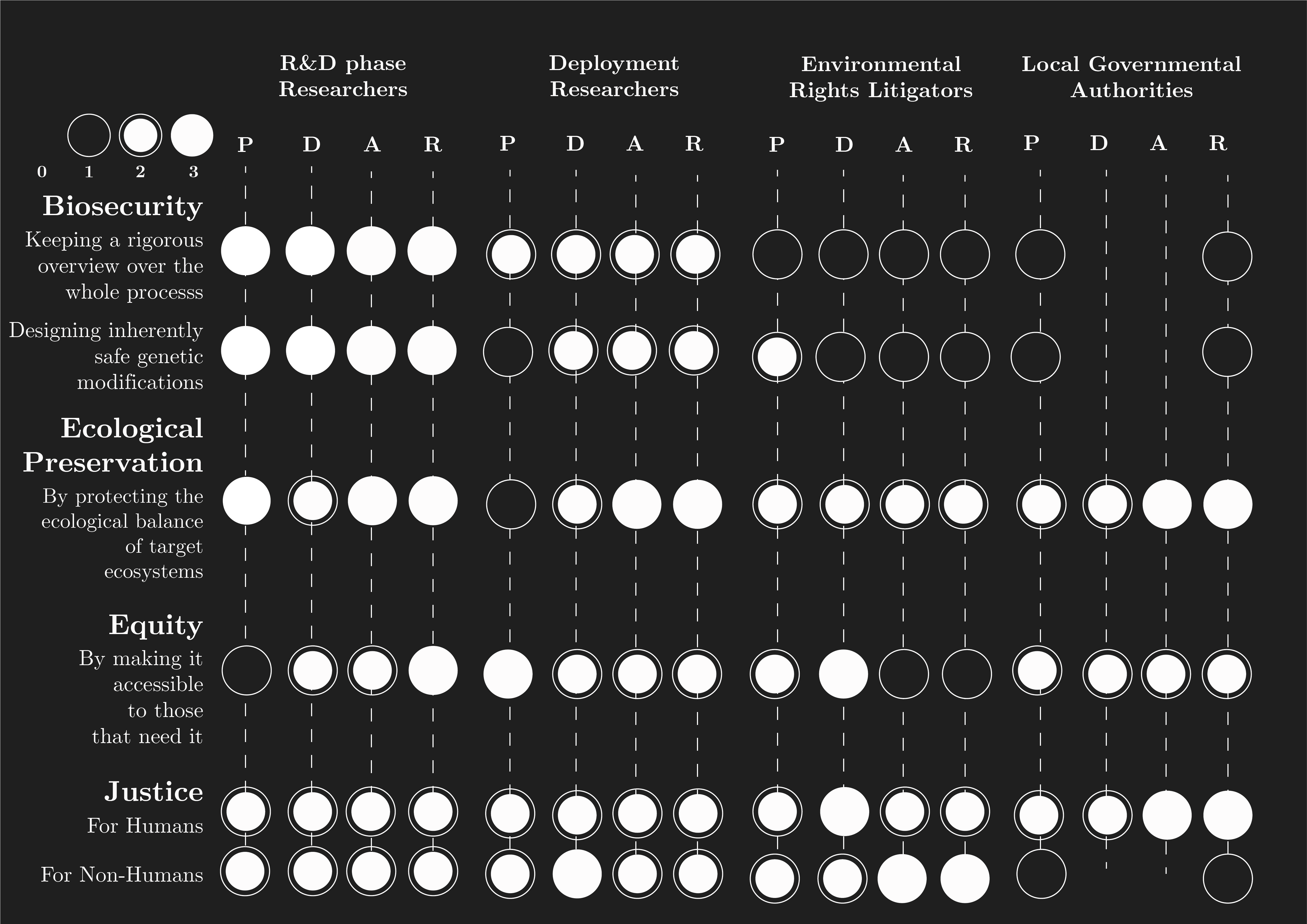
Based on this scoring, perhaps due to personal bias, the core values of this project lie with environmental justice. The protection of the people and organisms affected by PFAS pollution should be prioritised. This includes both within the bounds of this project as well as the context that it resides in. Environmental issues like PFAS pollution are complex and multifaceted, so only focussing on the pure biological aspects would not do it justice.
In general, a combination of ‘R&D Phase Researchers’ strategies and ‘Deployment Researchers’ seems to be the most effective strategy to use. For example: abiding by pre-existing policy standards like ERAs and PMEM mandates, would be effective for goals across the spectrum.
While Local Governmental Authorities might seem like the least effective category for governance actions within the scoring, its importance should not be underestimated. Especially in cases like the one described in the introduction, where the Biesbosch and Alblasserwaard have been heavily polluted by PFAS originating from a local DuPont factory. Local communities have been kept in the dark for years about the realities and severity of the situation, while bearing the consequences to their health. This has led to a general sense of mistrust and a sense of anxiety towards their own ecological environment.
This indicates that the remediation needed should be aimed towards both biological and psychological problems. Engaging in and sustaining a relationship with local communities is not only a matter of ensuring the synbio project can be implemented, but also a contribution to a more holistic form of remediation.
SECTION 3: BACKGROUND
Background and Literature Context
Provide background research that explains the current state of knowledge and identifies the gap in knowledge or capability that your project addresses.
Briefly summarize two peer-reviewed research citations relevant to your research (minimum four sentences).
The first peer-reviewed research citation relevant to my research is “PFAS biodegradation by Labrys portucalensis F11: Evidence of chain shortening and identification of metabolites of PFOS, 6:2 FTS, and 5:3 FTCA’ by Wijahayena et al.
The study evaluated the biodegradation of three PFAS compounds (PFOS, 6:2 FTS, and 5:3 FTCA) by the aerobic bacterium Labrys portucalensis F11 using each compound as the sole carbon source. PFOS degradation produced multiple shorter-chain perfluorinated metabolites and, after extended incubation, several partially defluorinated products. The breakdown of 5:3 FTCA generated similar shorter-chain acids and unsaturated fluorotelomer intermediates, while 6:2 FTS degraded more slowly, showing only a modest reduction over 100 days. Overall, PFOS and 5:3 FTCA showed substantial degradation compared to 6:2 FTS, suggesting that L. portucalensis F11 has potential for bioremediation of certain PFAS-contaminated environments.
The second peer-reviewed research citation relevant to my research is “Genome sequence and silkomics of the spindle ermine moth, Yponomeuta cagnagella, representing the early diverging lineage of the ditrysian Lepidoptera”. This study investigates the silk composition of Yponomeuta cagnagella, an early-diverging lepidopteran species, to address the limited knowledge of silk genes in this group. Researchers generated a draft genome using Oxford Nanopore and Illumina sequencing, complemented by silk-gland transcriptomics and proteomics to identify key silk proteins and confirm their tissue-specific expression. They provide detailed annotations of major silk genes, including full sequences, exon–intron structures, and associated protein products, along with descriptions of silk gland and fiber morphology. The findings expand understanding of silk gene evolution and function while offering valuable genomic resources for future studies on the chemical ecology of Yponomeuta species.
Explain how your project is novel or innovative. (Minimum 3 sentences.)
As described in the prior question, not much is known about the silkomics and genomics of ermine moths and their silk production. Therefore exploring this material as a substrate would potentially give valuable insights into the silk as a novel biomaterial. Moreover, these caterpillar infestations are often seen as pests, but their presence in the ecosystem is not problematic by definition as healthy affected trees can regenerate relatively fast afterwards.
Within the field of biodesign, discourse is often centred around “collaboration with the more-than-human”, while more often than not final outcomes involve neutralising the living organism for applications. This project challenges this paradigm by not framing the organism as a product, but their bodily secretions as a product. In this sense, the ermine moth becomes a cocreator in this process.
Explain why your project matters and what impact it could have. (Minimum 5 sentences.)
The main real-world problem that the project attempts to solve is PFAS pollution, a problem that poses significant threats to human and ecological health. While source material focuses on detection of PFAS pollutants, the project envisions a mechanism that is able to detect and remediate in a single genetic circuit.
The project could improve scientific understanding and technical capability, in the way that it approaches biological systems and investigates natural substances as substrates for bacterial growth. This would allow for remediation strategies to be embedded directly into ecosystems.
If the aims of the project are achieved, it could shift the perspective we have on bioremediation strategies, in line with redirecting towards strategies that do not rely on organisms absorbing toxic chemicals into their body but rather directly metabolising them.
Describe the ethical implications associated with your project and identify relevant ethical principles (e.g., non-maleficence, beneficence, justice, or responsibility). (Minimum 2 paragraphs.)
The ethical implications associated with my project and the relevant ethical principles are most of my development for aim 3 of my project. As such, please find a reiteration of this documentation below:
I first explored the implication of my aim 3 early on in the course, focussing my governance research on this project proposal. The research was developed with “de Alblasserwaard", a region within the province of Zuid-Holland, the Netherlands, in mind. I grew up in this area. In recent years, it has become known that the entire area is heavily contaminated with PFAS originating from industrial emissions by a local DuPont factory.
I envisioned the following policy/governance goals in relation to this project:
Biosecurity
Keeping a rigorous overview over the whole process
Designing inherently safe genetic modifications
Ecological Preservation
By protecting the ecological balance of target ecosystems
Equity
By making it accessible to those that need it
Justice
For humans
For non-humans
As well as the following potential governance actions:
R&D phase researchers:
P - Ensuring the core mechanism of PFAS degradation by genetically engineered bacteria within a fine meshed network-substrate works efficiently and does not leave residual harmful chemicals.
D - Researching to what capacity the principle can be scaled up for real world deployment.
A - Ensuring that no harmful organisms are produced and released into the wild throughout laboratorial development.
R - Ensuring that potentially implemented engineered microorganisms are rigorously tested to meet EU ecological risk assessment regulations (ERAs) for genetically engineered microorganisms.
Deployment researchers:
P - Localising areas affected by airborne PFAS pollution.
D - Finding suitable host sites for the caterpillar-biofilter within affected areas throughout the development process, allowing for efficient application.
A - Assess potential environmental factors that could undermine the efficacy of implementation.
R - Conducting post-market environmental monitoring (PMEM)* as mandated by the EU.
Environmental Rights litigators
P - Use legal tools to safeguard responsible application of the biofilter.
D - Draft funding mandates for known local major polluters to fund local bioremediation pilots like this biofilter.
A - Advocating for the legal recognition of the trees targeted for the application under the Right of Nature, protecting them from overexploitation through caterpillar infestation. building towards legally protecting the entire local ecosystems they reside in.
R - Compose long term Risk and Success assessment based on legal precedents in relation to ‘Right of Nature’ and funding mandate rulings.
Local Governmental Authorities
P - Spreading informative campaigns to local communities that transparently communicates the project and informs people not to destroy the webs
D - Forming a recognised mediative contact point between the executive parties and impacted communities, allowing communities to voice potential concerns.
A - Assist interested members of the public to find ways to get involved, incentivising people to (re)connect with their local ecosystems and giving back a sense of agency in their own wellbeing.
R - Formulate contingency plans to manage misinformation and potential community protest, as well as rapid response protocols to pause activities in case community concerns escalate.
*Post-market environmental monitoring (PMEM) is a mandatory, legally required process in the EU for GMOs and authorized products, designed to detect direct, indirect, or unforeseen adverse effects on human health or the environment after market release. It consists of Case-Specific Monitoring (CSM) to verify assumptions in the Environmental Risk Assessment (ERA) and General Surveillance (GS) to identify unexpected effects.
Based on this scoring, perhaps due to personal bias, the core values of this project lie with environmental justice. The protection of the people and organisms affected by PFAS pollution should be prioritised. This includes both within the bounds of this project as well as the context that it resides in. Environmental issues like PFAS pollution are complex and multifaceted, so only focussing on the pure biological aspects would not do it justice.
In general, a combination of ‘R&D Phase Researchers’ strategies and ‘Deployment Researchers’ seems to be the most effective strategy to use. For example: abiding by pre-existing policy standards like ERAs and PMEM mandates, would be effective for goals across the spectrum.
While Local Governmental Authorities might seem like the least effective category for governance actions within the scoring, its importance should not be underestimated. Especially in cases like the one described in the introduction, where the Biesbosch and Alblasserwaard have been heavily polluted by PFAS originating from a local DuPont factory. Local communities have been kept in the dark for years about the realities and severity of the situation, while bearing the consequences to their health. This has led to a general sense of mistrust and a sense of anxiety towards their own ecological environment.
This indicates that the remediation needed should be aimed towards both biological and psychological problems. Engaging in and sustaining a relationship with local communities is not only a matter of ensuring the synbio project can be implemented, but also a contribution to a more holistic form of remediation.
SECTION 4: EXPERIMENTAL DESIGN, TECHNIQUES, TOOLS, AND TECHNOLOGY
Create a detailed experimental plan for your final project. Include a timeline for each part of your experimental plan (i.e., how long you would expect each step in your final project to take). (min. 15 lines/sentences—a numbered list is acceptable)
Complete Detailed Experimental Protocol: Ermine Moth Silk Degradation by Engineered E. coli*
Objective Demonstrate that E. coli expressing BmCoc (Bombyx mori cocoonase) can degrade sericin from wild ermine moth silk (weight loss), proliferate using sericin as a nutrient source (CFU count), and secrete functional His-tagged enzyme (protein quantification).
Experimental Design
15 wild silk fibers (n=3 per assay/condition):
Condition | Weight Loss Fibers | CFU Fibers | Supernatant Analysis |
BmCoc+ E. coli | Fibers 1-3 | Fibers 4-6 | 1 mL culture |
Uninduced Control | Fibers 7-9 | Fibers 10-12 | 1 mL culture |
No cells baseline | Fibers 13-15 | None | None |
Timeline:
Day 0: Preparation
1. Harvest and Sterilize Wild Silk
"Collect mature ermine moth (Yponomeuta padellus) silk webs or cocoons from infested trees. Carefully dissect ~1 cm silk fibers, removing debris. Place 15 fibers in a sterile 15 mL tube with 10 mL 70% ethanol and rock gently for 15 minutes at room temperature. Decant ethanol and rinse fibers three times with 10 mL sterile PBS (pH 8.0), 5 minutes each rinse. Transfer to a new sterile Eppendorf tube and air dry completely in a biosafety cabinet for 30 minutes."
2. Initial Weight Measurements
"Working in a biosafety cabinet, tare an analytical balance with pre-weighed filter paper and Eppendorf tube. Place one dry silk fiber on the filter paper and record the DRY_START weight (target 9-11 mg per fiber) for fibers 1-15. Store weighed fibers in a sterile tube."
3. Start E. coli Overnight Cultures (15 min)
"Pick a single colony of BmCoc+ E. coli (pET28-BmCoc-His, AmpR) and a single colony of empty vector control into separate 5 mL LB + 50 µg/mL ampicillin tubes. Incubate overnight at 37°C, 200 rpm shaking."
Day 1: Culture Preparation & Silk Inoculation
4. Grow Day Cultures
"The next morning, dilute each overnight culture 1:100 into 50 mL fresh LB + Amp in 250 mL flasks. Grow at 37°C, 200 rpm until OD600 = 0.5 (approximately 3 hours). For BmCoc+ culture only, add 0.5 mM IPTG and induce for 4 additional hours at 30°C (reduces inclusion bodies)."
5. Prepare Silk Incubation Medium
"Prepare sterile minimal medium: PBS pH 8.0 + 0.2% glucose + 0.1% casamino acids + 50 µg/mL ampicillin. This forces cells to use sericin as primary nutrient source during 48h incubation."
6. Inoculate Silk Fibers
"In a 24-well plate, add 2 mL OD600=0.5 culture per well containing one sterile silk fiber (fibers 1-12). For no-cell controls (fibers 13-15), add 2 mL minimal medium only. Save 1 mL culture from each condition in Eppendorf tubes for supernatant analysis. Incubate at 37°C, 200 rpm shaking for 48 hours."
Day 3: Sample Harvesting
7. Weight Loss Measurement (Fibers 1-15)
"Using sterile tweezers, carefully remove each silk fiber from its well. Transfer to a clean Petri dish and wash by dipping three times in 10 mL PBS (5 seconds each dip with gentle swirling). Perform one brief 3-second MilliQ water rinse to remove salts. Blot excess water on Kimwipe for 10 seconds. Transfer to pre-weighed filter paper + Eppendorf tube. Air dry 30 minutes at room temperature in a desiccator, then oven dry at 60°C for 1 hour. Cool 30 minutes in desiccator and record DRY_END weight."
8. CFU Proliferation Assay (Fibers 4, 6, 10, 12)
"Place one CFU fiber in a 1.5 mL Eppendorf with 900 µL PBS + 0.1% Tween20. Vortex vigorously for 2 minutes to detach biofilm cells. Transfer 100 µL to new tube with 900 µL PBS (10⁻¹ dilution). Repeat serial dilutions to 10⁻⁵. Plate 100 µL of 10⁻⁴ and 10⁻⁵ dilutions on LB + Amp agar plates using sterile spreaders. Incubate plates overnight at 37°C."
9. Supernatant Collection for Protein Analysis
"Centrifuge 1 mL saved cultures from each condition at 16,000g for 5 minutes. Transfer supernatant to new Eppendorf tubes and store at 4°C for His-tag purification."
Day 4: Analysis
10. Colony Counting
"Count colonies on plates with 30-300 colonies (ideal range). Calculate CFU per fiber = (colonies counted) × dilution factor × 10."
11. His-Tag Protein Purification
"To 1 mL supernatant, add 20 µL Ni-NTA slurry. Nutate 1 hour at room temperature. Spin 1 minute, discard supernatant. Wash beads three times with 500 µL wash buffer (50 mM phosphate pH 8.0, 300 mM NaCl, 20 mM imidazole). Elute twice with 50 µL elution buffer (same + 300 mM imidazole). Pool eluates for quantification."
12. Protein Quantification
"Bradford assay: Mix 2 µL eluate + 200 µL Bradford reagent. Read OD595 after 10 minutes vs BSA standards. A280 confirmation: Measure 1 µL eluate directly on NanoDrop using BmCoc extinction coefficient."
Day 5: Data Analysis & Imaging
13. Calculate Results
% Weight loss = 100 × (DRY_START - DRY_END) / DRY_START
Expected: BmCoc+ = 15-25%, Uninduced = 1-3%, No cells = <1%
14. Optional Imaging
"Image representative fibers pre/post treatment using stereo microscope. Stain one spare fiber with 0.1% Crystal Violet for total biomass visualization."
Transform into e.coli chassis. Experiments involve exposure to wild Ermine caterpillar moth silk. Silk samples are inoculated with both transformed and uninduced control, accompanied by non-cell baseline. Samples are then analysed in 3 ways:
Weight loss measurement of silk
CFU proliferation assay
Protein quantification through Bradford assay
We discussed and practiced various techniques related to synthetic biology throughout the semester. Place a check next to the techniques relevant to your project.
Pipetting
DNA Gel Art
Lab Automation
Protein Design
| Bioproduction
Cell-Free Systems
Gibson Assembly
CRISPR
|


Expand upon two techniques you checked in the previous question by describing how you would utilize those techniques in your final project. (min. 4 sentences)
In my final project I used Benchling to prepare my Twist order of the sequence for BmCoc (cocoonase). It allowed me to add a His-tag to the sequence and codon optimise it for my chosen chassis e.coli K12. I retrieved the amino acid sequence for BmCoc from the NCBI database, and reverse translated it to get the DNA sequence for it.
In my experiment design, pipetting is a key methodology in the lab protocol. Ultimately, the genetic circuit is a mechanism that has PFAS degrading enzymes as a bioproduction output.
SECTION 5: Results & Quantitative Expectations
You are required to validate at least one aspect of your final project aims. This is to ensure that you are able to successfully apply a relevant synthetic biology technique to your project. Include figures if you have them—accuracy is critical in figures, tables, and graphs
Here is a non-exhaustive list of acceptable validations:
Designing DNA relevant to your final project
Performing a PCR reaction using primers relevant to your final project
Performing a Gibson assembly relevant to your final project
Creating and performing a cell-free assay related to your final project
Creating and running code to validate an aspect of your final project
Developing a model or completing a computational analysis relevant to your project
Designing DNA construct(s) that can express at least one gene of interest, ordering it (via Twist), and testing of the expression of the construct(s) (potentially using an Opentrons robot)
What aspect of your final project did you choose to validate? (min. 2 sentences)
I chose to validate the DNA design I made for a hlFABP/split-TetR sensor for the presence of PFAS particles. This aspect builds on two main papers found in my research. Firstly, a paper detailing a biosensor for PFAS, through a hlFABP/GFP fusion protein. Secondly, a paper describing a split transcriptional repressor that links protein solubility to an orthogonal genetic circuit.
The second paper lists a set of three fissured TetR fragments after residue 180, 184 and 193. I combined the hlFABP sequence with the sequence for each of these fragments and validated them in AlphaFold.
Write down a detailed protocol of how you validated this aspect of your final project. (Numbered list or paragraph is fine)
Retrieve TetR AA sequence from uniprot
Reverse translate the TetR AA sequence
Retrieve hlFABP sequence from uniprot
Reverse translate the TetR AA sequence
Upload both in Benchling
Create new Benchling files for each fragment
Create new Benchling files for the fused sequences, make sure to remove the start codons from the TetR sequences.
Retrieve the AA sequence for hlFABP and all three of the fused proteins.
Upload all to AlphaFold
Document PTM scores and position the hlFABP component similarly to one another for comparison.
Comparatively investigate the structural integrity
Draw conclusions on the potential further development of the sensor.
What synthetic biology techniques did you utilize in validating this aspect of your final project? You can refer to the list of techniques in question 8. (min. 4 sentences)
I consulted the online database UniProt for the sequences. I edited DNA. I designed protein. I used AlphaFold to model the proteins.
You must present data as part of your final project and include some analysis of that data. The data may be collected experimentally in the lab or generated as simulated data (e.g., using the Asimov Kernel or another simulation method). (min. 2 sentences)
The data I presented is the predicted form of the biosensor proteins simulated in AlphaFold. The data was then analyzed visually and by comparison of PTM scores.
Did you encounter any unexpected challenge(s) when performing your validation? If so, describe the challenge(s) and strategies to overcome it. If not, discuss potential problems, difficulties, limitations, and/or alternative strategies to overcome challenges in your final project. (min. 4 sentences).
It was a challenge to analyse and compare the protein structures of the hlFABP protein. This problem arose due to the fact that properly situating the models to compare was rather difficult. I attempted to solve this problem by placing them in a similar position to one another, taking an image and comparing them in Illustrator. While this probably was not the most accurate, I did find it helpful to better understand the protein structure.
SECTION 6: ADDITIONAL INFORMATION
12. List all references cited in this assignment (bullet-point list)
Kafatos F, Tartakoff A, Law J Cocoonase Journal of Biological Chemistry, 1967; 242, 1477-1487
Preeti Anand, Jay Prakash Pandey, Dev Mani Pandey, Study on cocoonase, sericin, and degumming of silk cocoon: computational and experimental, Journal of Genetic Engineering and Biotechnology, Volume 19, Issue 1, 2021, 32, ISSN 1687-157X, https://doi.org/10.1186/s43141-021-00125-2.
Volenikova, A., Nguyen, P., Davey, P. et al. Genome sequence and silkomics of the spindle ermine moth, Yponomeuta cagnagella, representing the early diverging lineage of the ditrysian Lepidoptera. Commun Biol 5, 1281 (2022). https://doi.org/10.1038/s42003-022-04240-9
J. Garcia-Ojalvo, M.B. Elowitz, & S.H. Strogatz, Modeling a synthetic multicellular clock: Repressilators coupled by quorum sensing, Proc. Natl. Acad. Sci. U.S.A. 101 (30) 10955-10960, https://doi.org/10.1073/pnas.0307095101 (2004).
Mann, M.M., Berger, B.W. A genetically-encoded biosensor for direct detection of perfluorooctanoic acid. Sci Rep 13, 15186 (2023). https://doi.org/10.1038/s41598-023-41953-1
Bois, J. BE 150: Design Principles of Genetic Circuits Caltech (2018)
Adamala, K. P., Martin-Alarcon, D. A., Guthrie-Honea, K. R., & Boyden, E. S. (2017). Engineering genetic circuit interactions within and between synthetic minimal cells. Nature chemistry, 9(5), 431–439. https://doi.org/10.1038/nchem.2644
Zeng Y, Jones AM, Thomas EE, Nassif B, Silberg JJ, Segatori L. A Split Transcriptional Repressor That Links Protein Solubility to an Orthogonal Genetic Circuit. ACS Synth Biol. 2018 Sep 21;7(9):2126-2138. doi: 10.1021/acssynbio.8b00129. Epub 2018 Aug 23. PMID: 30089365; PMCID: PMC6858789.
Beal, J., Nguyen, T., Gorochowski, T. E., Goñi-Moreno, A., Scott-Brown, J., McLaughlin, J. A., Madsen, C., Aleritsch, B., Bartley, B., Bhakta, S., Bissell, M., Castillo Hair, S., Clancy, K., Luna, A., Le Novère, N., Palchick, Z., Pocock, M., Sauro, H., Sexton, J. T., Tabor, J. J., … Wipat, A. (2019). Communicating Structure and Function in Synthetic Biology Diagrams. ACS synthetic biology, 8(8), 1818–1825. https://doi.org/10.1021/acssynbio.9b00139
Han, J., Fu, J., Sun, J., Hall, D. R., Yang, D., Blatz, D., Houck, K., Ng, C., Doering, J., LaLone, C., & Peng, H. (2021). Quantitative Chemical Proteomics Reveals Interspecies Variations on Binding Schemes of L-FABP with Perfluorooctanesulfonate. Environmental science & technology, 55(13), 9012–9023. https://doi.org/10.1021/acs.est.1c00509
Addgene (n.d.) Gibson assembly protocol. Available at: https://www.addgene.org/protocols/gibson-assembly/ (Accessed: 27 May 2026).
Bornhorst, J.A. and Falke, J.J. (2000) ‘Purification of proteins using polyhistidine affinity tags’, Methods in Enzymology, 326, pp. 245–254.
Bradford, M.M. (1976) ‘A rapid and sensitive method for the quantitation of microgram quantities of protein utilizing the principle of protein-dye binding’, Analytical Biochemistry, 72(1–2), pp. 248–254.
European Commission (n.d.) Environmental risk assessment of genetically modified organisms. Available at: https://food.ec.europa.eu/plants/genetically-modified-organisms/environmental-risk-assessment_en (Accessed: 27 May 2026).
European Commission (n.d.) Post-market environmental monitoring of GMOs. Available at: https://food.ec.europa.eu/plants/genetically-modified-organisms/post-market-environmental-monitoring_en (Accessed: 27 May 2026).
Gibson, D.G., Young, L., Chuang, R.-Y., Venter, J.C., Hutchison, C.A. III and Smith, H.O. (2009) ‘Enzymatic assembly of DNA molecules up to several hundred kilobases’, Nature Methods, 6(5), pp. 343–345.
PubMed (2015) Purification of His-tagged proteins. Available at: https://pubmed.ncbi.nlm.nih.gov/26096499/ (Accessed: 27 May 2026).
13. Create a supply list and budget for your project (bullet-point list)
Focussing on my aim 1 for this project, the supply list would be as follows:
Raw silk: £0 (wild-sourced)
LB powder: £5 (100g makes 5L)
Agar powder: £3 (100g makes 50 plates)
Bradford dye: £7 (Coomassie R-250, 1g)
Ampicillin: £2 (100 mg stock)
IPTG: £1 (10 mg stock)
Eppendorfs: £2 (50 tubes)
24-well plates: £2 (2 plates)
Glucose/PBS: £0 (lab staples)