Week 9 HW: Cell Free Systems
Cell-free protein synthesis essentially uses biology as an engineering tool without needing living cells. Traditional in vivo systems require cells to stay alive, meaning you constantly need to maintain the correct conditions such as nutrients, water, gases, temperature, pressure, and energy supply. In contrast, cell-free systems remove many of these constraints, giving much greater flexibility and control over experimental variables. Since there are no living cells, researchers can directly tune reaction conditions, add or remove components easily, and rapidly test biological circuits or protein designs without worrying about cell survival or toxicity.
Another major advantage is portability and stability. Cell-free systems can be freeze-dried and stored for long periods, sometimes up to a year, then simply activated again by adding water. This makes them extremely useful for therapeutics on demand, rapid manufacturing, and applications where maintaining living cells would be difficult. They also have improved biosafety because there is less risk of engineered organisms escaping into the environment.
Cell-free expression is especially beneficial in environments such as space, where sustaining living cell cultures is difficult and resources are limited. It is also useful in developing regions or disaster zones where supply chains and laboratory infrastructure may not be reliable. Other important applications include rapid protein engineering, biosensors, metabolic engineering, and testing CRISPR or synthetic biology systems in a highly controlled environment.
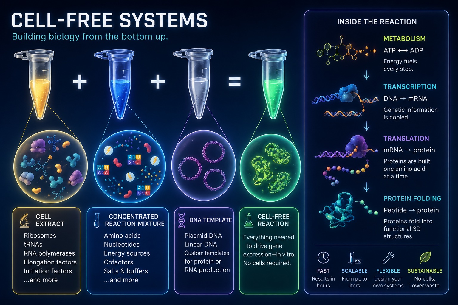
A cell-free expression system contains all the molecular machinery needed for transcription and translation without requiring living cells. One of the main components is the whole cell extract, which contains ribosomes, tRNAs, aminoacyl-tRNA synthetases, translation factors, and often RNA polymerase. Together, these provide the machinery required to transcribe mRNA and translate it into protein.
Another key component is the DNA template, usually in the form of a plasmid or linear PCR product. This acts as the blueprint for the desired protein because it contains the coding sequence as well as a promoter, such as a T7 promoter, which allows RNA polymerase to initiate transcription. Amino acids and nucleotides (NTPs) are also required because they serve as the building blocks for proteins and mRNA respectively.
Since there is no living metabolism present, the system also requires an external energy source such as ATP, GTP, phosphoenolpyruvate, and pyruvate kinase to power protein synthesis and regenerate ATP. In addition, salts and buffers are needed to maintain the correct chemical environment. For example, magnesium stabilises ribosomes and supports polymerase activity, potassium helps maintain ionic strength for enzyme activity and protein folding, and buffers such as HEPES maintain a stable pH. Finally, chaperones and protease inhibitors are often included to help proteins fold correctly and prevent them from being degraded during synthesis.
Energy provision and regeneration are critical in cell-free systems because there is no living cell metabolism to continuously produce ATP. Unlike living cells, cell-free systems do not contain mitochondria or other metabolic pathways that naturally regenerate energy, yet processes such as transcription and translation require large amounts of ATP and GTP. Without a continuous energy supply, protein synthesis would quickly stop.
One common method for maintaining ATP levels is using phosphoenolpyruvate (PEP) together with the enzyme pyruvate kinase (PK). PEP acts as a high-energy phosphate donor, while pyruvate kinase catalyses the transfer of a phosphate group from PEP onto ADP, regenerating ATP. As the ribosomes and other molecular machinery consume ATP during protein synthesis, ADP accumulates in the reaction mixture. Pyruvate kinase then converts this ADP back into ATP using the energy stored in PEP, allowing the system to continue functioning until the PEP supply is depleted.
Prokaryotic and eukaryotic cell-free expression systems each have different advantages depending on the type of protein being produced. Prokaryotic systems, most commonly based on E. coli extracts, are typically faster, lower cost, and capable of producing very high protein yields. They are therefore ideal for simple proteins and high-throughput screening applications. In contrast, eukaryotic systems such as rabbit reticulocyte, wheat germ, HeLa, or CHO cell extracts are slower, more expensive, and generally produce lower yields, but they are much better at handling complex protein folding and post-translational modifications such as glycosylation.
A good example of a protein suited for a prokaryotic cell-free system is GFP. GFP is a relatively robust and simple protein that does not require major post-translational modifications in order to function, making it ideal for rapid and inexpensive production in E. coli-based systems.
In contrast, a protein such as human erythropoietin (EPO) is much better suited to a eukaryotic cell-free system. Although EPO is not extremely large, it is a glycoprotein hormone that requires glycosylation to become biologically active and stable in the human body. Around 40% of its mass consists of carbohydrate chains. Standard prokaryotic systems cannot naturally perform these modifications, meaning the resulting protein would be non-functional in a medical context. Eukaryotic systems contain the necessary enzymes and endoplasmic reticulum-derived vesicles required for glycosylation and proper folding, allowing complex proteins like EPO to be produced correctly.
To design a cell-free experiment for optimizing membrane protein expression, the main challenge is dealing with the hydrophobic parts of the protein. In a normal cell, these transmembrane regions are stabilised by the phospholipid bilayer, but in a cell-free extract there is no natural membrane environment. This means the protein can easily misfold, aggregate, or become insoluble.
To address this, I would add synthetic membrane-like systems directly into the cell-free reaction. For example, liposomes could be used to provide a membrane compartment for the protein to insert into, while nanodiscs could help keep the membrane protein soluble and properly stabilised. I would then test different concentrations and types of liposomes or nanodiscs to see which gives the highest yield of correctly folded protein.
I would also add molecular chaperones to help newly synthesised proteins fold into their correct 3D structure and reduce aggregation. Finally, I would optimize variables such as temperature, magnesium concentration, reaction time, and DNA template concentration, then check expression and folding using a fluorescence tag, Western blot, or activity assay. Overall, the goal would be to recreate enough of a membrane-like environment that the protein can fold and function properly outside of a living cell.
A low yield in a cell-free system could happen for several reasons. One common issue is energy depletion. Protein synthesis uses a lot of ATP and GTP, and once these energy sources are used up, translation slows down or stops. Some energy systems also create inhibitory byproducts such as inorganic phosphate, which can disrupt the reaction. To troubleshoot this, I would switch to a cleaner energy regeneration system such as glucose or pyruvate, or use dialysis so fresh substrates can diffuse in while inhibitory byproducts diffuse out.
Another possible reason is template instability or poor template quality. If the DNA or mRNA template is degraded by nucleases in the extract, the ribosomes will not have enough time to produce the target protein. To fix this, I would use a circular plasmid instead of a linear PCR product, since plasmids are generally more resistant to nuclease degradation. I could also add RNase inhibitors or protect linear DNA using GamS protein or phosphorothioate-modified primers.
A third issue could be protein folding or solubility. The protein may be synthesised but then misfold, aggregate, or become insoluble, especially if it has hydrophobic regions or needs disulfide bonds. To troubleshoot this, I would lower the reaction temperature to slow down translation and give the protein more time to fold properly. I would also add chaperones such as DnaK/J or GroEL/ES, include mild detergents if needed, and adjust the redox environment with GSH/GSSG if the protein requires disulfide bond formation. Finally, I would check basic reaction conditions such as magnesium, potassium, pH, and codon usage, since poor tuning of these variables can also reduce yield.
I would design a magnetically guided synthetic minimal cell that can sense a disease-like environment and produce a signal or therapeutic output. The input could be a small molecule associated with cancer or inflammation, such as high lactate. The output would first be something easy to measure, like sfGFP fluorescence, but later this could be replaced with a therapeutic protein.
Partly yes, but encapsulation makes it more useful because the membrane gives the system a cell-like boundary. It protects the reaction, allows communication with the environment, and makes the system behave more like a programmable artificial cell rather than just a test-tube reaction.
Yes, but a synthetic minimal cell is safer and more controllable because it is not alive and cannot replicate. This is useful for therapeutic or environmental applications where you do not want engineered cells spreading.
The desired outcome is that the synthetic cell only produces a fluorescent or therapeutic output when it detects the correct disease-associated signal. Ideally, it could also be guided or concentrated using magnetic particles.
The membrane could be made from a simple lipid vesicle using POPC and cholesterol, with a small amount of DOTAP to tune membrane charge and stability.
Inside, I would encapsulate an E. coli cell-free Tx/Tl system, DNA templates, ribosomes, tRNAs, amino acids, NTPs, ATP/GTP, an energy regeneration system, salts, buffer, and magnetic nanoparticles.
A bacterial E. coli Tx/Tl system would be fine for the first version because it is fast, cheap, and high-yield. Since the output is sfGFP or a simple protein, we do not need a mammalian system unless the protein requires complex folding, glycosylation, or mammalian promoters like Tet-ON.
Small molecules could enter through a membrane pore. A good example is α-hemolysin, encoded by the hla gene, which forms pores that allow small molecules to pass into the vesicle and activate the internal expression system.
The lipids would be POPC, cholesterol, and possibly DOTAP. The genes would include sfGFP as the reporter gene, a sensor-controlled promoter for the disease-associated input, and hla if I wanted the vesicle to express or contain α-hemolysin membrane pores.
I would measure sfGFP fluorescence over time using a plate reader or fluorescence microscope. I would compare vesicles with and without the input signal, and also compare vesicles with and without α-hemolysin pores. If the system works, only vesicles exposed to the correct input should become fluorescent.
I would design a soft robotic skin embedded with freeze-dried cell-free systems that allows robots to chemically sense and respond to their environment like a form of synthetic biological touch.
The idea would involve integrating freeze-dried cell-free biosensors directly into the flexible outer layer of a soft robot. When exposed to moisture or environmental chemicals, the embedded cell-free systems would activate and detect specific signals such as toxins, pH changes, bacterial contamination, or stress-related molecules. Depending on the detected input, the system could generate fluorescent outputs, trigger enzymatic reactions, or even alter the physical properties of the robotic material itself, such as stiffness, adhesion, or permeability. For example, a search-and-rescue robot could detect dangerous gas leaks or bacterial contamination in environments where traditional electronic sensors struggle. I think the exciting part is that instead of just giving robots electronic sensors, you are essentially giving them a programmable biochemical layer inspired by living tissue.
This could help address the need for safer and more adaptable robots in hazardous environments such as disaster zones, chemical spills, industrial sites, or healthcare settings. Traditional sensors are often rigid, power-intensive, and limited in the types of molecules they can detect. A biologically integrated robotic skin could allow robots to sense subtle chemical changes in real time while remaining lightweight and flexible. It could also reduce reliance on expensive sensor hardware and open up new possibilities for soft robotics and human-robot interaction.
The cell-free systems could be freeze-dried into hydrogel compartments or microcapsules embedded throughout the robotic skin, allowing them to remain stable until activated by water or environmental moisture. To address one-time use limitations, the robotic skin could contain replaceable sensing patches or layered compartments that activate sequentially over time. Stability could be improved using protective polymer coatings, antioxidants, and UV-resistant materials to protect the biological components during long-term operation.
Freeze-dried cell-free reactions have great potential in space, where resources are constrained. As described in my talk, the Genes in Space competition challenges students to consider how biotechnology, including cell-free reactions, can be used to solve biological problems encountered in space. While the competition is limited to only high school students, your assignment will be to develop your own mock Genes in Space proposal to practice thinking about biotech applications in space!
For this particular assignment, your proposal is required to incorporate the BioBits® cell-free protein expression system, but you may also use the other tools in the Genes in Space toolkit (the miniPCR® thermal cycler and the P51 Molecular Fluorescence Viewer). For more inspiration, check out https://www.genesinspace.org/ .
Spaceflight exposes astronauts to microgravity and radiation, both of which can stress human cells and disrupt normal biological function. One major concern is DNA damage, since long-duration missions to the Moon or Mars will involve greater radiation exposure than life on Earth. Understanding how cells respond to DNA damage in space is important for astronaut health, cancer risk, and future space medicine. It is also scientifically interesting because space acts like an extreme biological environment, revealing how fundamental repair pathways behave when normal gravity and environmental conditions are removed.
The DNA damage response protein p53, encoded by the TP53 gene, using a p53-responsive fluorescent reporter in the BioBits® cell-free system.
p53 is a key regulator of the cellular response to DNA damage. When DNA damage occurs, p53 helps activate repair pathways, cell-cycle arrest, or apoptosis depending on the level of stress. Since radiation in space can damage DNA, studying p53-related activity provides a useful way to model how human cells might respond to spaceflight conditions. In a BioBits® cell-free system, a p53-responsive fluorescent reporter could provide a simplified, safe way to measure whether DNA-damage signalling is being activated without needing to culture living human cells in space.
My research goal is to test whether a BioBits® cell-free system can be used as a simple biosensor for space-like DNA damage stress. I hypothesize that DNA templates exposed to radiation or simulated damage will produce a stronger fluorescent output from a p53-responsive reporter compared with undamaged controls. The reasoning is that p53 is one of the most important proteins involved in sensing and responding to DNA damage in human cells. If this pathway can be modelled in a freeze-dried cell-free reaction, it could become a portable tool for monitoring biological stress during space missions. This would be useful because cell-free systems are lightweight, stable, and do not require living cells, making them well-suited for constrained environments like spacecraft.
I would test BioBits® reactions containing a p53-responsive fluorescent reporter. Samples would include an undamaged DNA template control, a radiation-exposed DNA template, and a positive control designed to strongly activate fluorescence. After adding water to activate the freeze-dried reactions, samples would be incubated using the miniPCR® thermal cycler if temperature control is needed. Fluorescence would be measured using the P51 Molecular Fluorescence Viewer. The main data collected would be fluorescence intensity over time, comparing damaged versus undamaged samples to determine whether the system can detect DNA-damage-related signalling.