Week 9 Lab: Protein Purification
This lab introduced the fundamentals of protein extraction and purification workflows commonly used in synthetic biology and bioengineering. It was particularly valuable for my final project, since our system required growing and extracting GFP protein before conjugating it to magnetic microparticles. It was also useful to understand how magnetic separation and purification methods can be integrated into biological systems, as my project similarly uses magnets both to purify ligand-conjugated particles and to actively control their interactions with cells. To isolate our protein of interest, we first grew the cells and then lysed them using a combination of B-PER (Bacterial Protein Extraction Reagent) and sonication, producing a lysate solution containing the total protein content of the cells.
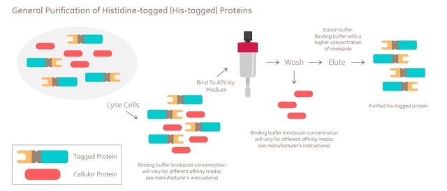
To purify the fluorescent proteins from the lysate, we explored two different purification strategies: magnetic bead-based purification and Ni-NTA spin column purification. Both methods selectively isolate His-tagged proteins from the complex lysate mixture, but they rely on different physical mechanisms for separation and recovery.
The first method used functionalized magnetic beads, where magnets were used to immobilize bead-bound proteins during washing and elution steps. The second method used Ni-NTA spin columns, where centrifugal force was instead used to move buffers through a nickel resin that selectively binds His-tagged proteins. Comparing both approaches was particularly valuable for my final project, since it highlighted how magnetic particle-based systems can integrate purification, localization, and external control into a single experimental framework.
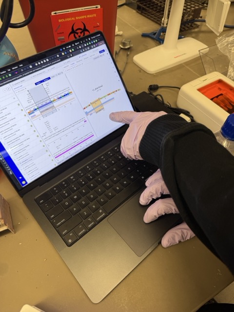
This image shows us inspecting the plasmid construct in Benchling, including the location of the His₆-tag (histidine tag) attached to the fluorescent protein coding sequence. The His-tag is important because it acts as a molecular handle that enables protein purification. During purification, the string of histidine amino acids strongly binds to nickel ions on Ni-NTA resin or functionalized magnetic beads, allowing the target protein to be selectively isolated from the rest of the cell lysate through washing and elution steps.
Procedure:
1. Magnetic beads were added to the lysate solution, allowing the tagged proteins to bind to the bead surface.
2. A 500 μL sample of the mWatermelon lysate mixed with magnetic beads was placed onto a magnetic rack, causing the bead-bound proteins to collect tightly against the magnet.
3. The remaining supernatant, containing excess buffer and unbound proteins, was carefully removed by pipetting.
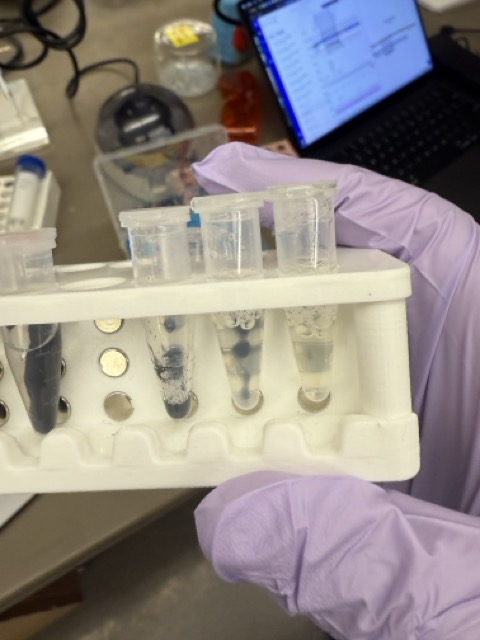
4. The beads were washed with 500 μL of wash buffer containing a low concentration of imidazole (20 mM) to remove non-specific and weakly bound proteins. The sample was mixed and returned to the magnetic rack before the wash solution was removed.
5. To release the purified protein, 200 μL of elution buffer containing a high concentration of imidazole (500 mM) was added to the beads.
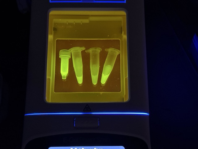
6. Once the beads recollected against the magnet, the fluorescent protein-containing liquid was removed and collected as Solution 4.
7. A second elution step was performed with an additional 200 μL of elution buffer to recover remaining fluorescent protein, producing Solution 5.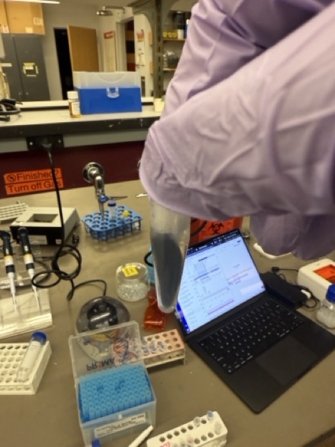
Procedure:
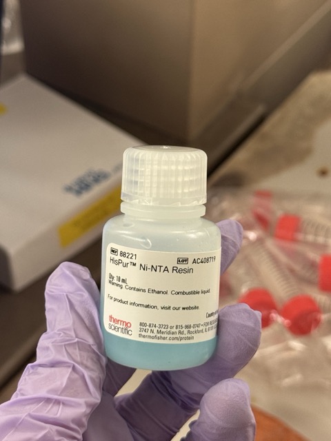
1. We combined 200 μL of Ni-NTA bead solution with 2 mL of cell lysate and incubated the mixture for approximately 30 minutes, allowing the His-tagged fluorescent proteins to bind to the nickel resin.
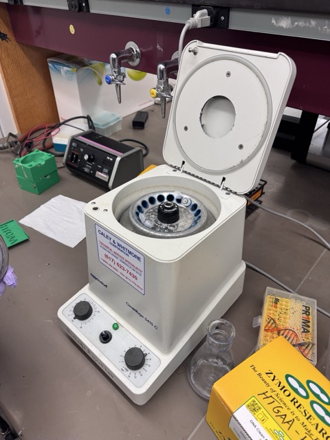
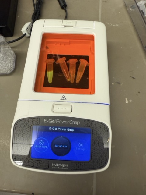
2. The mixture was transferred into a spin column and centrifuged at 8,000 RPM for 1 minute, producing a flow-through fraction that was collected for observation.
3. The resin was then washed with 500 μL of wash buffer and centrifuged again at 8,000 RPM for 1 minute to remove unbound and non-specific proteins.
4. To recover the purified protein, 200 μL of elution buffer was added to the column followed by a final centrifugation step at 8,000 RPM for 1 minute.
5. The final eluted fraction was analyzed to confirm the successful purification of the fluorescent protein.