Week 10 Lab: Mass Spectrometry
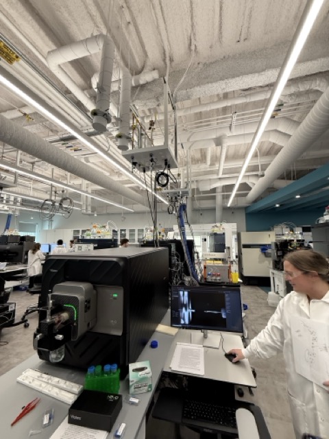
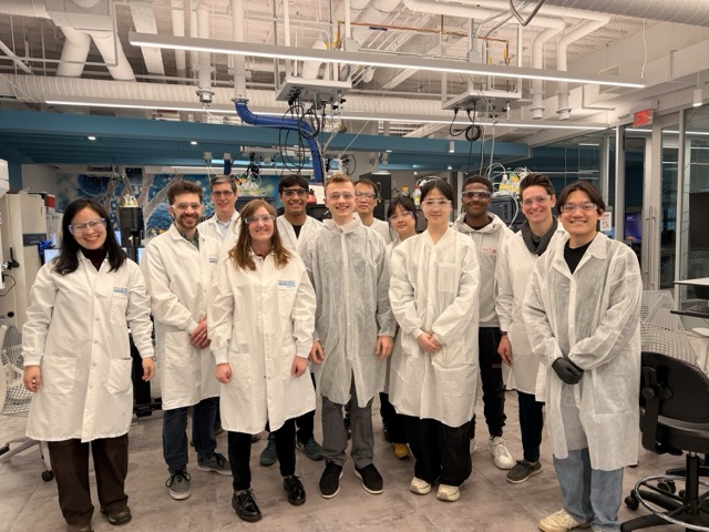
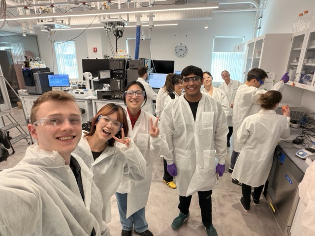
This week we did a lab at Waters Corp on Liquid Chromatography Mass Spectrometry, one of the core technologies used for modern protein characterization. Their lab was so cool!!
Using enhanced Green Fluorescent Protein (eGFP) as the model system, the lab showed how proteins can be analyzed at multiple levels ranging from overall molecular weight and folding state to their exact amino acid sequence. I found it especially interesting because the workflow progressively “breaks down” the protein from an intact structure into smaller peptide fragments, revealing different layers of biological information at each stage. We also briefly explored Charge Detection Mass Spectrometry (CDMS), which can analyze extremely large biological complexes that are too massive for conventional mass spectrometry techniques.
The lab was split into four rotating stations, each focused on a different stage of protein analysis:
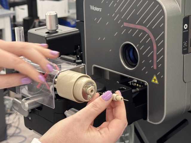
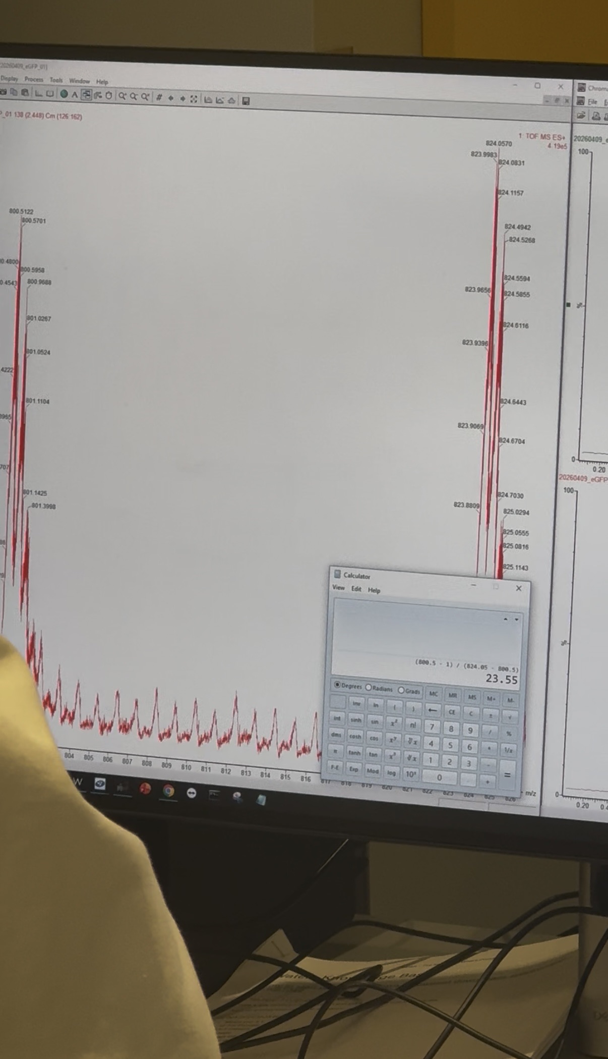
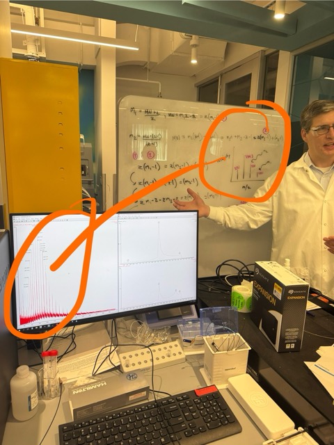
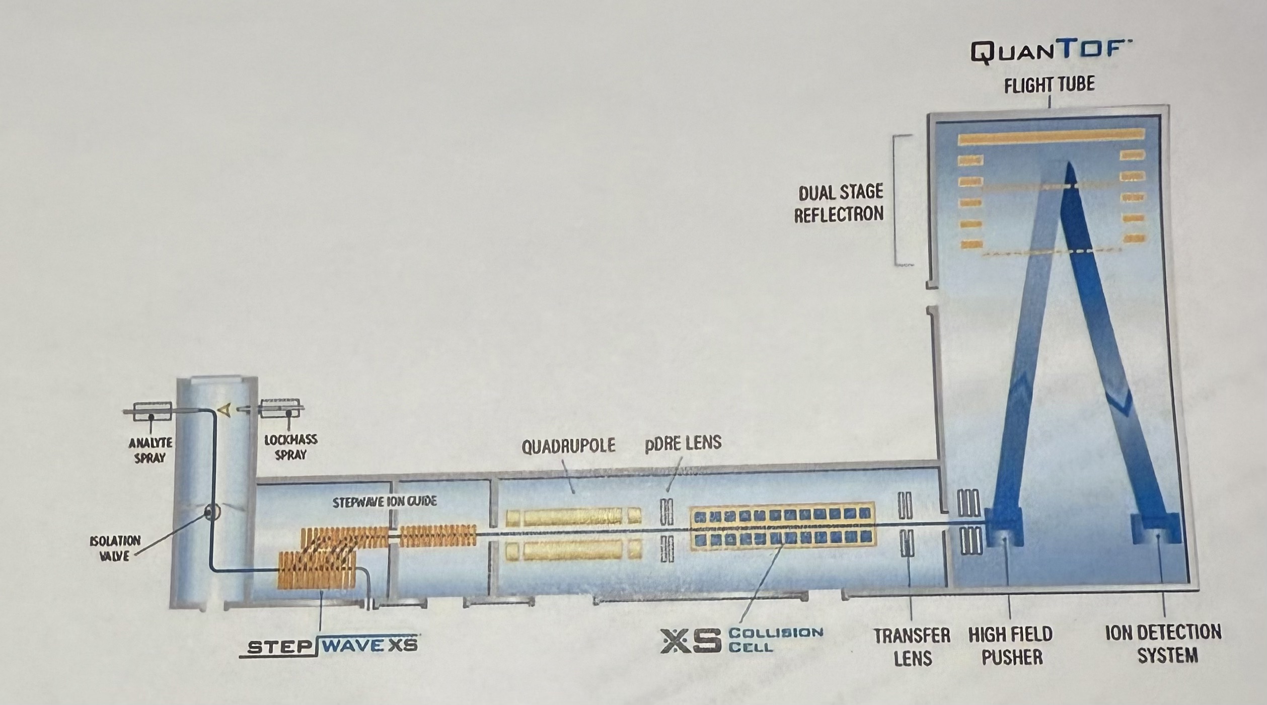
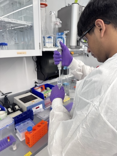
In the first station, we used the Waters Xevo G3 QTof LC–MS system to analyze intact enhanced Green Fluorescent Protein (eGFP). The protein was first buffer-exchanged into ammonium acetate using spin columns before being run under denaturing LC conditions, allowing the mass spectrometer to determine its molecular weight from its mass-to-charge ratio (m/z) and charge states. I thought it was fascinating that proteins can essentially be “weighed” with such extreme precision using electrospray ionization and time-of-flight measurements.
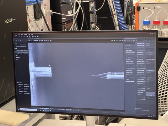
The second part of the station explored how protein folding changes mass spectrometry behaviour. Instead of using chromatography, we directly infused eGFP into the Xevo G3 QTof using a syringe pump so the protein could remain in its native folded state. We then compared this against a denatured version created using formic acid. Folded proteins generate lower charge states because they are compact, while unfolded proteins expose more surface area and produce broader, higher charge state distributions. I found this especially interesting because it showed how mass spectrometry can probe protein structure and conformation, not just molecular weight.
In the second station, we moved into bottom-up proteomics using the Waters BioAccord LC–MS system. The eGFP protein was denatured, reduced, and digested with trypsin, which cuts proteins at lysine and arginine residues to generate smaller peptide fragments. These peptides were then fragmented further inside the mass spectrometer, allowing sections of the amino acid sequence to be reconstructed through peptide mapping. This was probably the most hands-on station and felt almost like molecular reverse engineering, rebuilding the protein sequence from fragmented spectral data.
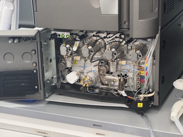
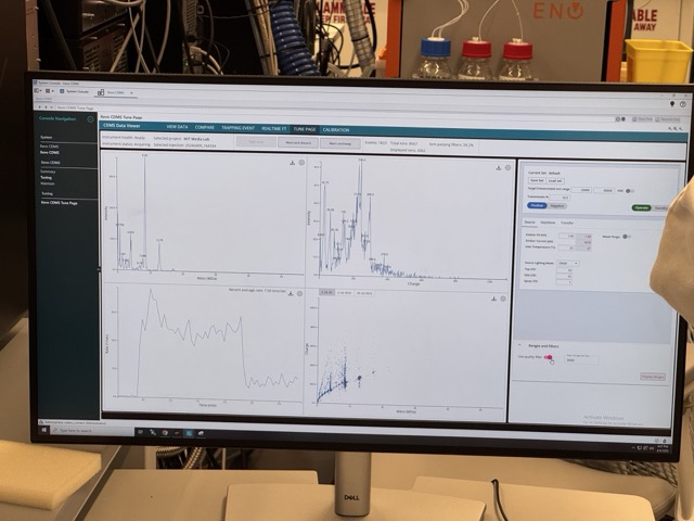
The final station introduced Charge Detection Mass Spectrometry (CDMS) using the Waters Xevo CDMS system. Unlike conventional mass spectrometry, CDMS can directly measure both the charge and mass-to-charge ratio of individual ions, making it possible to analyze enormous biological assemblies that are too large for standard MS techniques. We used it to analyze Keyhole Limpet Hemocyanin (KLH), a huge multi-megadalton protein complex that exists in different oligomeric states. I thought this was one of the coolest stations because it showed how the same underlying physics can scale from relatively small proteins like GFP all the way up to molecular structures approaching the complexity of biological machines.