Week 11 HW: Bioproduction & Cloud Labs
Part A: The 1,536 Pixel Artwork Canvas | Collective Artwork
1) Contribute at least one pixel to this global artwork experiment before the editing ends on Sunday 4/19 at 11:59 PM EST. 2) Make a note on your HTGAA webpages including: what you contributed to the community bioart project (e.g., “I made part of the DNA on the bottom right plate”); what you liked about the project; and what about this collaborative art experiment could be made better for next year.
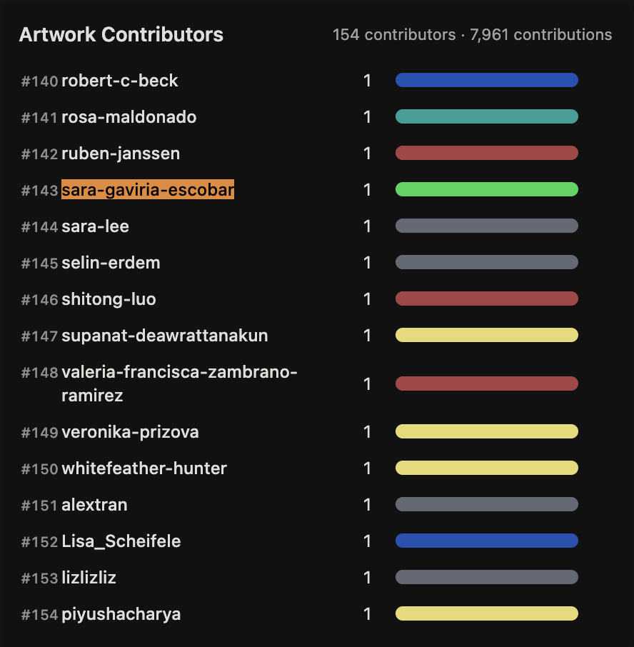
For the first Global Pixel Art, I contributed one green pixel (with GFP) in a corner of the artwork:
And for the second Global Pixel Art, I contributed four blue wells on the plate located at
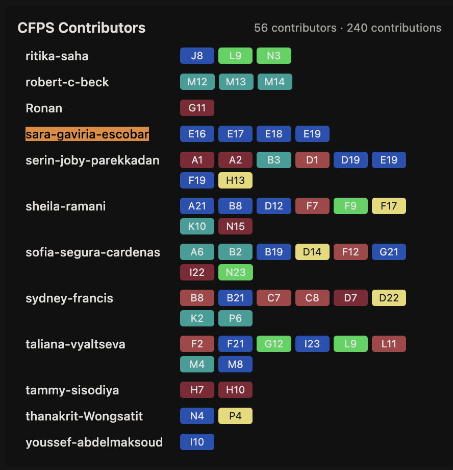
Since this contribution required a hypothesis with the different provided chemical concentrations and DNA template concentrations for every well, my hypothesis would be that increasing HEPES‑KOH from 45 mM to 67.5 mM will increase fluorescence by better maintaining neutral pH throughout the reaction, which is important for folding fluorescent proteins slower like the ones used in the artwork.
Here are the final values I adjusted for every well:
| Well | Condition | HEPES concentration | DNA Template |
|---|---|---|---|
| Well Q4 - E16 | Baseline | 45 mM (standard) | 50 nM |
| Well Q4 - E17 | Baseline replicate | 45 mM | 50 nM |
| Well Q4 - E18 | High buffer | ~67.5 mM (1.5x) | 50 nM |
| Well Q4 - E19 | High buffer replicate | ~67.5 mM (1.5x) | 50 nM |
What I liked about this activity:
I really enjoyed the collaborative aspect and the idea that everyone in the class could contribute to a single shared artwork, even if just by changing one pixel (like I did). I also appreciated that you could only edit one pixel at a time between set time periods. I liked that the process was gradual. What could be better for next year:
What could be improved:
The Part 2 instructions about concentrations and wells were a bit confusing, I wasn’t sure of what I had to do exactly even after watching the recitation.
Part B: Cell-Free Protein Synthesis | Cell-Free Reagents
1. Referencing the cell-free protein synthesis reaction composition (the middle box outlined in yellow on the image above, also listed below), provide a 1-2 sentence description of what each component’s role is in the cell-free reaction.
a) E. coli Lysate
- BL21 (DE3) Star Lysate (T7 RNA Polymerase included) Provides ribosomes , tRNAs and enzymes for transcription and translation . The mutation “Star” reduces the degradation of mRNA and the T7 RNA polymerase allows high level transcription from the DNA template .
b) Salts/Buffer
Potassium Glutamate: Contributes to the ionic strength and the potassium ions needed for activity of the ribosome and protein synthesis.
HEPES-KOH pH 7.5: buffer to keep the pH of the reaction at ~pH 7.5 to avoid acidification from metabolic by-products during long incubations.
Magnesium Glutamate: Magnesium ions are important cofactors for the stability of ribosomes, activity of RNA polymerase and NTP-requiring enzymes.
Potassium phosphate monobasic & dibasic: Provides inorganic phosphate for ATP formation through glycolysis. Also provides a secondary buffer system.
c) Energy / Nucleotide System
Ribose & Glucose: Are energy sources converted into ATP and GTP by the lysate’s metabolic pathways over 20+ hours.
AMP, CMP, GMP & UMP: Act as recyclable nucleotide precursors, which the lysate converts into active NTPs (ATP, CTP, GTP, UTP) for transcription.
Guanine: A nucleobase that the salvage pathway converts into GMP and then GTP when GMP is not supplied directly.
d) Translation Mix (Amino Acids)
17 Amino Acid Mix: Supplies the required building blocks for polypeptide chain assembly during protein synthesis.
Tyrosine: Added separately as it has low solubility at neutral pH and would precipitate if mixed with the other amino acids.
Cysteine: Added separately as it is prone to oxidation which can form unwanted disulphide bonds
e) Additives
- Nicotinamide: Is a precursor for NAD⁺/NADH cofactors, necessary for metabolic energy flux over long incubations.
f) Backfill
- Nuclease Free Water: Brings the reaction to final volume without adding RNases or DNases that could degrade DNA or RNA templates.
2. Describe the main differences between the 1-hour optimized PEP-NTP master mix and the 20-hour NMP-Ribose-Glucose master mix shown in the Google Slide above. (2-3 sentences)
The PEP-NTP mix is ready to churn out proteins in its short time, providing pre-made NTPs and high-energy phosphoenolpyruvate for quick transcription and translation. The NMP-Ribose-Glucose mix is optimized for longer periods of time, using cheaper nucleotide precursors and sugars that are slowly converted to energy and NTPs by the lysate’s own metabolic pathways over many hours.
3. Bonus question: How can transcription occur if GMP is not included but Guanine is? Transcription still occurs because the E. coli lysate has salvage pathway enzymes that add a phosphoribosyl group to convert Guanine to GMP. This GMP is then phosphorylated to GTP, which is the actual substrate for RNA polymerase.
Part C: Planning the Global Experiment | Cell-Free Master Mix Design
1. Given the 6 fluorescent proteins we used for our collaborative painting, identify and explain at least one biophysical or functional property of each protein that affects expression or readout in cell-free systems. (Hint: options include maturation time, acid sensitivity, folding, oxygen dependence, etc) (1-2 sentences each)
- sf-GFP: It matures fast and folds well in cell-free systems, but needs molecular oxygen to form the chromophore.
- mRFP1: Slow maturation time (less than 1h) means fluorescence lags translation and it is moderately acid sensitive so pH drops over a few hours can quench the signal.
- mKO2: Bright orange protein, relatively fast maturation, but moderate acid sensitivity (to help manage it some use glycolysis driven acidification but this can gradually reduce its fluorescence).
- mTurquoise2: Good pH stability and high quantum yield with fast maturation, it is one of the most forgiving reporters in cell-free systems even though it is oxygen dependent.
- mScarlet-I: Bright red protein, faster maturation than previous red FPs, but very oxygen-dependent and needs maintained energy input during long incubations.
- Electra2: Achieves efficient folding in E. coli cytoplasm and high stability, but needs two-step oxygen-dependent maturation.
2. Create a hypothesis for how adjusting one or more reagents in the cell-free mastermix could improve a specific biophysical or functional property you identified above, in order to maximize fluorescence over a 36-hour incubation. Clearly state the protein, the reagent(s), and the expected effect.
I picked mRFP1 because of its sensitivity to pH changes, so the target improvement would be to slow its maturation time and moderate its sensitivity to acidic conditions. This could be achieved by experimenting with HEPES-KOH and Magnesium Glutamate. So over approximately 36 hours, HEPES-KOH will be increased to pH 7.5, and Magnesium Glutamate will also increase from 6.975 mM to 8.5 mM. What I expect to see is a maintained pH near 7.5 for longer times, preserving fluorescence of matured mRFP1 protein, so it also folds properly. The Magnesium Glutamate increase can allow for chelation to keep the ribosomes active and maturation can continue.
3. The second phase of this lab will be to define the precise reagent concentrations for your cell-free experiment. You will be assigned artwork wells with specific fluorescent proteins and receive an email with instructions this week (by April 24). You can begin composing master mix compositions here.
Based on my hypothesis for mRFP1, the master mix compositions would be increasing HEPES-KOH from 45 mM to 80 mM and Magnesium Glutamate from 6.975 mM to 8.5 mM.
4. The final phase of this lab will be analyzing the fluorescence data we collect to determine whether we can draw any conclusions about favorable reagent compositions for our fluorescent proteins. This will be due a week after the data is returned (date TBD!). The reaction composition for each well will be as follows: 6 μL of Lysate 10 μL of 2X Optimized Master Mix from above 2 μL of assigned fluorescent protein DNA template 2 μL of your custom reagent supplements Total: 20 μL reaction
!! The final fluorescence data has not yet been returned, so analysis of whether my hypothesis was correct is not possible yet.