Week 6: Genetic Circuits Part I: Assembly Technologies
Assignment: DNA Assembly Answer these questions about the protocol in this week’s lab:
- What are some components in the Phusion High-Fidelity PCR Master Mix and what is their purpose? The Phusion Master Mix is a convenient 2X concentrated solution designed for high-performance PCR. Key components include:
- Phusion DNA Polymerase: A high-fidelity enzyme with a processivity-enhancing domain, ensuring extremely low error rates and fast extension times.
- dNTPs: The essential building blocks (dATP, dCTP, dGTP, dTTP) required for new DNA strand synthesis.
- Reaction Buffer: Maintains the optimal pH and provides necessary ions.$
- MgCl2: Acts as a vital cofactor for the polymerase enzyme activity.
What are some factors that determine primer annealing temperature during PCR? The Ta is critical for primer specificity and yield. It is primarily determined by:
- Melting Temperature (Tm): Based on the primer’s length and GC content (higher GC means higher stability).
- Salt Concentration: The concentration of monovalent cations (K,Na) in the buffer affects DNA duplex stability.
- Primer Concentration: Higher concentrations can slightly shift the kinetics of annealing.
There are two methods from this class that create linear fragments of DNA: PCR, and restriction enzyme digests. Compare and contrast these two methods, both in terms of protocol as well as when one may be preferable to use over the other. Both methods generate linear DNA, but they are used in different contexts:
PCR: Used to amplify a specific sequence from a template. It is preferable when you have a small amount of starting material or need to add “overhangs” (homology arms) for Gibson Assembly.
Restriction Digest: Used to cut an existing plasmid at specific recognition sites. It is preferable when you already have the DNA in a vector and want to isolate a part without the risk of PCR-induced mutations.
Comparison: PCR is more versatile but requires careful primer design; Digestion is simpler but limited by the location of available restriction sites.
- How can you ensure that the DNA sequences that you have digested and PCR-ed will be appropriate for Gibson cloning? To ensure fragments are “Gibson-ready,” I must:
Incorporate Overlaps: Each fragment must share a 15–40 bp homologous sequence with its neighbor. This is usually achieved by designing PCR primers with 5’ extensions.
Ensure Purity: After PCR or digestion, fragments must be purified (e.g., via column purification) to remove salts, primers, and enzymes that could interfere with the Gibson exonuclease.
- How does the plasmid DNA enter the E. coli cells during transformation? During chemical transformation (Heat Shock):
- Competency: Cells are treated with CaCl2 to neutralize the negative charge of the DNA and the cell membrane.
- Heat Shock: Briefly heating the cells to 42°C creates a pressure imbalance that generates temporary pores in the cell wall/membrane.
- Uptake: The plasmid DNA enters the cell through these pores. Once returned to ice, the pores close, trapping the DNA inside.
- Describe another assembly method in detail (such as Golden Gate Assembly)
- Explain the other method in 5 - 7 sentences plus diagrams (either handmade or online).
- Model this assembly method with Benchling or Asimov Kernel!
Golden Gate Assembly is a molecular cloning method that allows for the simultaneous assembly of multiple DNA fragments using Type IIS restriction enzymes and T4 DNA Ligase.
Mechanism: Unlike standard enzymes, Type IIS enzymes (like BsaI) cut outside their recognition sites, creating unique 4-bp overhangs.
The Process: Because the recognition sites are removed during the cut, the reaction is irreversible and “scarless.” All components are added to a single tube, and the reaction undergoes thermocycling (alternating between cutting and ligating temperatures). This makes it highly efficient for complex, multi-part constructs.
Assigment: Asimov Kernel
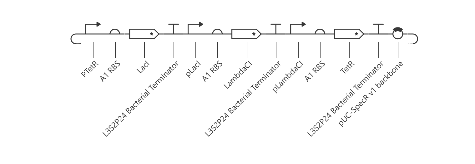
The goal of this section was to reconstruct the classic Elowitz-Leibler Repressilator—a synthetic biological clock governed by a cyclic, three-gene negative feedback loop.
Upon running the deterministic ODE simulator, the circuit failed to exhibit the expected sinusoidal oscillations and instead rapidly settled into a flat, static steady-state equilibrium.
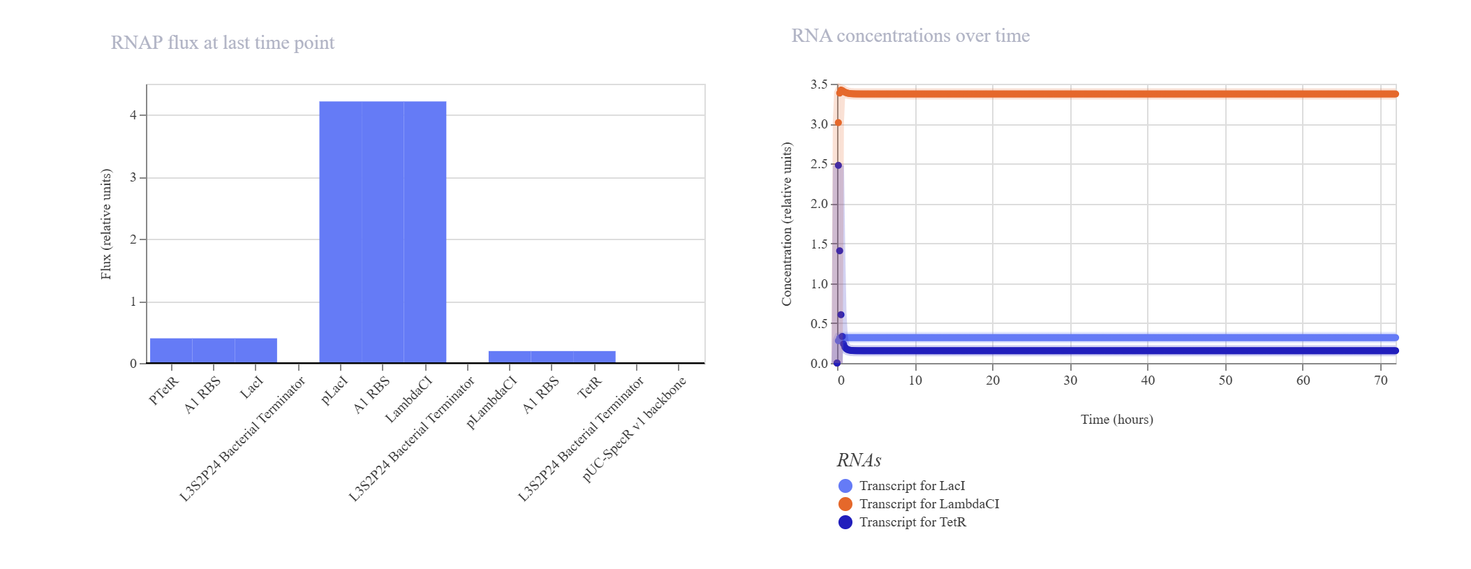
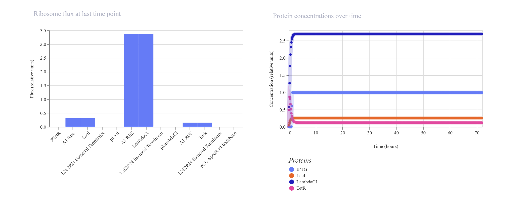
- Kinetic Analysis of Failure: The lack of oscillation is a clear result of a mathematical and biological “kinetic lock-in.” At T=0, the absolute absence of TetR causes the initial pTetR promoter to fire at maximum capacity, generating a rapid surge of LacI. This cascade immediately triggers an overabundance of LambdaCI transcript (~3.4) and protein (~2.7).Consequently, this intense concentration of LambdaCI completely overwhelms and shuts down the downstream pLambdaCI promoter, dropping its RNAP flux to near-zero (~0.2). Because TetR production is permanently blocked, the negative feedback loop cannot be completed to repress the initial pTetR promoter, trapping the entire network in a static steady-state.
- Proposed Resolution: To break this symmetry and trigger dynamic oscillations, the simulator parameters would require an asymmetrical starting condition (e.g., introducing an initial concentration of TetR protein at $T=0$ to initiate the delay cascade) or a weaker pLacI promoter efficiency.
Part 3: Custom Synthetic Biology Construct Designs
- Inducible ON Switch Layout
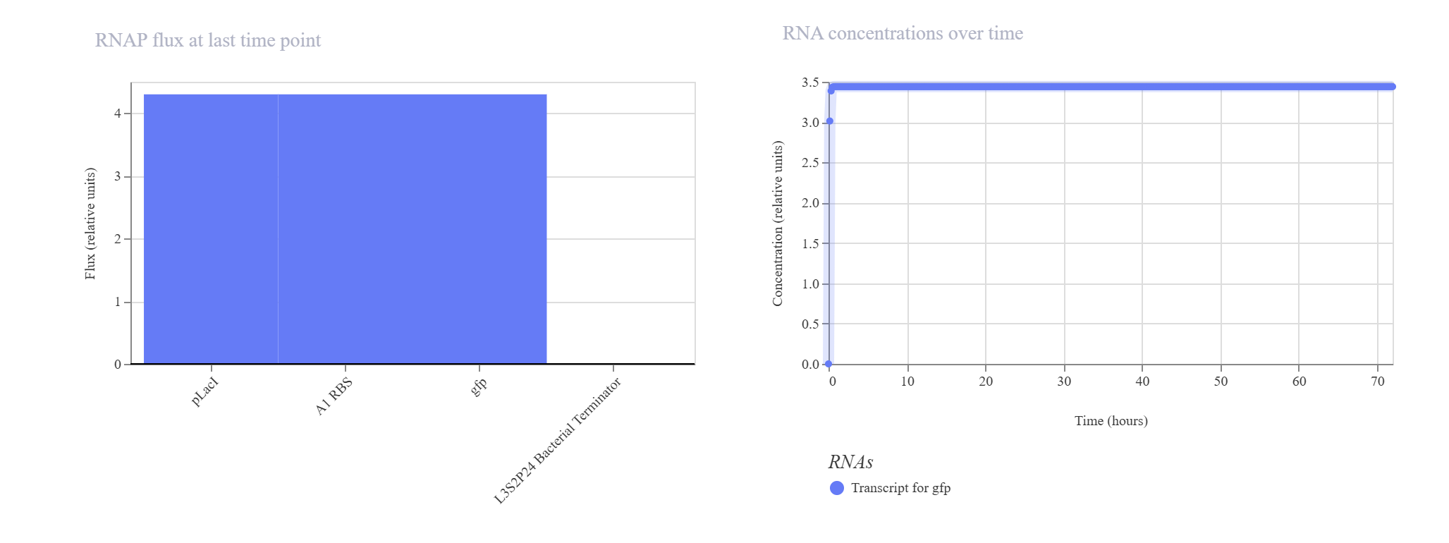
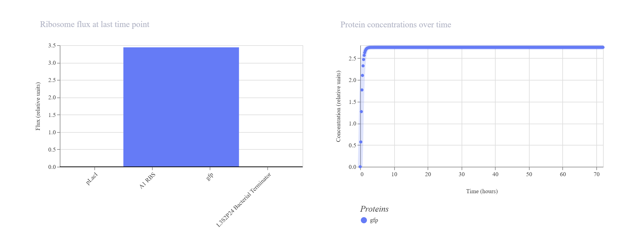
Hypothesis & Logic: This construct acts as a basic constitutive/inducible reporter system. Driven by the single pLacI promoter, the downstream gfp should be expressed linearly from zero until it hits a balanced equilibrium dictated by default synthesis and degradation rates.

The simulation perfectly validated the hypothesis. The gfp transcript and protein curves rise sharply from 0 hours and settle into a robust steady-state plateau at approximately 2.7 relative units. Unhindered RNAP flux (~4.3) and Ribosome flux (~3.4) confirm a fully functional, active ON-switch configuration.
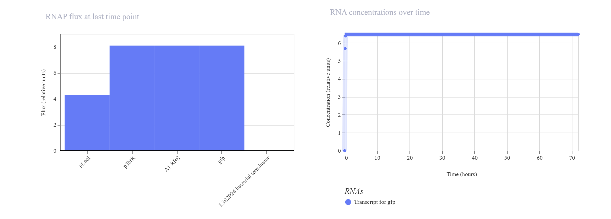
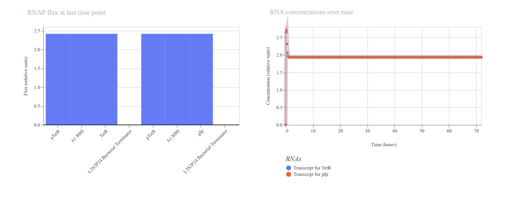
- Negative Feedback Loop for Homeostasis Hypothesis & Logic: The objective of this circuit is to establish a self-regulating, homeostatic mechanism. The pTetR promoter drives the expression of its own repressor, TetR. As TetR protein builds up, it physically binds back to the pTetR promoter, throttling further transcription to cap maximum output. A downstream gfp reporter under the same promoter framework tracks the overall expression dynamics.
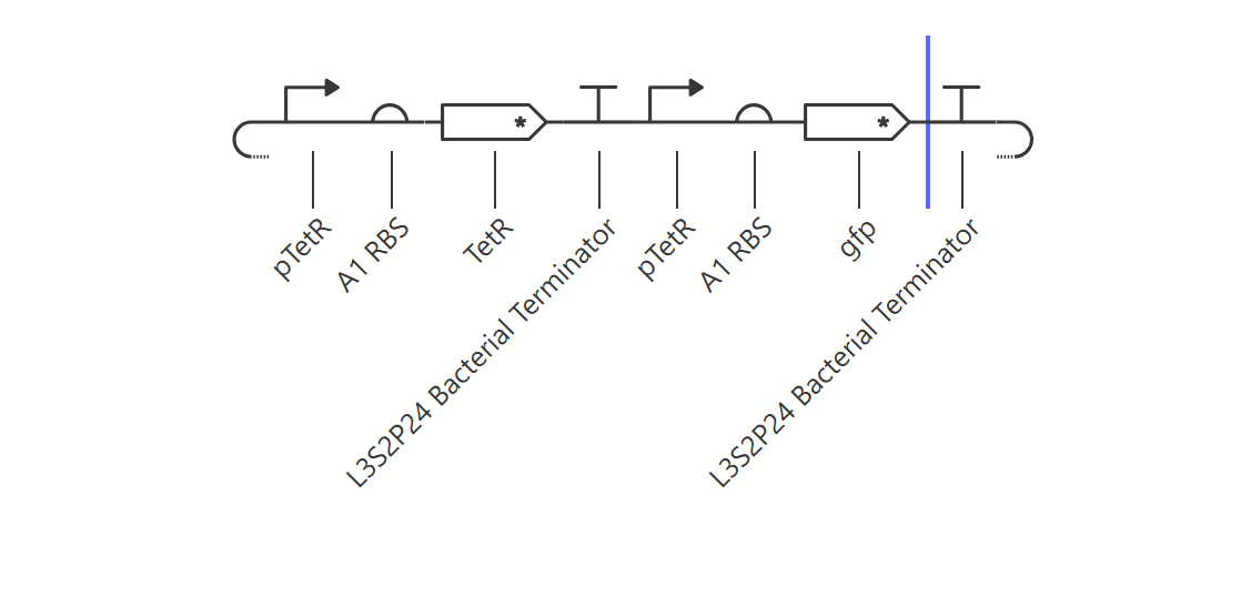
The simulation successfully verified homeostatic dampening. The gfp transcript initially spikes but quickly undergoes a controlled downward shift, balancing out at a plateau of ~1.9. Crucially, the final protein concentration is successfully capped at ~1.5 relative units. Compared to the unrepressed Design 1 (which reached 2.7), this significant reduction proves that the negative feedback loop is actively regulating and stabilizing the system’s output.
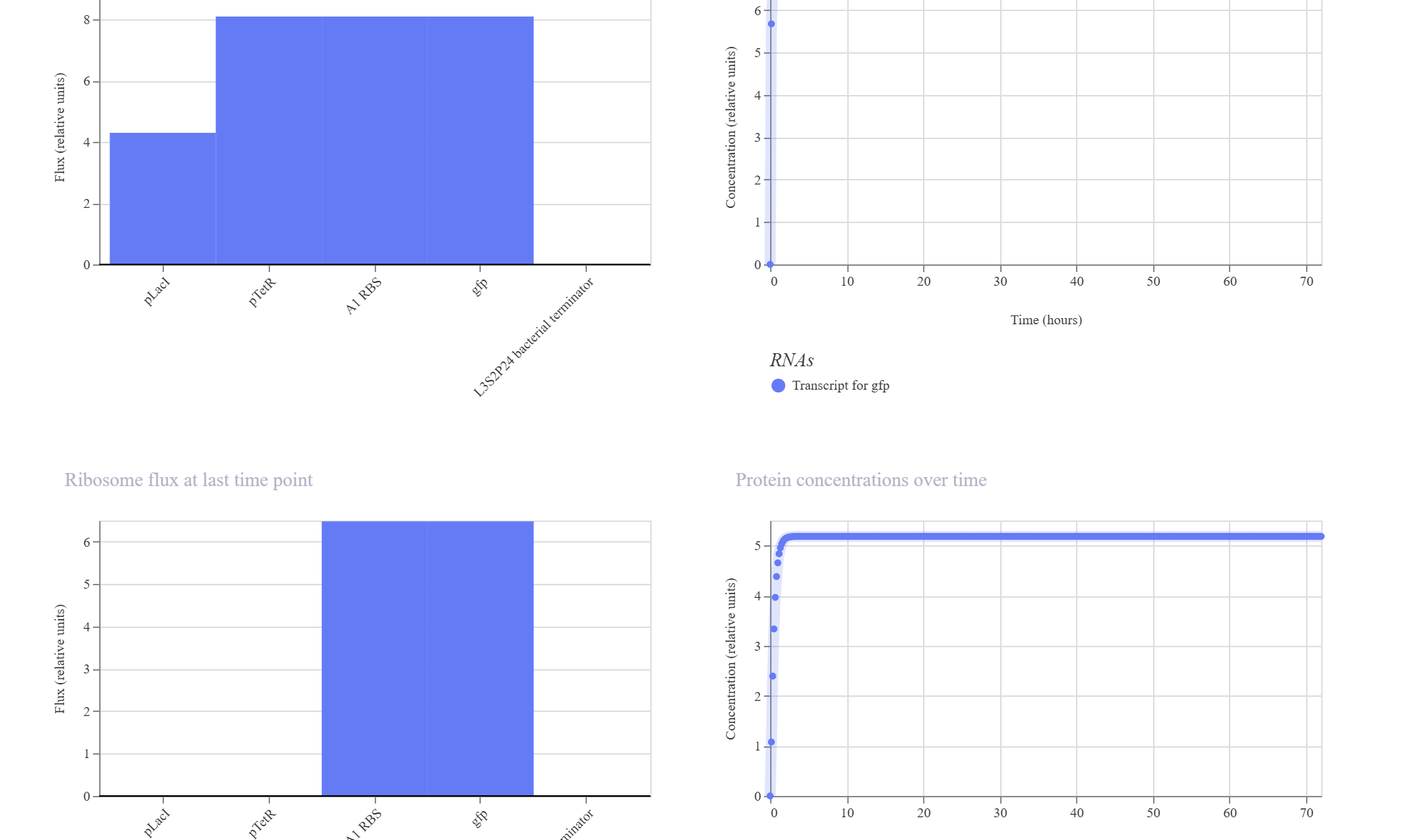
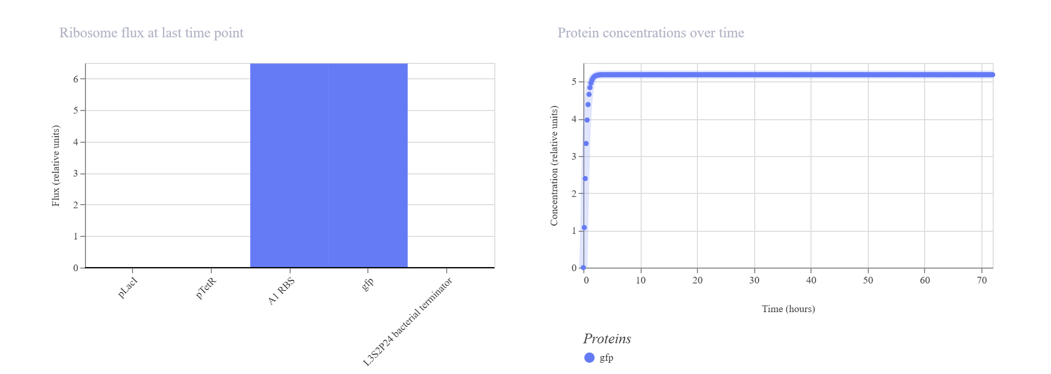
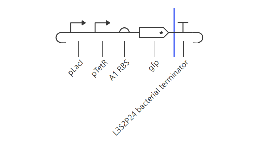
- Simple Synthetic AND Gate Layout Hypothesis & Logic: This design aims to construct a biological logic gate by placing two distinct regulatory promoter checkpoints (pLacI and pTetR) in tandem immediately upstream of a single gfp reporter. Transcription of the reporter gene strictly requires both gates to be clear and active simultaneously, allowing RNA Polymerase to read through the consecutive sequence into the coding region.
The simulation results confirmed the tandem gate topology. The RNAP flux shows a distinctive step-like cumulative increase, scaling from ~4.2 relative units at the pLacI boundary up to ~8.0 relative units past the pTetR threshold, pouring smoothly into the gfp domain. Unhindered by active repressors in this state, the expression climbs rapidly to hit an incredibly robust, stable plateau at approximately 5.2 relative units.