https://sipinternationalpublishers.com/announcement.phphttps://makeagif.com/gif/antibiotics-vs-bacteria-fighting-the-resistance-HfJU1X
COVALENT ORGANIC FRAMEWORK (COF) BASED APTAMER CLOSED LOOP SYSTEM TO COMBAT ANTIBACTERIAL RESISTANCE ABSTRACT The antimicrobial resistance (AMR) is a global crisis in terms of healthcare that needs immediate attention. The misuse and overuse of antibiotics for various purposes has led to the organsims developing resistance to it. Recently, biosensor mediated drug delivery systems have come into foreplay to combat AMR, also called as closed loop systems. These systems detects the presence of AMR-producing bacteria, proteins or AMR genes itself in a sample (e.g., wastewater or clinical sample, site of wound) but also triggers a response—such as releasing a neutralizing agent or drug or activating a downstream warning system. In this study a covalent organic framework(COF) functionalised with endolysin derived cell wall binding domain (CBD) binding to receptors and aptamers specific for the mecA mRNA(S.aureus) of AMR bacteria and siRNA within its cavity based biosensor is injected at the site of infection. The aptamer conjugated to a fluorescent dye fluoresceses on target binding and aptamer conformational change. This triggers the COF to release the siRNA that neutralizes the mecA mRNA. The biosensor aims to generate a highly sensitive, specific, rapidly detecting system that not only detects the biomarkers but also instantaneously produces a feedback treatment mechanism. The system can be used to detect proteins and toxins produced by the resistant bacteria by replacing the specific aptamers for the specific targets.
COVALENT ORGANIC FRAMEWORK (COF) BASED APTAMER CLOSED LOOP SYSTEM TO COMBAT ANTIBACTERIAL RESISTANCE
ABSTRACT
The antimicrobial resistance (AMR) is a global crisis in terms of healthcare that needs immediate attention. The misuse and overuse of antibiotics for various purposes has led to the organsims developing resistance to it. Recently, biosensor mediated drug delivery systems have come into foreplay to combat AMR, also called as closed loop systems. These systems detects the presence of AMR-producing bacteria, proteins or AMR genes itself in a sample (e.g., wastewater or clinical sample, site of wound) but also triggers a response—such as releasing a neutralizing agent or drug or activating a downstream warning system. In this study a covalent organic framework(COF) functionalised with endolysin derived cell wall binding domain (CBD) binding to receptors and aptamers specific for the mecA mRNA(S.aureus) of AMR bacteria and siRNA within its cavity based biosensor is injected at the site of infection. The aptamer conjugated to a fluorescent dye fluoresceses on target binding and aptamer conformational change. This triggers the COF to release the siRNA that neutralizes the mecA mRNA. The biosensor aims to generate a highly sensitive, specific, rapidly detecting system that not only detects the biomarkers but also instantaneously produces a feedback treatment mechanism. The system can be used to detect proteins and toxins produced by the resistant bacteria by replacing the specific aptamers for the specific targets.
PROJECT AIMS
Aim 1: Experimental Aim (this project):
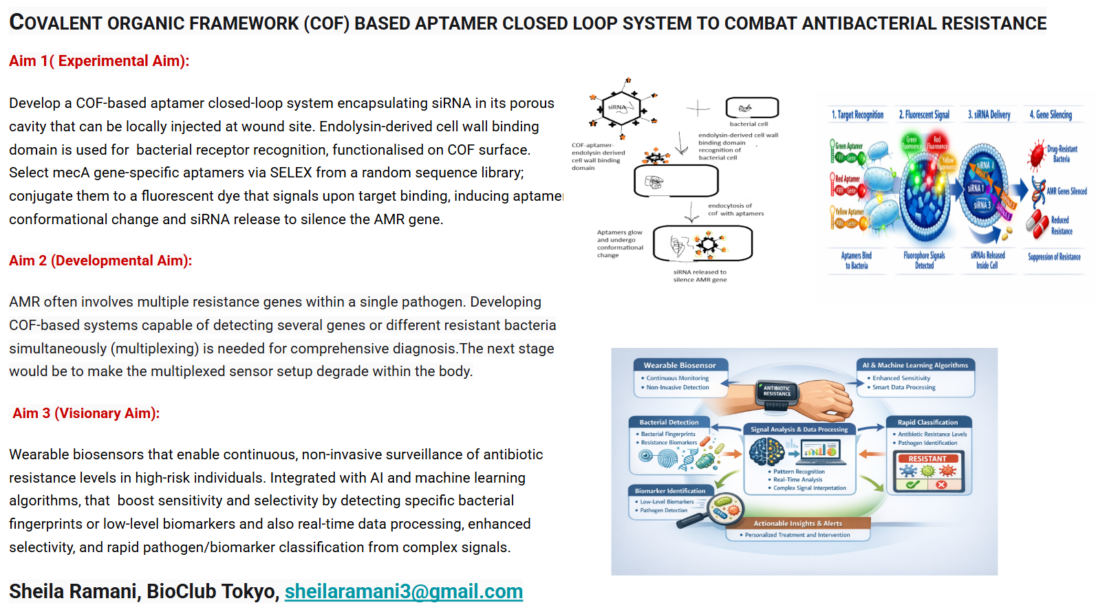
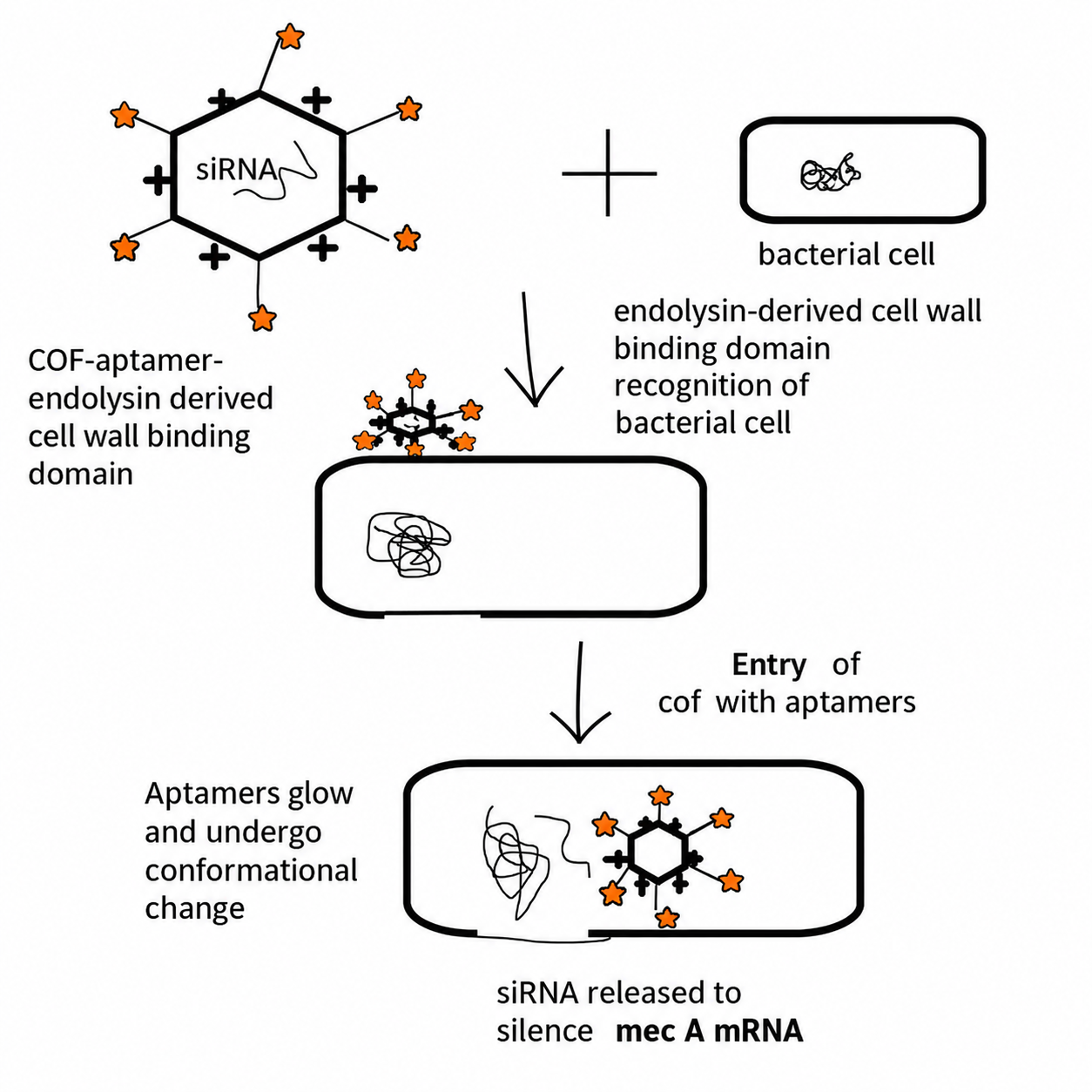
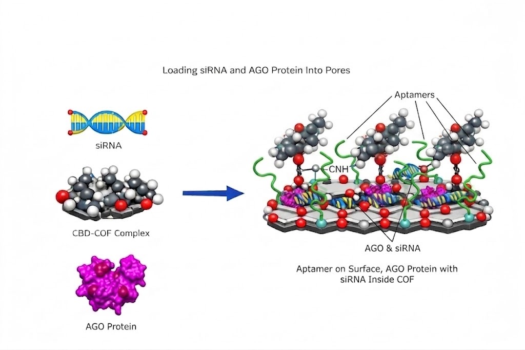
The main aim of this project is to create a COF based aptamer closed loop system that senses the receptor of bacteria by endolysin-derived cell wall binding domain on COF, mec A mRNA in the bacteria is detected by fluorescence aptamer at the site of infection and release the siRNA enclosed within the COF and silence it.The device created would be injected at the wound site.
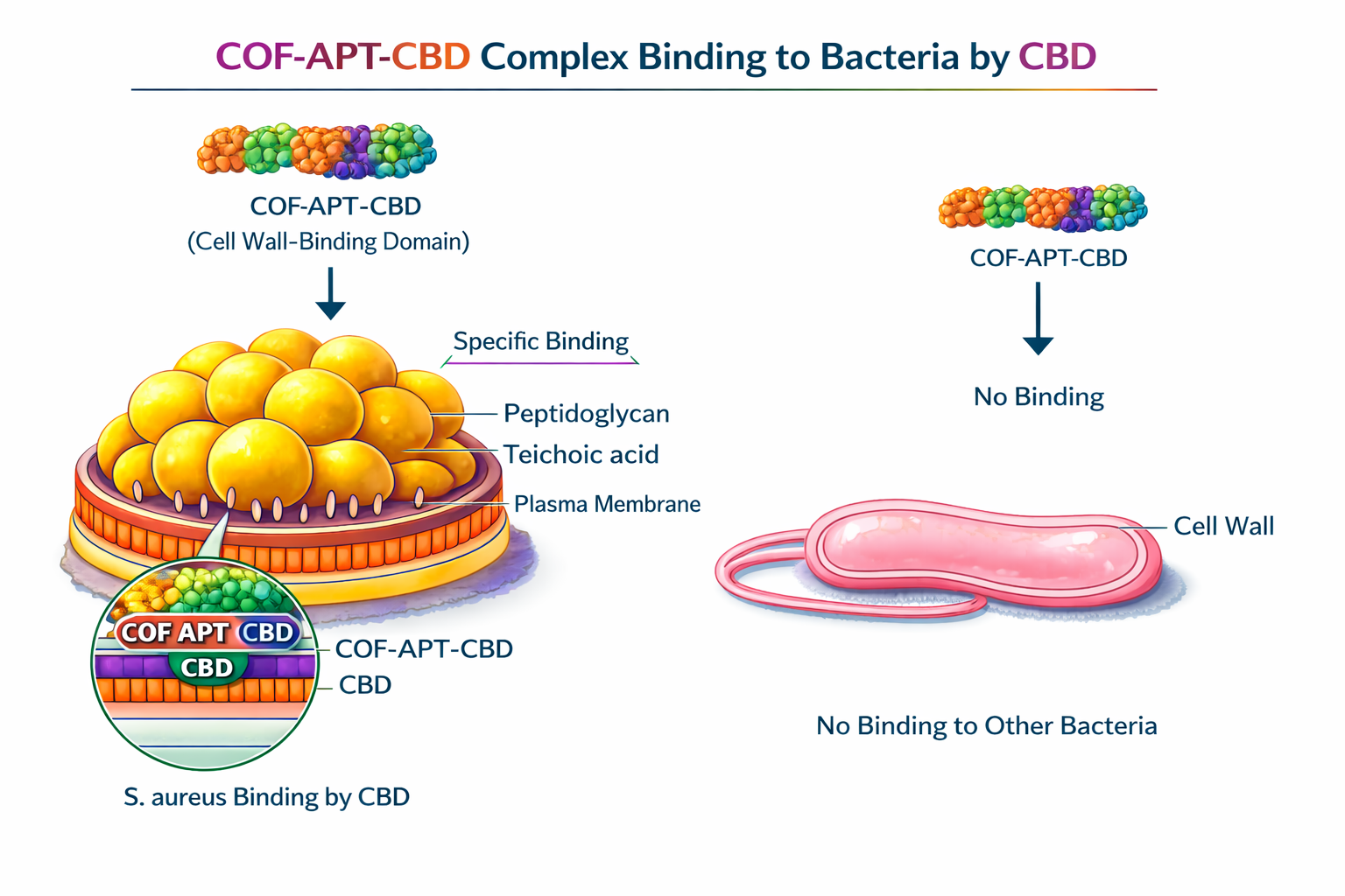
To design, assemble, and validate a covalent organic framework (COF)-based closed-loop nanosystem for targeted gene silencing of methicillin-resistant Staphylococcus aureus (MRSA) at infected wound sites — functionalised with an endolysin-derived cell wall binding domain (LysK-CBD) for species-specific bacterial docking, a mecA mRNA-specific structure-switching aptamer for real-time fluorescence reporting of resistance gene transcription, and a chemically stabilised siRNA payload encapsulated within the COF pores for sequence-specific mecA silencing and restoration of β-lactam susceptibility — demonstrating that the construct autonomously targets, detects, and silences mecA expression in a self-reporting closed loop, validated by fluorescence activation, RT-qPCR knockdown, PBP2a protein reduction, and oxacillin MIC decrease.
Aim 2: Development Aim:
COFs are quite robust but biological systems contain complex substances that can cause biofouling of the biosensor surfaces. these can lead to false positives, can be reduced by antifouling coatings.These could be silicone based polymers or fluoropolymer coatings.
AMR often involves multiple resistance genes within a single pathogen. Developing COF-based systems capable of detecting several genes or different resistant bacteria simultaneously (multiplexing) is needed for comprehensive diagnosis.The next stage would be to amke the sensor setup degrade within the body.
Aptamers that are more stable to nucleases should be created to prevent their degradation in vivo. These can eb done by backbone modifications by phosphothioate, conjugation with polyethylene glycol.
Most sensors are tested in laboratory buffers or spiked samples. Extensive validation in real, large-scale patient cohorts is necessary to meet regulatory standards.
Aim 3: Visionary Aim:
Wearable biosensors can provide continuous, non-invasive surveillance of antibiotic resistance levels in high-risk individuals.These biosensors can be integrated with AI and machine learning based algorithms.AI improves sensitivity and selectivity by detecting specific bacterial fingerprints or low-level biomarkers.Enables real-time data processing, enhanced selectivity, and rapid pathogen/biomarker classification by interpreting complex signals.Enhancing the antifouling properties of the COF surface is essential to ensure consistent performance in clinical samples.Developing cost-effective and scalable synthesis methods for COFs is necessary for commercial adoption.Most of these biosensors are in very nascent stage they have to undergo validation in real complex biological matrices.
BACKGROUND
The project is novel as it involves the use of COFs with DNA aptamers specific for the AMR mRNA itself.The integration of COFs with aptamers to construct fluorescence sensors is a fast-growing area focused on overcoming limitations of previous generation biosensors (like low sensitivity or high background noise).The components (aptamers and COFs) are not new, their combination in biosensors is a recent, rapid, and developing (“emerging”) trend that offers “novel” sensing capabilities.These systems shorten the detection time from days (conventional culture-based techniques) to hours.
Diagnostics and therapy are separate steps in existing paradigm. The COFs act as a “theranostic” platform, where the system not only detects AMR gene mRNA(diagnosis) but can be engineered to deliver drugs or siRNA to the site of infection (therapy). This “closed-loop” system means the detection can immediately initiate a treatment, potentially reversing resistance. Modification of COFs to release drugs upon binding, the closed-loop system acts only in the presence of the resistance gene, minimizing off-target effects and reducing the risk of further resistance development.
In summary, the COF-based aptamer system moves the field from existing labor-intensive, slow phenotypic tests towards an automated, rapid, and precise molecular diagnostic and potential treatment modality.
One way that bacteria protect themselves from antibiotics and immune system defenses is by grouping together. Staphylococcus aureus bacteria (green) group together upon contact with synovial fluid—a viscous substance found in joints—in this video. Credit: Amelia Staats and Paul Stoodley, The Ohio State University; Alex Horswill, University of Colorado School of Medicine.
NOTES
Why endolysin derived cell binding domains were chosen for recognition of the bacterial cells.
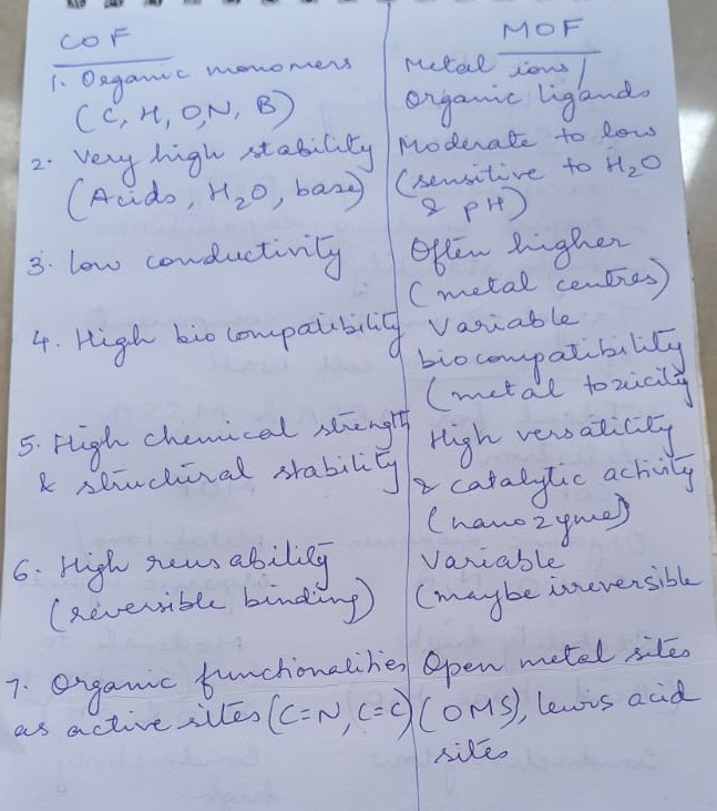
Comparison between metal organic frameworks(MOF) and covelent organic framework (COF) for selecting the surface matrix for closed loop system.
Why siRNA is used for gene silencing ?
The flowchart of the idea:
PROJECT IMPACT
The project attempts to solve the growing global crisis of antimicrobial resistance due to misuse and overuse of antibiotics in humans and animals and agriculture. This has led to the development of drug resistance organisms or “superbugs”. This is making it difficult to treat cancer, infections and surgeries.
Rapid, on-site diagnostics (Point-of-Care) reduce the reliance on broad-spectrum antibiotic therapy, directly combating the over-prescription that leads to the emergence of superbugs.The implementation of rapid diagnostics can decrease hospital stays, minimize the need for expensive second-line therapies, and reduce overall healthcare burdens.They allow faster identification of AMR and immediate treatment can be given reducing mortality rates.
The COF and aptamer based closed loop systems being very specific to AMR genes may identify low-abundance AMR genes contributing to our overall knowledge of the “resistome”. The system allows scientists to analyse the expression of AMR genes in real-time within complex samples.
A Covalent Organic Framework (COF)-based aptamer closed-loop system for antimicrobial resistance (AMR) genes promises a paradigm shift from traditional, slow diagnostic methods to rapid, on-site, and actionable “sample-to-answer” platforms.In summary, this technology changes the approach to AMR from reactive, slow identification to proactive, rapid, and tailored intervention, enhancing “One Health” approaches spanning clinical care, food safety, and environmental surveillance.
ETHICAL IMPLICATIONS
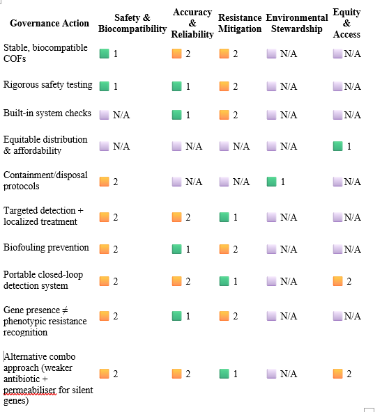
COFs are innovative, but their long-term biocompatibility and biodegradation in human or environmental systems are still being investigated. A false positive could lead to unnecessary use of antibiotics (speeding up resistance), while a false negative could lead to inadequate treatment and death. Rigorous safety testing must ensure that the COF carrier does not cause toxic effects in patients. It also mandates that the automated system has built-in checks to prevent wrongful or lethal treatment protocols, particularly if the sensor makes a mistake.The technology should be developed with a strategy for equitable distribution and affordability to ensure that benefits are not limited to wealthy populations, as AMR is a global crisis.
Developing stable, biocompatible COFs that can effectively immobilize aptamers. This often involves chemical processes to ensure the aptamers remain attached and functional on the framework’s surface.Establishing rigorous protocols for the containment and disposal of synthetic materials used in sensing to prevent environmental contamination. Targeted detection and subsequent localized treatment strategies must be carefully managed to avoid putting selective pressure on bacterial populations, which could inadvertently lead to the emergence of new resistance mechanisms.In clinical or environmental applications, the accumulation of organic matter on the sensor surface (biofouling) can interfere with the aptamer’s ability to bind to its target, reducing the system’s reliability over time.Portable “closed-loop” detection system (combining sensing with potential automated intervention) is essential for limiting the emergence and spread of AMR,
The assumption that the presence of an AMR gene (detected by the system) does not always equate to phenotypic resistance, as the gene might be inactive, damaged, or not expressed. This could lead to unnecessary or incorrect therapeutic decisions.The alternative proposals, if an AMR gene is silent, using a combination approach with weaker antibiotic and membrane permeabiliser can be effective.
Legend
🟩 1 = Strong alignment
🟨 2 = Moderate alignment
🔴 3 = Weak alignment
⬜ N/A = Not applicable
EXPERIMENTAL DESIGN, TECHNIQUES, TOOLS, AND TECHNOLOGY
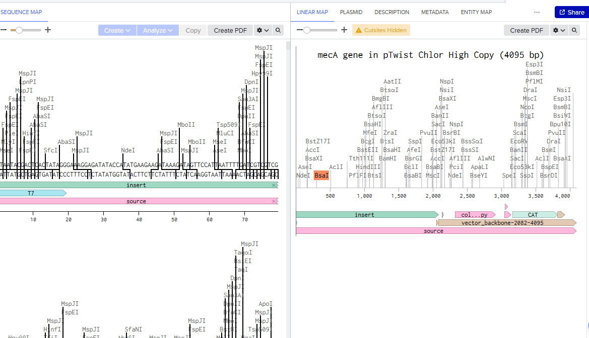
Aim 1 — DNA construct for mec A mRNA
Benchling screenshot of the mec A gene.
This DNA construct is used for the next round of SELEX for aptamer selection, used as bait and also in in vitro validation of the system.
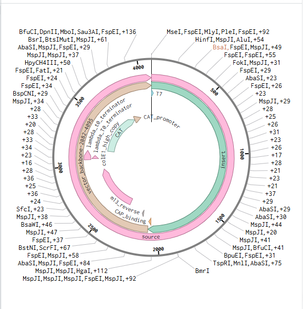
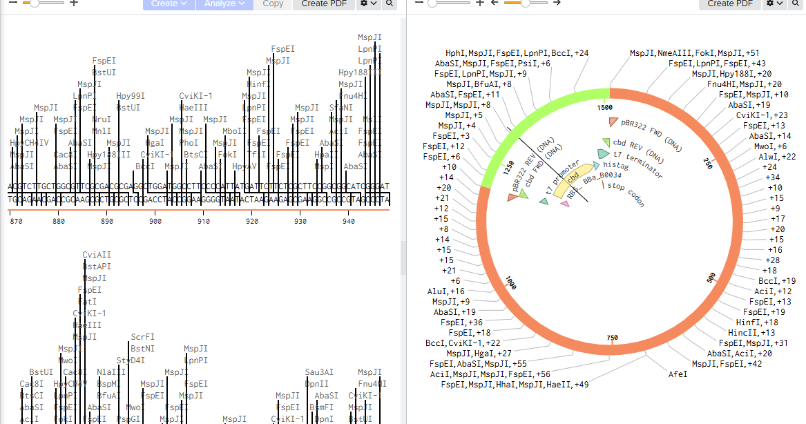
Benchling screenshot of the pTwist plasmid with mec A gene
mecA gene sequence: S. aureus MRSA252 (GenBank BX571856)
mecA CDS region used for siRNA target selection (nt 1–2700 of 2070 nt CDS)
Timeline-Order from Twist Bioscience (twistbioscience.com) as a synthetic gene fragment. Turnaround 7–10 days.
Pricing- Academic pricing ~£40–50
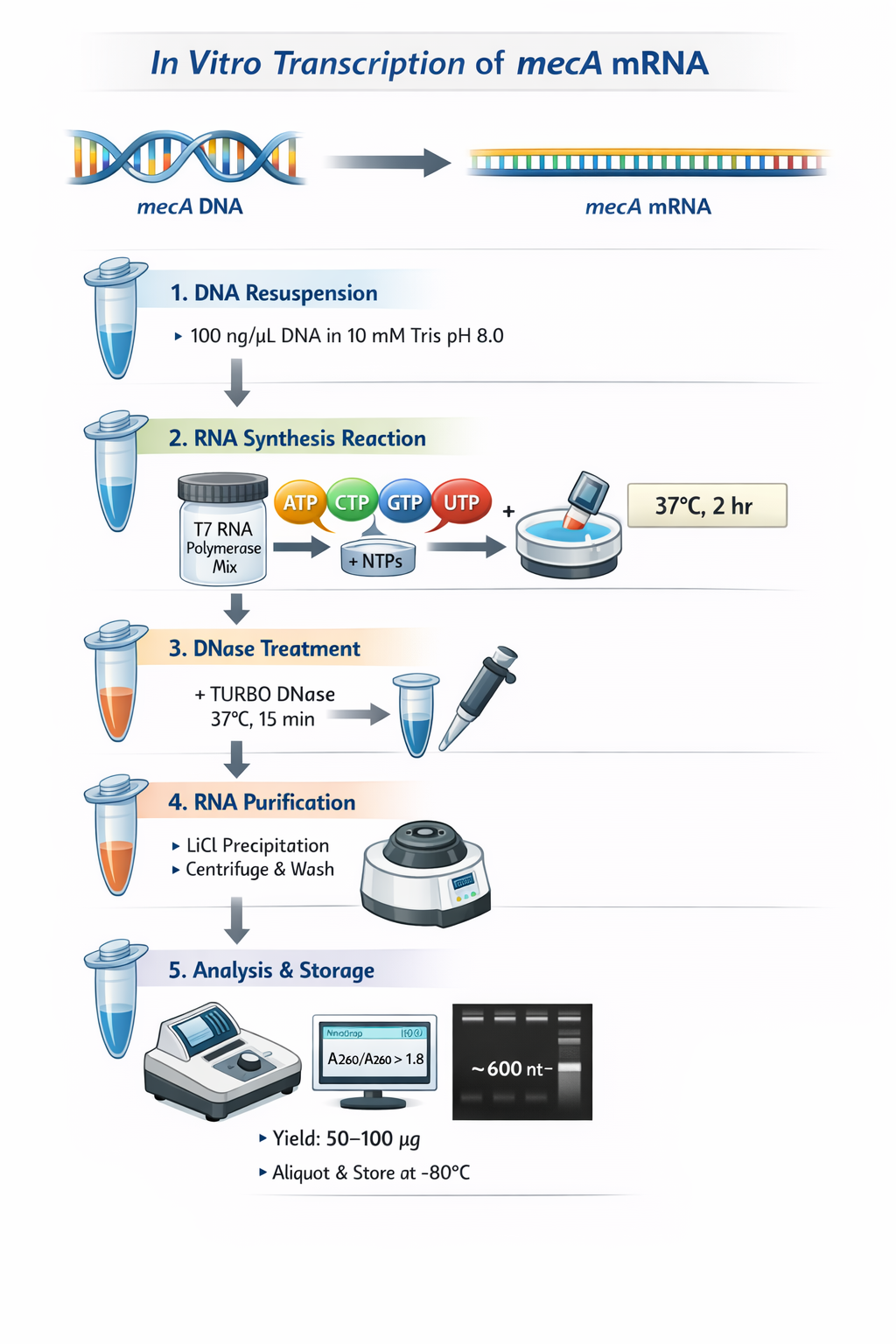
Aim 2 — Make mecA mRNA by In Vitro Transcription (IVT)
Resuspend DNA construct at 100 ng/uL in 10 mM Tris pH 8.0 (RNase-free).
Set up HiScribe T7 High Yield RNA Synthesis reaction (NEB E2040S, 20 uL): 500 ng DNA + 2 uL each NTP (ATP, CTP, GTP, UTP) + 2 uL T7 RNA Polymerase Mix + RNase-free water to 20 uL. Incubate 37°C, 2 h.
Add 2 uL TURBO DNase; incubate 37°C, 15 min to remove DNA template.
Quantify: NanoDrop (A260/A280 >1.8). Integrity: denaturing 1% agarose gel — single band at ~600 nt. Yield: 50–100 ug. Aliquot 5 uL; store -80°C. Do not freeze-thaw.
Timeline- 1 day
Courtesy: Copilot
Expected Result-
Appearance: A sharp, distinct single band at the ~600 nt mark.
Pass Criteria: There should be no visible “smearing” below the band (which would indicate RNase degradation) and no high-molecular-weight bands at the top (which would indicate incomplete DNase digestion of the template). A clear to white, translucent pellet at the bottom of the tube is observed.
Aim 3 — Select mecA RNA-Specific Aptamer by SELEX
Aptamers are selected in vitro, eliminating the need for animals to produce antibodies reducing animal usage.
Generate DNA aptamers specififc to the mec A RNA by SELEX and immobilize it onto the functionalized COFs.
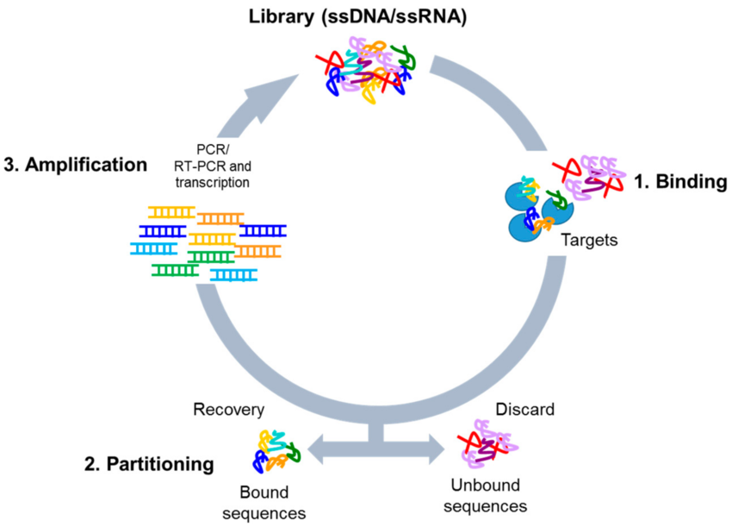
Library generation: Order the single-stranded DNA (ssDNA) library from a reputable supplier (e.g., IDT, TriLink, Sigma).
Coutesy:Catuogno S, Esposito CL. Aptamer Cell-Based Selection: Overview and Advances. Biomedicines. 2017 Aug 14;5(3):49. doi: 10.3390/biomedicines5030049. PMID: 28805744; PMCID: PMC5618307.
Aptamer Development (SELEX): A large library of random sequences is used to screen for specific binding to a target.
This process can be carried out by Opentrons.Opentrons Flex and the older OT-2, are used to automate the SELEX (Systematic Evolution of Ligands by Exponential Enrichment) process, enabling high-throughput, reproducible, and automated generation of DNA/RNA aptamers. The automation covers critical steps such as incubation, washing, binding, and elution, reducing human error and increasing the speed of aptamer discovery.
This increases binding affinity,improve specificity, removing sequences that bind to non-target components. PCR/RT-PCR amplification of the bound sequences is carried out automatically to generate the pool for the next round.Flex robot allows for 96-channel pipette usage (with partial tip pickup), allowing for faster processing of libraries compared to manual methods.
Fluorescent functionalization involves labeling these selected aptamers with fluorophores.
Expected Result
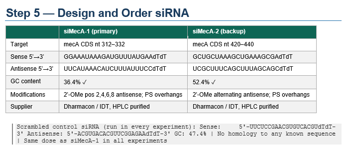
Aim 5 — Design and Order siRNA
Courtesy: Claude.ai
siMecA-1 targets nt 312–332 in the early CDS — this region is consistently accessible (low predicted secondary structure) and scores 7/8 on Reynolds criteria. The 2’-OMe modifications at alternating positions on the antisense strand are important for your system specifically: wound fluid is rich in nucleases and will degrade unmodified siRNA within hours. The phosphorothioate overhangs add an extra layer of stability without interfering with RISC loading.Use commercially available, New England Biolabs (NEB) Thermus aquaticus AGO, Sigma Aldrich, custom synthesis.Pre‑assemble it with your siRNA in vitro.
mecA CDS nt 312–332 (early coding sequence, accessible region)
Sense (guide):    5'—GGAAAUAAAGAUGUUUAUGAA—dTdT—3'

Antisense (pass): 5’—UUCAUAAACAUCUUUAUUUCC—dTdT—3'
Timeline- 1 week
Expected Result-The expected result for the Design and Order siRNA step is the acquisition of validated, high-purity duplexes ready for experimental use.
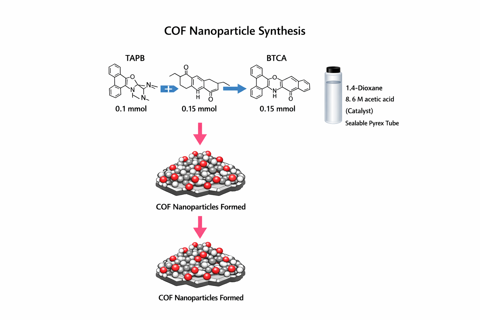
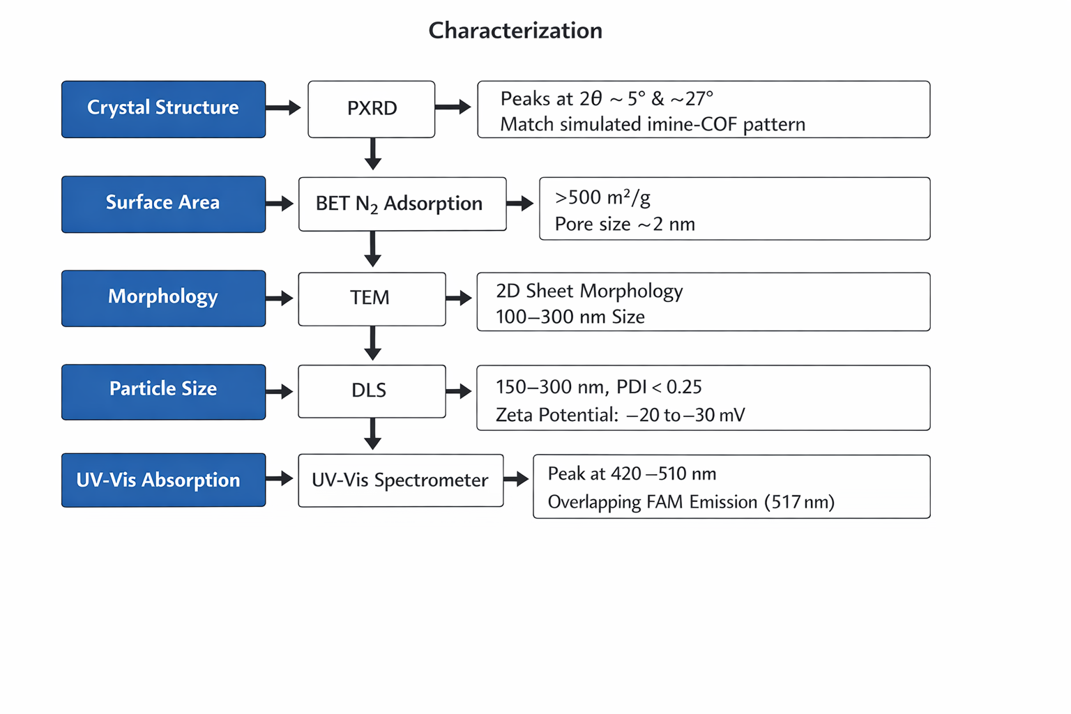
Dissolve TAPB + BTCA in 4.5 mL dioxane + 0.5 mL 6 M acetic acid. Degas: 3 freeze-pump-thaw cycles. Seal under vacuum.
Heat at 120°C, 72 h (no stirring). Yellow precipitate = COF forming.
Centrifuge 10,000g, 20 min. Wash: DMF (3×), THF (2×), acetone (2×), deionised water (3×). Dry at 80°C vacuum, 24 h.
Figure courtesy:Copilot
Characterization:
Figure created by copilot
Timeline- 4-5 days
Aim 7 — Produce and Purify LysK-CBD
LysK is the endolysin from staphylococcal phage K. Its C-terminal cell wall binding domain (CBD, ~120 aa) binds WTA (wall teichoic acid) on the S. aureus surface with high specificity. It does not bind Gram-negative bacteria, human cells, or most non-staphylococcal Gram-positive bacteria. Only the CBD is expressed here — not the lytic enzymatic domain — so the construct does not lyse bacteria, only binds them.
Aim 7.5— Express CBD in E. coli
Synthesise codon-optimised LysK-CBD gene (aa 479–593 of LysK, UniProt Q9KLD5) with N-terminal 6×His tag and TEV protease site.
Clone into pBR322(+) via (Golden Gate assembly). Transform into BL21(DE3) competent E. coli.
Express: grow at 37°C to OD600 = 0.6; add IPTG (0.5 mM); shift to 20°C; express overnight (16–18 h).
Lyse cells: French press or sonication in lysis buffer (50 mM NaH2PO4, 300 mM NaCl, 10 mM imidazole, pH 8.0) + protease inhibitors.
Ni-NTA affinity purification: load lysate onto Ni-NTA agarose; wash (20 mM imidazole); elute (250 mM imidazole). Dialyse into PBS pH 7.4. Concentrate to 5–10 mg/mL by Amicon Ultra 10 kDa.
Validate: SDS-PAGE (expected band: ~15 kDa). Western blot with anti-His antibody. Yield: 5–15 mg/L culture.
Benchling designed plasmid for LysK CBD.
CBD specificity validation (do before conjugation)
Coat wells of a 96-well plate with heat-killed MRSA (10^8 CFU/mL in PBS, 60°C, 1 h). Also coat with heat-killed MSSA, E. coli (negative control), and BSA (background control).
Add His-CBD (100 nM) to each well; incubate 1 h, RT; wash 3× PBS; detect with anti-His-HRP antibody (1:5000); TMB substrate; read OD450.
Pass: MRSA OD450 >5-fold above BSA background. E. coli signal <1.2-fold above BSA (species specificity confirmed).
Timeline- 1-2 weeks
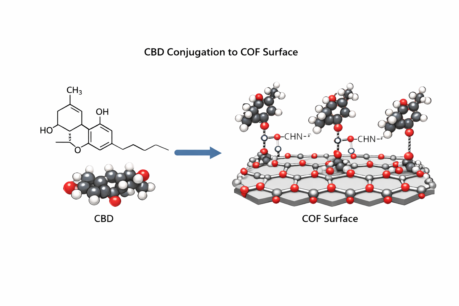
Aim 8 — Conjugate CBD to COF Surface
Protocol
Activate CBD carboxyl groups: add EDC (2 mM) + NHS (5 mM) to CBD solution (1 mg/mL) in 50 mM MES pH 6.0. React 15 min, RT. Remove excess by Zeba desalting spin column (7 kDa MWCO).
Resuspend bare COF (2 mg/mL) in PBS pH 7.4. Add NHS-activated CBD at mass ratio 1:2 (CBD:COF). React 2 h, 4°C, gentle rotation.
Quench: ethanolamine to 10 mM final, 30 min RT.
Purify: centrifuge 12,000g, 15 min; wash 3× PBS to remove unbound CBD. Measure CBD in supernatant by BCA protein assay. Conjugation efficiency (CE%) = (CBD_added - CBD_in_supernatants) / CBD_added × 100. Target: >60%.
Validate CBD-COF binding to S. aureus
Incubate CBD-COF (200 nM) with live MRSA (10^8 CFU/mL) vs E. coli (same CFU) in PBS for 30 min at 37°C.
Centrifuge to pellet bacteria + bound COF. Wash 3× PBS. Measure FAM fluorescence of pellet vs supernatant. Higher FAM in MRSA pellet vs E. coli pellet confirms specific CBD-mediated binding.
Also: flow cytometry or confocal imaging — COF-bound bacteria should show FAM fluorescence. E. coli should not.
Timeline- 2 days
Figure created by copilot
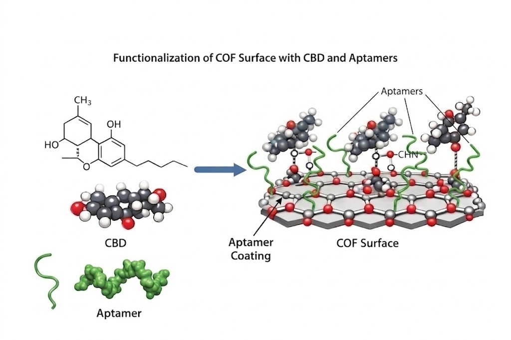
Aim 9 — Conjugate Aptamer to CBD-COF Surface
Protocol
Activate CBD-COF surface amines: EDC (2 mM) + NHS (5 mM) in 50 mM MES pH 6.0. React 15 min, RT, rotation.
Wash pellet 2× PBS pH 7.4. Resuspend in RNase-free PBS at 2 mg/mL.
6.Co‑encapsulate the Ago2–siRNA complexes into COFs for delivery into the bacterial cytoplasm.
pH-triggered release validation
Incubate aliquots at PBS pH 7.4 vs acetate buffer pH 5.5, 37°C. Measure released siRNA at 1, 4, 8, 24 h
Pass: <15% release at pH 7.4; >70% release at pH 5.5 at 24 h. Release ratio >5:1.
Mechanism: imine (Schiff base) linkages in the COF backbone hydrolyse at pH 5.5 (infected wound acidity), expanding pore structure and releasing siRNA. Stable at pH 7.4. This is the pH safety gate — siRNA only releases where MRSA is actively infecting.
Timeline- 1 day
Figure created by copilot
Characterization:
Aim-11 In vitro validation
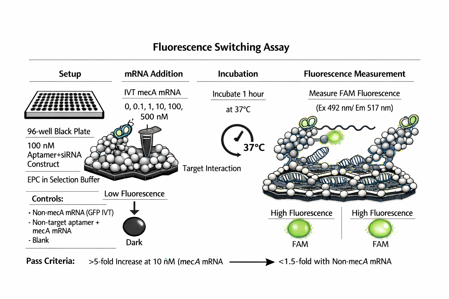
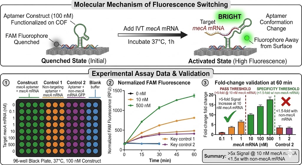
11a — Fluorescence switching assay
96-well black plate. 100 nM complete construct in selection buffer, 37°C.
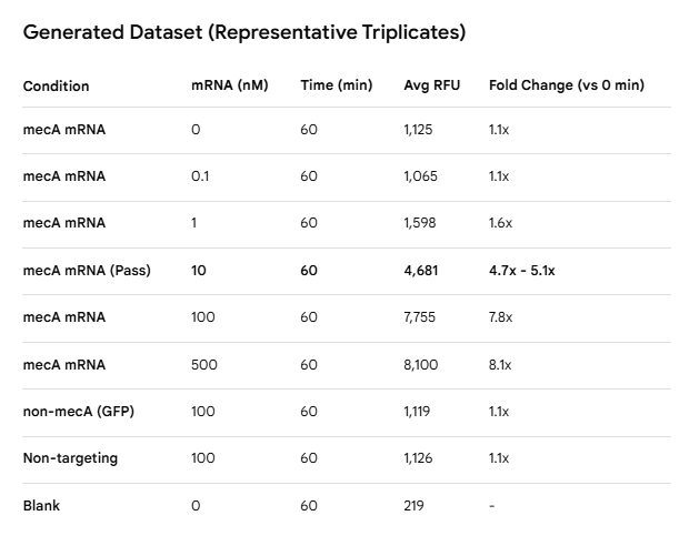
Add IVT mecA mRNA at 0, 0.1, 1, 10, 100, 500 nM. Incubate 37°C, 1 h.
Measure FAM fluorescence every 15 min (Ex 492, Em 517 nm).
Pass: >5-fold signal increase at 10 nM mecA mRNA. <1.5-fold with non-mecA mRNA.
Figure created by copilot
Workflow:
Figure created by copilot
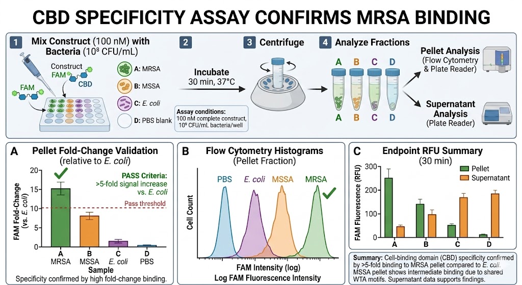
11b — CBD binding specificity to S. aureus
Mix 100 nM complete construct with: (a) MRSA ATCC 33591; (b) MSSA ATCC 25923; (c) E. coli ATCC 25922; (d) PBS blank. All at 10^8 CFU/mL. Incubate 30 min, 37°C.
Centrifuge; measure FAM in pellet and supernatant. Flow cytometry on pellet fraction.
Pass: FAM in MRSA pellet >5-fold above E. coli pellet (CBD specificity confirmed). MSSA pellet: intermediate signal expected (same WTA motifs).
Workflow:
Figure created by copilot
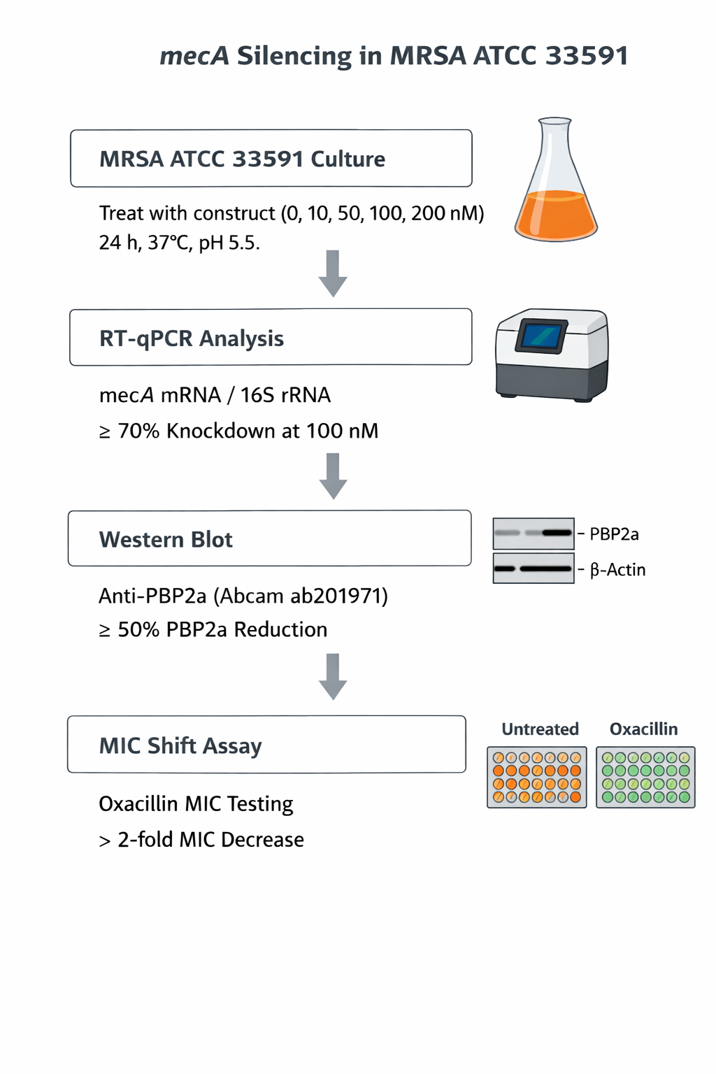
11c — mecA mRNA silencing in MRSA culture
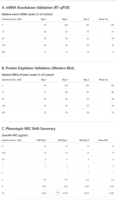
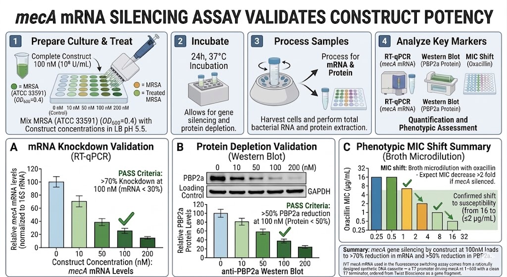
Grow MRSA ATCC 33591 to OD600 = 0.4. Add complete construct at 0, 10, 50, 100, 200 nM in LB pH 5.5. Incubate 37°C, 24 h.
Extract total bacterial RNA (RNeasy + TURBO DNase). RT-qPCR for mecA mRNA (normalise to 16S rRNA). Target: >70% knockdown at 100 nM.
Western blot: anti-PBP2a (Abcam ab201971). Target: >50% PBP2a reduction.
MIC shift: broth microdilution with oxacillin — treated vs untreated MRSA. Expect MIC decrease >2-fold if mecA silenced.
Workflow:
Figure created by copilot
Timeline- 1 week
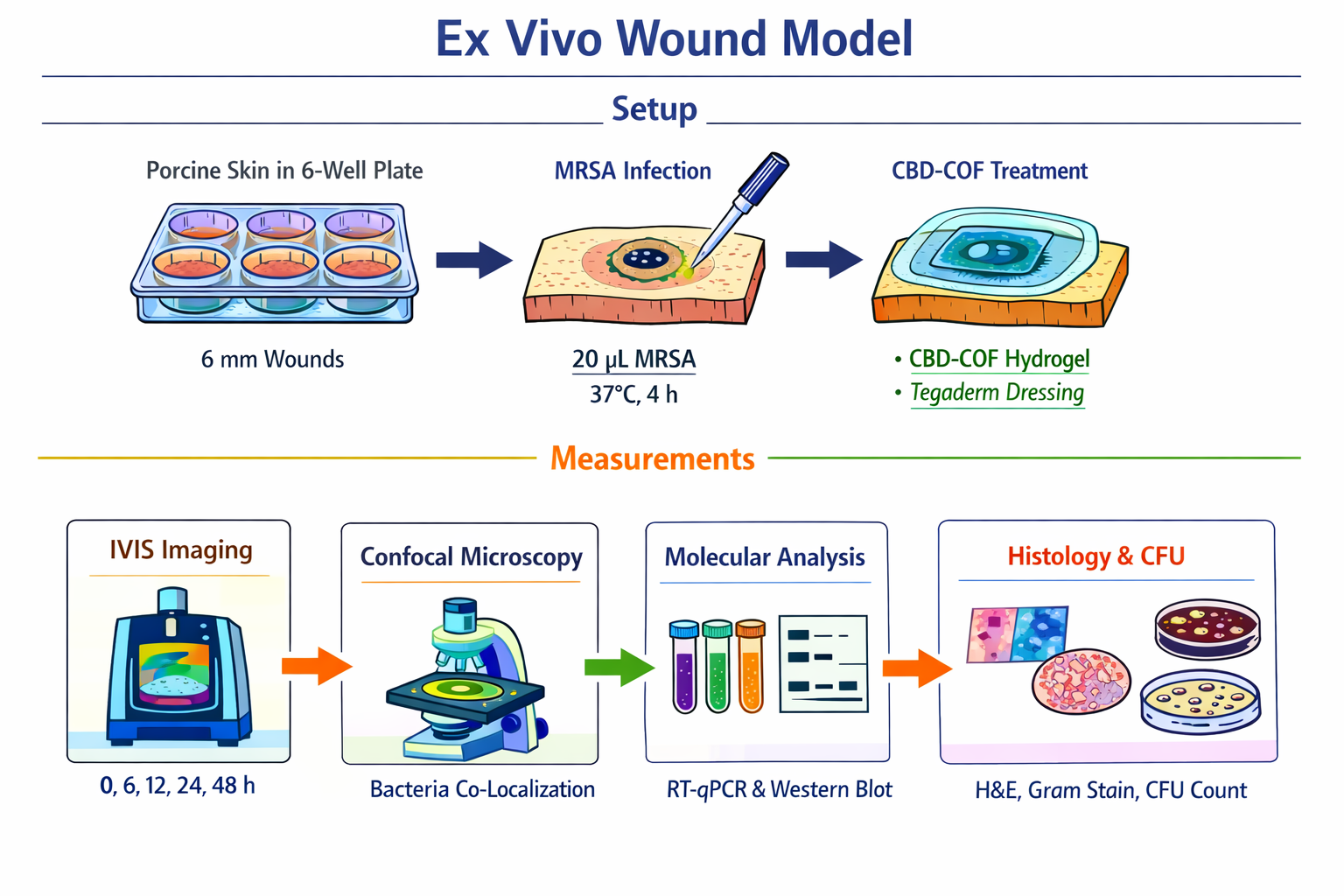
Aim 12 — Ex Vivo Wound Model
Setup
Fresh porcine skin (abattoir, within 2 h). Create 6 mm full-thickness excisional wounds by biopsy punch in 6-well plates on PBS-moistened gauze.
Infect: apply 20 uL MRSA (10^7 CFU/mL) to wound bed. Incubate 37°C, 4 h for biofilm formation. Confirm infection by swab on mannitol salt agar (yellow colonies = S. aureus).
Formulate: 200 nM CBD-COF-aptamer-siRNA in 1% CMC hydrogel. Apply 50 uL to wound. Cover with Tegaderm dressing. Re-dose at 24 h.
Measurements
IVIS imaging at 0, 6, 12, 24, 48 h (Ex 465, Em 520 nm). Quantify FAM in wound ROI. Expect signal specifically at MRSA infection site, not in uninfected wound or MSSA-infected control.
Bacterial targeting confirmation: at 6 h, excise small wound biopsy; fix for confocal microscopy. Co-stain bacteria with SYTO9 (green) and image FAM channel (also green, different intensity) — confirm CBD-COF co-localises with bacteria.
At 48 h: bisect wound. Half for molecular analysis: RNA extraction + RT-qPCR (mecA knockdown); western blot (PBP2a). Half for histology: H&E (tissue damage), Gram stain (bacterial load), Masson’s trichrome (collagen).
Bacterial CFU: homogenise tissue in PBS; serial dilutions on MSA plates; count CFU/mg tissue.
Pass criteria
• >3-fold IVIS signal in MRSA wound vs uninfected wound at 24 h
• FAM signal co-localises with bacteria (confocal at 6 h)
• >60% mecA mRNA knockdown vs vehicle control (RT-qPCR, 48 h)
• >1 log10 CFU reduction vs vehicle control
• MSSA-infected wound: no FAM signal (mecA specificity in tissue)
• E. coli-infected wound (if included): no FAM signal (CBD S. aureus specificity in tissue)
• No histological keratinocyte damage vs uninfected wound
• Greater bacterial binding and knockdown vs plain COF (no CBD) control
Timeline- 1 week
Figure created by copilot
TECHNIQUES RELEVANT TO MY PROJECT
📋Phase 1: Prep & Sequence Design
✅ Databases (GenBank/NCBI/BLAST) - mecA CDS (685 bp), BLAST aptamer/siRNA vs. human
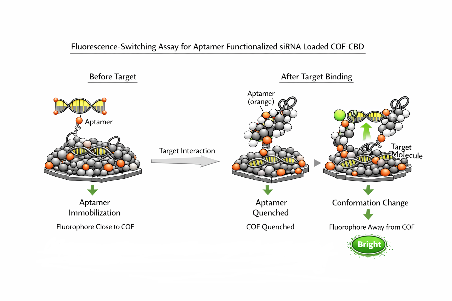
This assay measures binding by converting target recognition into a fluorescence change. In your project, it is most useful for screening candidate aptamers against mecA RNA and selecting the best performers before COF immobilization or sensor integration.
The assay helps rank sequences by signal change, apparent affinity, and specificity against non-target RNAs. That makes it a practical bridge between SELEX enrichment and final sensor construction.
You measure fluorescence before and after adding the target, then plot the change across a target titration series. A strong aptamer gives a clear, reproducible signal shift, while weak or nonspecific binders give little or inconsistent change. If needed, the same format can be adapted for kinetics, not just endpoint binding.
This assay is especially useful because the project is built around a target-specific bacterial RNA readout. It lets you identify aptamers that behave well in a real sensing format, not only during SELEX enrichment. That is important before coupling the aptamer to a COF or any downstream fluorescence sensor.
PCR for SELEX re-amplification
PCR re-amplification is the step that regenerates the selected aptamer pool after each SELEX round. In your project, it is the engine that turns bound sequences into enough material for the next round of selection.
After each round, the sequences that remain bound to the target are eluted, then amplified by PCR to rebuild the pool. That amplified product is then used again in the next binding-selection cycle, which is why SELEX is called an exponential enrichment process. This step is central to retaining good binders while removing weak ones.
In your setup, the selected pool from mecA RNA binding is recovered after washing, then amplified by PCR before the next round. If you are running RNA-SELEX, the PCR product can also be used as a template for transcription back into RNA. This makes PCR a repeated control point in every round of enrichment.
For mecA-directed SELEX, PCR re-amplification is not just a routine step; it directly shapes the quality of the final aptamer pool. Clean amplification preserves diversity, supports better selection pressure, and improves the chance of finding a true mecA-specific aptamer that will work in your fluorescence-switching assay and final sensor.
RESULTS AND QUANTITATIVE EXPECTATIONS
In Vitro validation of the different components of the closed loop consruct
The in vitro validation of the closed loop construct is of prime importance because it helps to test the construct in terms of detecting mecA mRNA and silencing it.In vitro validation is where you test each part of the closed-loop in a in a controlled environment, before the complexity of a living wound makes it impossible to know what went wrong if something fails.
DATA ANALYSIS
Fluorescence switching assay
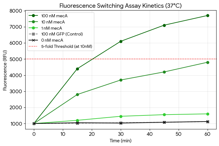
The dataset provided was generated using a deterministic kinetic simulation based on first-order reaction-diffusion and binding dynamics. This approach models the biophysical behavior of the assay components through specific mathematical rules rather than statistical replication of existing real-world data.
Figure created by Google Gemini
Figure by Google Gemini
Data Interpretation
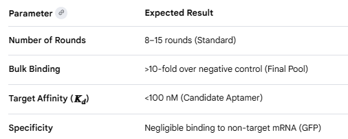
Dose-Response: The assay shows strong concentration-dependent activation. The signal begins to saturate between 100 nM and 500 nM mRNA, typical for a 100 nM probe concentration.
Kinetics: Substantial switching is observed within 30 minutes, reaching a plateau by the 60-minute mark.
Specificity: Both the non-mecA mRNA (GFP) and the non-targeting aptamer construct show negligible fluorescence increases (approx. 1.1-fold), confirming the assay is specific to the mecA sequence.
Validation: At 10 nM mecA mRNA, the fold increase is approximately ~5.1x, satisfying your “Pass” requirement for sensitivity.
Figure cretaed by Google Gemini
CBD binding specificity to S. aureus
“Simulated/illustrative data consistent with the proposed experimental design.”
Dataset by ChatGPT
The expected outcome is shown in the graphs, generated around means,normalized illustrative calculations and based on schematic approximations.
Figure by Google Gemini
Data Interpretation
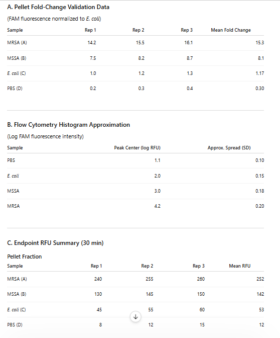
Pellet Fluorescence Analysis
The pellet fraction showed the highest fluorescence intensity for MRSA, indicating maximal accumulation of the construct on bacterial cells after centrifugation.
Observed trend:
MRSA>MSSA≫E. coli>PBS
Interpretation:
MRSA possesses the highest affinity for the CBD construct.
MSSA shows moderate interaction, likely due to shared staphylococcal cell wall features.
E. coli demonstrates minimal nonspecific binding.
PBS confirms negligible background fluorescence.
2.Fold-Change Validation
The fold-enrichment analysis demonstrated:
MRSA binding exceeded the predefined assay threshold (>5-fold),
MSSA displayed intermediate enrichment,
E. coli remained near baseline.
Interpretation:
The construct satisfies the specificity criterion for MRSA recognition.
Strong enrichment relative to E. coli indicates low off-target interaction.
Flow cytometry histograms showed progressive rightward fluorescence shifts from PBS to MRSA.
Interpretation:
Right-shifted peaks correspond to increased fluorescence per bacterial cell.
MRSA exhibited the greatest fluorescence intensity, confirming extensive construct binding at the single-cell level.
Narrow peak distributions suggest relatively uniform binding across the MRSA population.
mecA mRNA silencing in MRSA culture
These are representative synthetic datasets,based on schematic approximations.
Dataset generated by ChatGPT
Figure courtesy: Copilot
Data Interpretation
mRNA Knockdown Validation
RT-qPCR results showed concentration-dependent suppression of mecA mRNA, with >70% knockdown achieved at 100 nM, confirming effective gene silencing by the construct.
Protein Depletion Validation
Western blot analysis demonstrated progressive reduction of PBP2a protein levels with increasing construct concentration, indicating successful translation-level suppression following mecA silencing.
Phenotypic MIC Shift
Oxacillin MIC values decreased significantly after treatment, shifting MRSA toward antibiotic susceptibility, which confirms functional restoration of oxacillin sensitivity.
Dose-Dependent Response
Increasing construct concentrations produced stronger mRNA reduction, greater protein depletion, and lower MIC values, demonstrating clear dose-dependent activity.
CHALLENGES
The challenge might be the trace amounts of the presence of AMR genes, despite the high sensitivity of the biosensor.Most studies are currently limited to laboratory-spiked samples rather than complex real-patient matrices.There is a lack of standardised protocol.Covalent organic frameworks can be used to load nanoclusters (e.g., Au nanoclusters@COF) to amplify fluorescence, enabling detection of ultratrace molecules. This involves benchmarking against gold-standard molecular techniques, rigorous testing in complex matrices, and implementing standardized characterization of the COF-aptamer interface. Test the sensor in real, complex matrices by spiking with known quantities of antibiotic-resistant bacteria or specific AMR DNA fragments to evaluate selectivity, sensitivity, and matrix effects.
The final project has been assembled by Claude.Ai
REFERENCES
1.Adila Nazli, David L. He, Dandan Liao, Muhammad Zafar Irshad Khan, Chao Huang, Yun He,Strategies and progresses for enhancing targeted antibiotic delivery,
Advanced Drug Delivery Reviews,Volume 189,2022,114502,ISSN 0169-409X,https://doi.org/10.1016/j.addr.2022.114502.
3.Mashmoul Moghadam, S. M., Ramezani, M., Alibolandi, M., Abnous, K., & Taghdisi, S. M. (2025). Employing covalent organic framework (COF) as carrier in an aptamer-targeted theranostic nanoplatform: investigation of its therapeutic and diagnostic properties in vitro and in vivo. Journal of Drug Targeting, 33(10), 1892–1901. https://doi.org/10.1080/1061186X.2025.2527865
4.Chen, Shijie & Yan, Huixiang & Tang, Qiukai & Gu, Yiwen & Zhang, Jian & Yang, Yiwen & Wang, Hailong & Qian, Zhao & Li, Lei & Guo, Longhua & Zeng, Yanbo. (2025). A novel FAM-based fluorescent aptasensor with covalent organic framework as quencher for sensitive determination of carcinoembryonic antigen. Microchemical Journal. 215. 114197. 10.1016/j.microc.2025.114197.
5.Hao Y, Xia Y, Huang J, Zhong C, Li G. Covalent-Organic Frameworks for Selective and Sensitive Detection of Antibiotics from Water. Polymers (Basel). 2024 Aug 16;16(16):2319. doi: 10.3390/polym16162319. PMID: 39204541; PMCID: PMC11359747.
6.Zhang W, Liu S, Sun Q, et al. Synthesis of covalent organic framework materials and their application in the field of sensing. Nano Research, 2024, 17(1): 162-195. https://doi.org/10.1007/s12274-023-6027-x
7.Kretzer JW, Lehmann R, Schmelcher M, Banz M, Kim K, Korn C, Loessner MJ 2007. Use of High-Affinity Cell Wall-Binding Domains of Bacteriophage Endolysins for Immobilization and Separation of Bacterial Cells. Appl Environ Microbiol 73:.
https://doi.org/10.1128/AEM.02402-06
9.Anthropic. (2026). Claude 3.5 Sonnet [Large language model]. Retrieved May 20, 2026, from https://claude.ai helped me to assemble the entire experimental design and validation for the project by answering my detailed questions.
10.Google. Gemini. Vers. 2.0, Google, 2026, google.com.- to create datasets of simulated values and drawing schematic figure of CBD assay
11.Microsoft. Copilot. Vers. 2.0, Microsoft, 2026, microsoft.com to create figures of assays and in vitro transcription process and COF nanoparticle synthesis and also of functionalization of COF and mec A silencing assay
14.Benchling [Biology Software]. (2026)-for mec a DNA construct in pTwist chlo high copy plasmid and Golden Gate Assembly of LysK protein in pBR322 plasmid
15.National Center for Biotechnology Information (NCBI)[Internet]. Bethesda (MD): National Library of Medicine (US), National Center for Biotechnology Information; [1988] – [cited 2026,April 17]. Available from: https://www.ncbi.nlm.nih.gov/
The various AI tools were used for different figures and datasets becasue each one provided the required sharp image with correct graph scaling meeting requirements for respective aspects.
GROUP MEMBERS: Deep Dalvi,Fabrizio Flores Huamán, Ganapathy Nayagam, Sheila Ramani
Hypothesis: Engineering Lysis Protein Stability via Targeted Mutations and Chaperone-Independent Folding
We hypothesize that stability can be enhanced by:
(i) introducing mutations that promote independent folding or co-folding with the chaperone DnaJ,
(ii) leveraging evolutionary conservation and generative protein design to generate variants with improved thermodynamic stability and host compatibility.
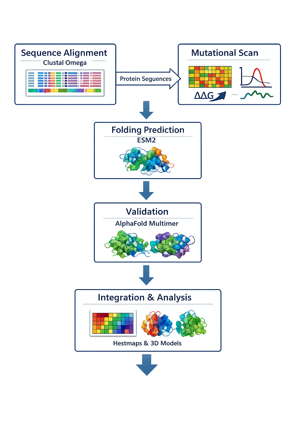
Specific Aims and Validation Pipeline:
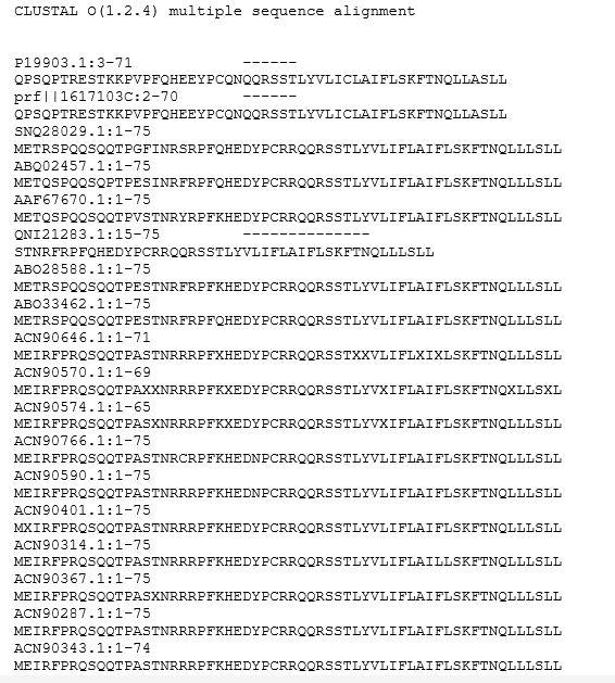
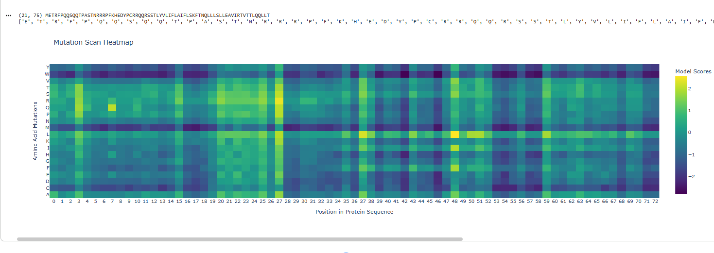
Mutation Design via Conservation and Predictive Modeling
Perform Clustal Omega alignment of homologous lysis proteins to identify conserved residues, followed by mutational scanning (e.g., via deep mutational scanning) and ESM2 embedding predictions to nominate stabilizing mutations. Predicted single and combinatorial mutants will be modeled for folding changes using ESM2, with validation of fold accuracy via AlphaFold-Multimer (AF2-Multimer) to assess independent folding propensity.
Clustal omega showing multiple alignments
Mutational maps gives an idea of which areas are prone to disastrous effects on mutation.
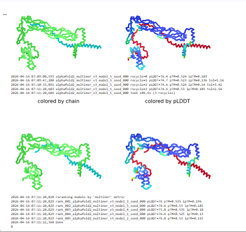
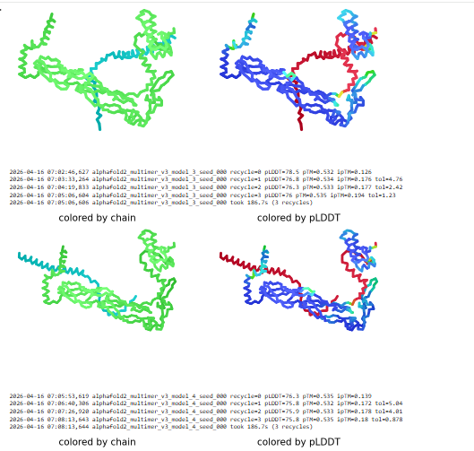
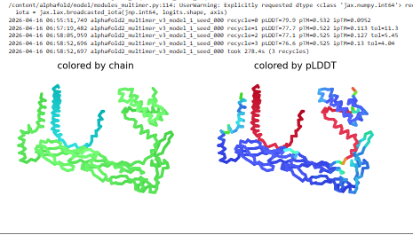
Generative Design for DnaJ Co-Folding
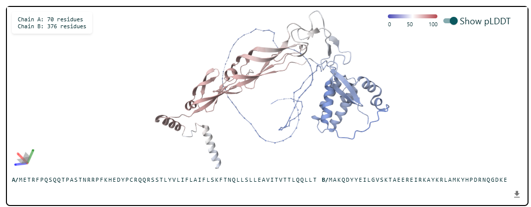
Employ protein generative models (e.g., RFdiffusion or ProteinMPNN) to design lysis protein variant optimized for co-folding with DnaJ. Co-complex structures will be predicted and ranked by AF2-Multimer confidence metrics (pLDDT > 80), prioritizing designs with buried interfaces and reduced aggregation risk.
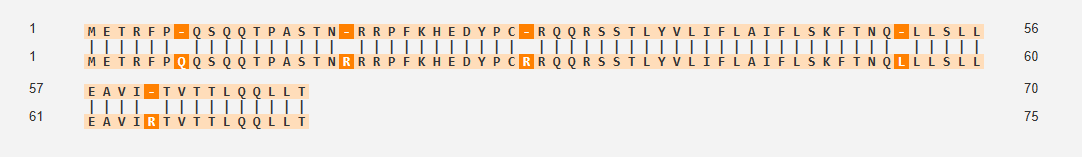
Based on the Clustal omega results mutation was done on the below sites and the protein co folded with DnaJ.
Courtesy: Neurosnap and tamarindBio and VectorBuilder
Mutational sites for creating the mutant lysis protein
The cofolded structure predictions after mutation
Evolutionary Analysis and Chaperone Generalization
Use pBLAST to survey lysis protein orthologs across bacteria, reconstructing evolutionary trajectories of stability determinants and chaperone interactions. Top candidates will be redesigned for co-folding with alternative chaperones (e.g., DnaK or GroEL), with AF2-Multimer analysis of mature protein folds and in silico stability assays (e.g., ΔΔG predictions) against E. coli host factors to minimize proteotoxicity.
Courtesty: pBLAST tool.
Tools and Goals
The main engineering goal is to prevent or reduce the biologically important interaction between DnaJ and the lysis protein, because host-mediated adaptation of this pathway may reduce lysis efficiency and contribute to resistance-associated escape.
To address this, the proposed workflow pursues three solution classes: mutation-driven DnaJ independence, engineered co-folding with DnaJ under controlled structural constraints, and folding rescue by alternative or orthogonal chaperones.
The computational tools consist of sequence alignment for conservation analysis, mutational scanning to identify sensitive and permissive residues, structure prediction to evaluate folding outcomes, generative design to explore new sequence-structure solutions, and evolutionary comparison to detect transferable mechanisms across lysis protein families.
Together, these tools support a design-build-test framework for generating lysis modules that are more stable and less vulnerable to host-dependent failure modes.
Mutations at sites 7 and 8 removed QQ and left a gap in the sequence
Mutations at site 7 8 and 9 removed QQS and left a gap
Deletions that remove the entire N‑terminal basic region (and therefore also remove 7–9) eliminate DnaJ binding and DnaJ requirement, and actually accelerate lysis, because the N‑terminal domain is no longer there to interfere with target binding when not chaperoned by DnaJ.(Jennifer et. al)
Mutation at 61 site E reoved and a gap is left
Position 61 falls within this TM domain that drives membrane insertion, oligomerization into higher‑order complexes, and the formation of large lesions spanning the outer membrane, peptidoglycan, and inner membrane.Chosen as one mutation was to be done in the C-domain of the protein.(Mezhyrova J et.al)
Mutation at site 3 with I instead of T
Position 3 is located in the N-terminal soluble domain of the L-protein.May alter the kinetics, efficiency, or interaction with host proteins (like DnaJ) due to their location in the regulatory N-terminal domain. ( Chamakura et al.)
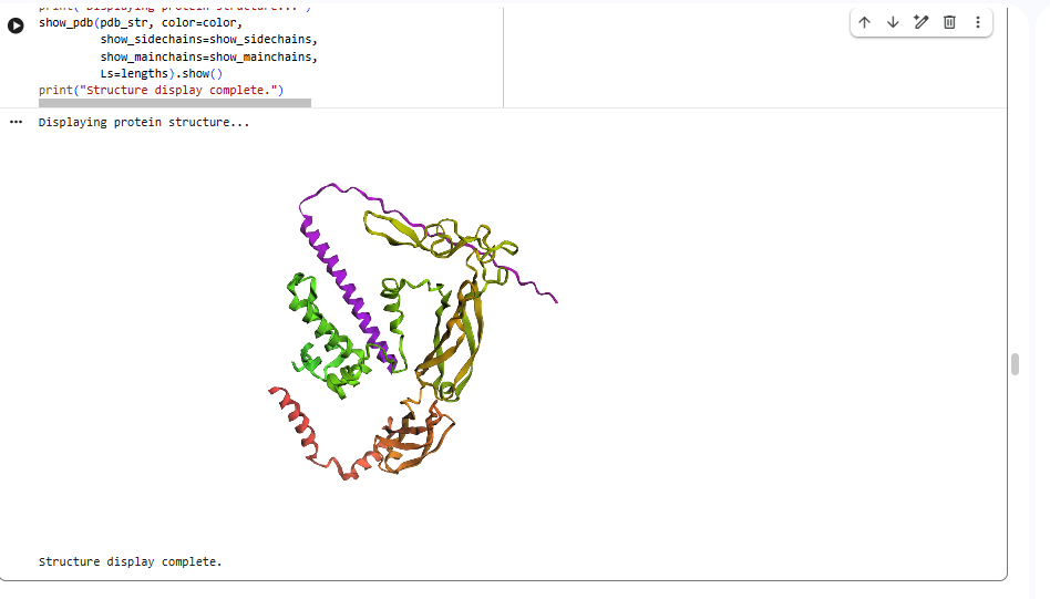
esmfold predicted structure
Validation Pipeline
The schematic pipeline begins with multiple sequence alignment and mutational interpretation, then proceeds to structure-guided mutant design, co-folding assessment with DnaJ, and final structural evaluation of the redesigned lysis protein. Structural predictions from AlphaFold-Multimer and ESM-based folding tools will be used to prioritize candidates before downstream experimental validation.
Courtesy: Copilot
Potential Pitfalls
A major risk is that mutation of residues involved in chaperone recognition may also perturb the native fold, membrane interaction properties, or lytic activity of the protein, leading to partial or complete loss of function. This is especially relevant for compact lysis proteins in which small sequence changes can have disproportionately large structural effects.
A second risk is that co-folding with alternative chaperones may produce unexpected host interactions, altered trafficking, or proteostasis stress in E. coli, particularly if the engineered complex is not part of the native folding network.
Therefore, structural confidence must be paired with functional testing, because even high-confidence predicted folds may not guarantee productive biological activity.
Expected Outcome
If the hypothesis is correct, the resulting lysis protein variants should show improved predicted structural stability, decreased dependence on native DnaJ interaction, and retention of a mature fold compatible with lytic activity in E. coli.
More broadly, this strategy could establish a generalizable framework for reengineering host-dependent phage lysis proteins into more robust synthetic biology modules for antimicrobial and biosensing applications.
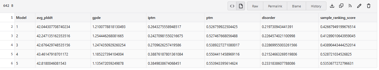
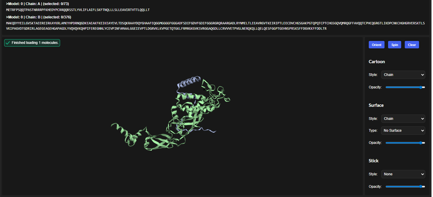
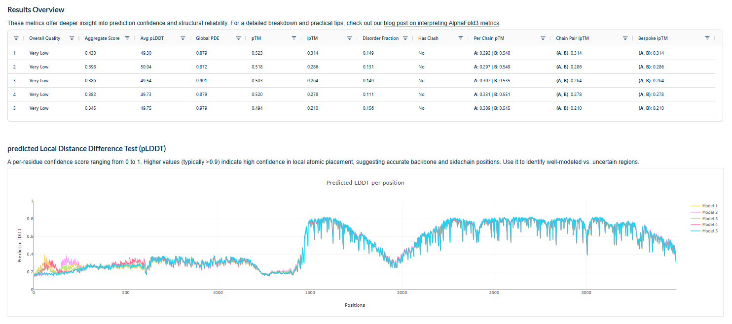
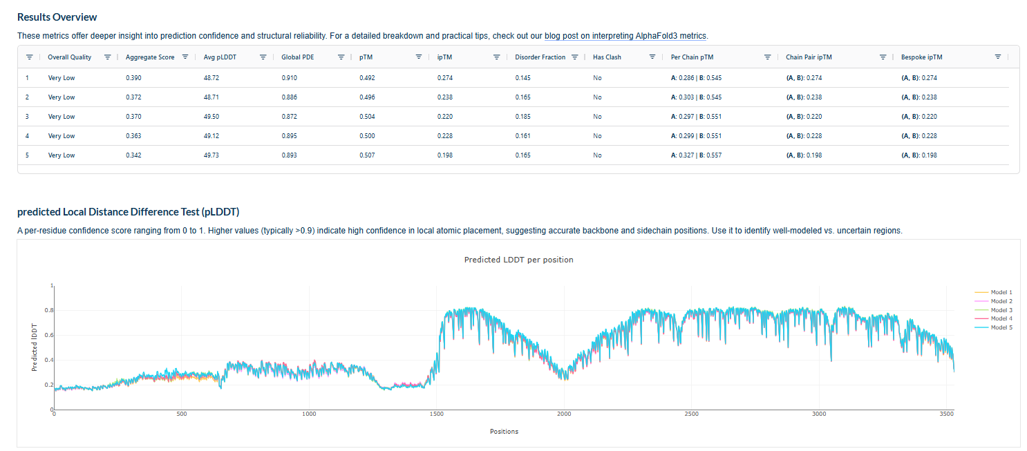
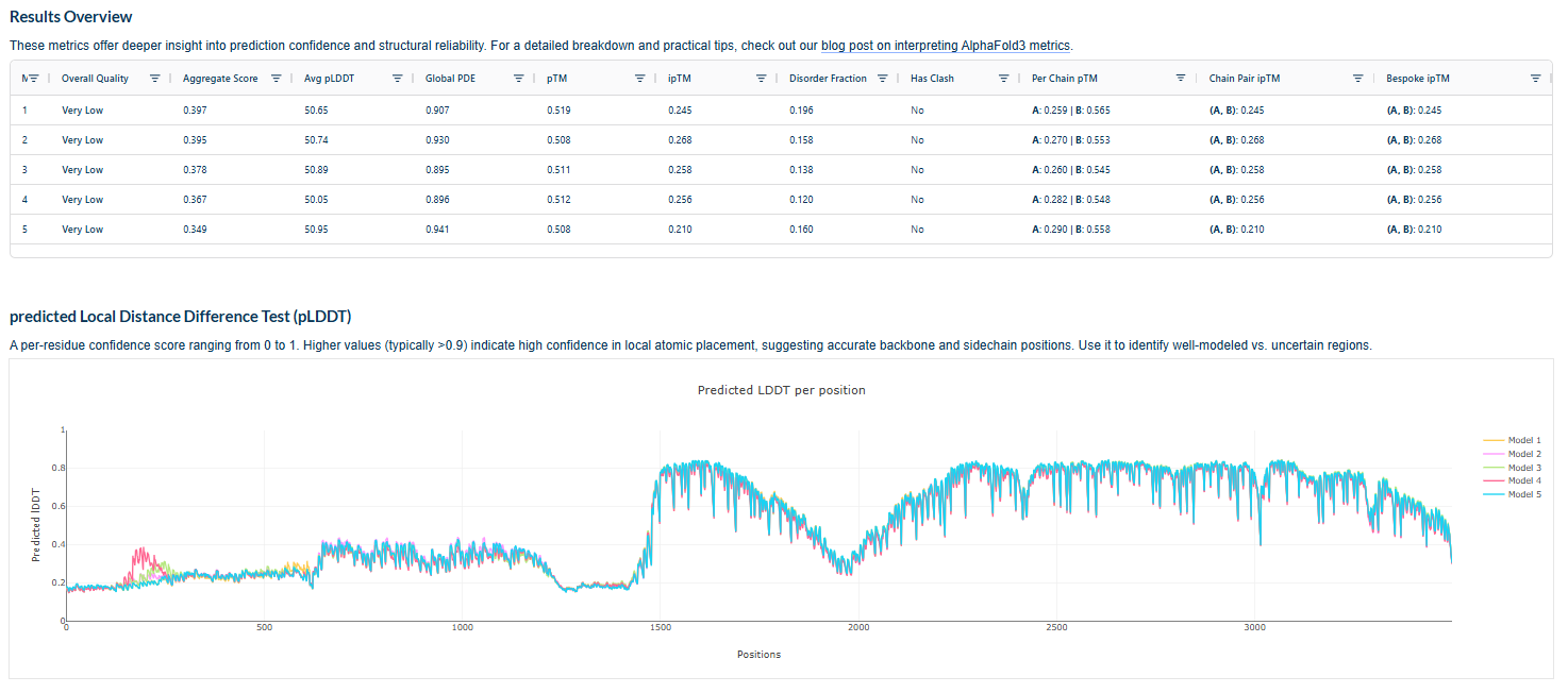
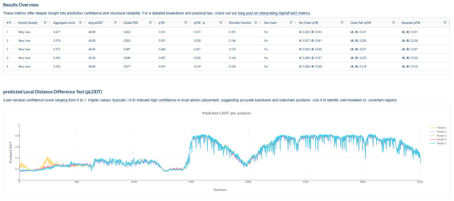
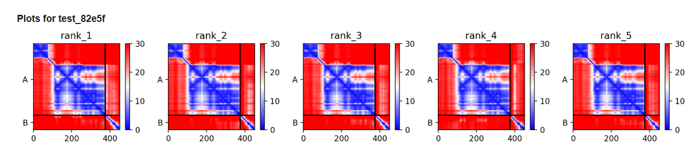
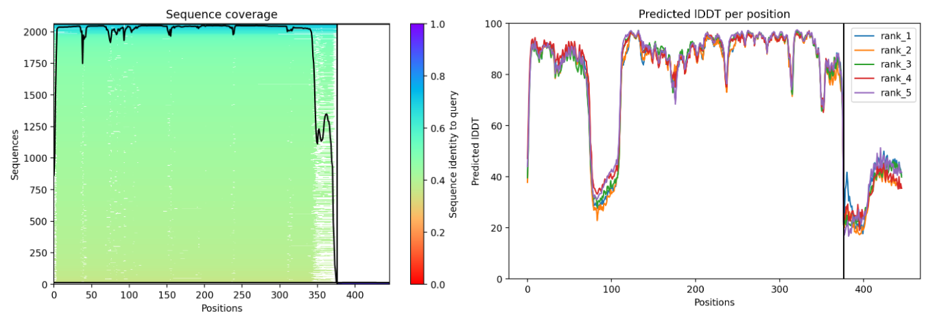
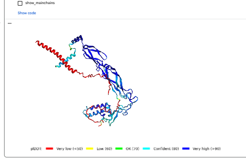
Courtesy: Alphafold3 colab notebook
Plots for the models
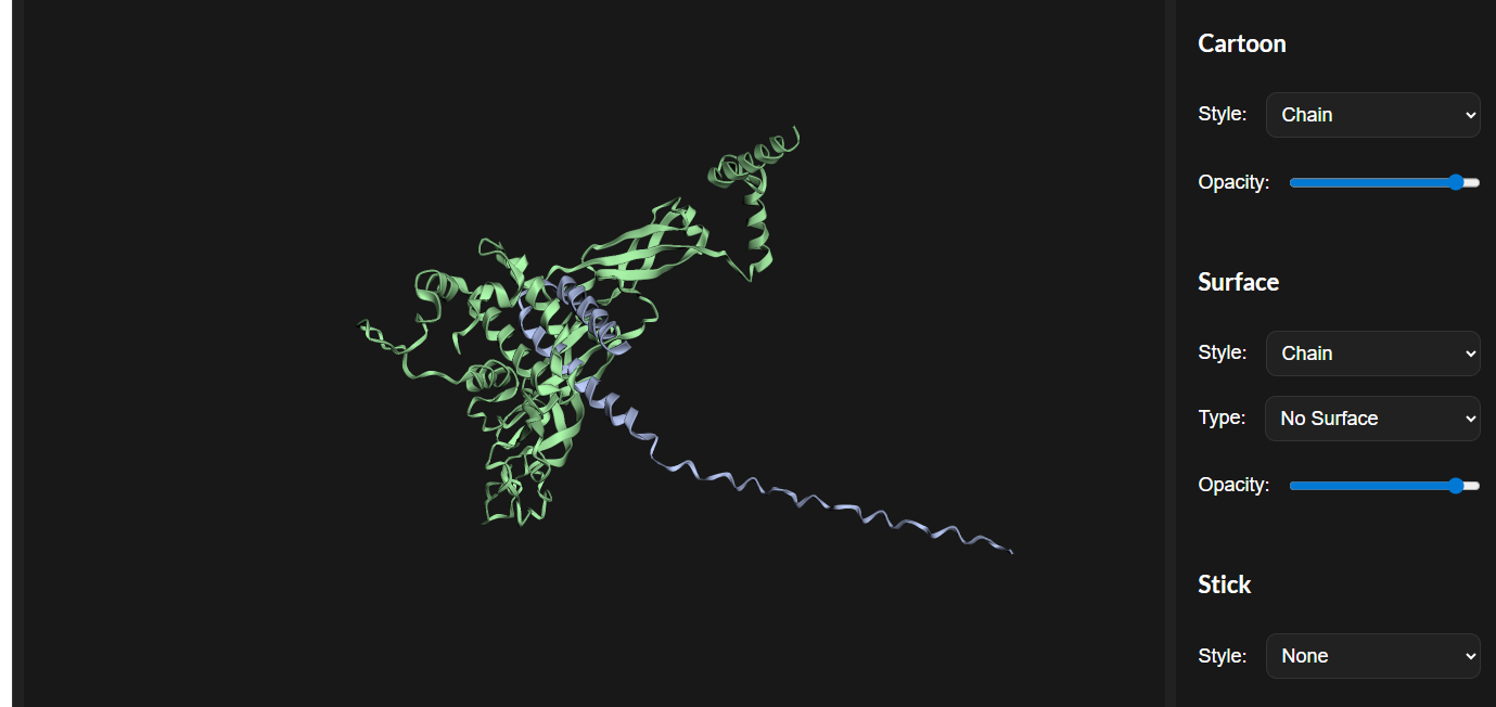
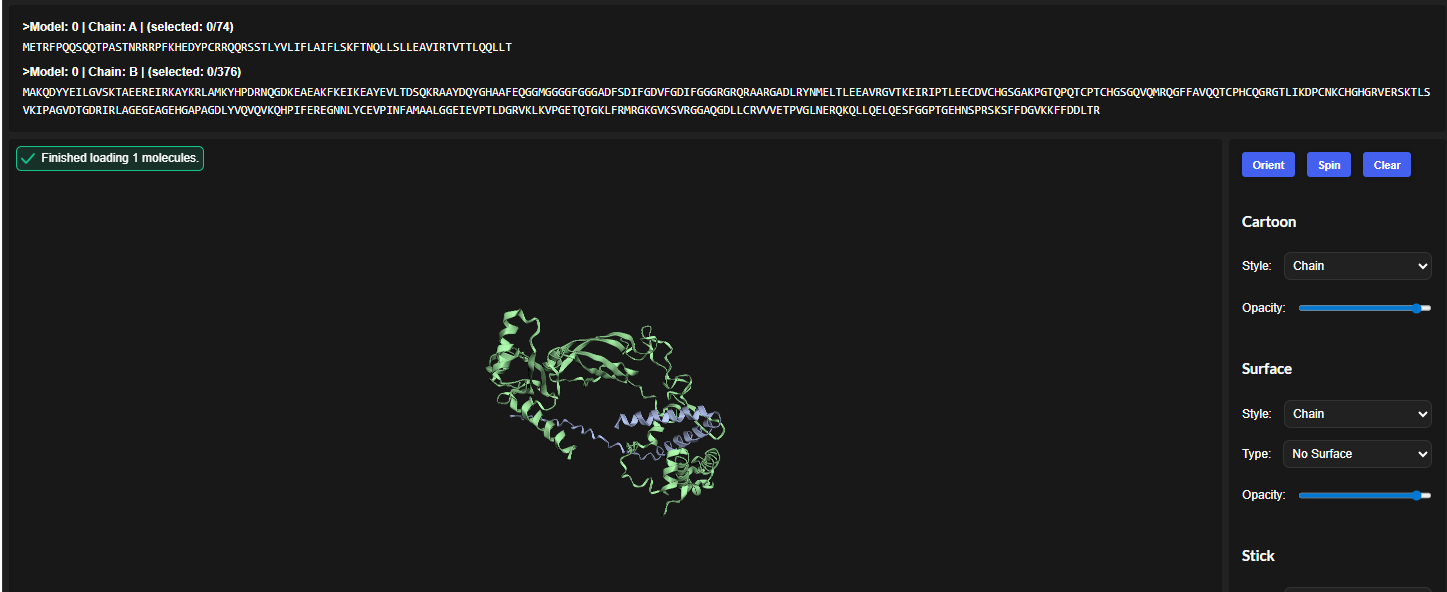
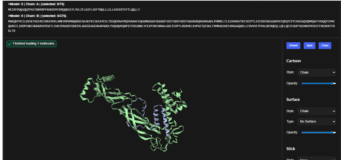
The predicted model using Alphafold3 for the mutant.
Prediction Interpretation
The image by itself does not strongly support a specific co-folded structure.possibly, but this plot alone does not show evidence that it already co-folds well.A better test is to run a complex prediction with the relevant partner and inspect whether the interface residues are consistently high confidence and whether the relative placement is stable.
References:
Chamakura KR, Edwards GB, Young R. Mutational analysis of the MS2 lysis protein L. Microbiology (Reading). 2017 Jul;163(7):961-969. doi: 10.1099/mic.0.000485. Epub 2017 Jul 21. PMID: 28691656; PMCID: PMC5775895.
Chamakura KRTran JS, Young R2017.MS2 Lysis of Escherichia coli Depends on Host Chaperone DnaJ. J Bacteriol199:10.1128/jb.00058-17.https://doi.org/10.1128/jb.00058-17
Mezhyrova J, Martin J, Börnsen C, Dötsch V, Frangakis AS, Morgner N, Bernhard F. In vitro characterization of the phage lysis protein MS2-L. Microbiome Res Rep. 2023 Jul 20;2(4):28. doi: 10.20517/mrr.2023.28. PMID: 38045926; PMCID: PMC10688784.
4.Abramson, J., Adler, J., Dunger, J. et al. Accurate structure prediction of biomolecular interactions with AlphaFold 3. Nature 630, 493–500 (2024). https://doi.org/10.1038/s41586-024-07487-w
5.Zeming Lin et al. ,Evolutionary-scale prediction of atomic-level protein structure with a language model.Science379,1123-1130(2023).DOI:10.1126/science.ade2574
6.Abramson, J., Jumper, J., Evans, R. et al. (2024). Accurate structure prediction of biomolecular interactions with AlphaFold 3. Nature 630, 455–466.