Homework
Weekly homework submissions:
Nature’s machinery for copying DNA is called polymerase. What is the error rate of polymerase? How does this compare to the length of the human genome. How does biology deal with that discrepancy? The error rate of polymerases depends on their type, as Human DNA has mechanisms for proofreading that other organisms’ DNA lacks. The error rate for DNA polymerase is 1 in every 107 base pairs. As compared to the human genome size of 6 X 10⁹ base pairs. The mechanisms include mismatch repair, base excision repair, nucleotide excision repair, NHEJ, HR, damage checkpoint, and some tolerance mechanisms. This is how biology deals with discrepancies via multiple mechanism before-during-after DNA replication and the cell cycle.
Week 01 HW: Principles and Practices
First, describe a biological engineering application or tool you want to develop and why. This could be inspired by an idea for your HTGAA class project and/or something for which you are already doing in your research, or something you are just curious about. As I have been thinking about the different ways synthetic biology can help with menstruation and its complexities (painful, irregular bleeding) when it comes to people with PCOS and endometriosis. I want to develop an autonomous endometrial gene circuit that senses estrogen or progesterone peaks and locally regulates endometrial growth. The goal is non-hormonal, reversible control of menstrual bleeding, which could prevent heavy bleeding or abnormal endometrial proliferation while minimizing systemic hormone exposure.
Week 02 HW: DNA Read Write & Edit
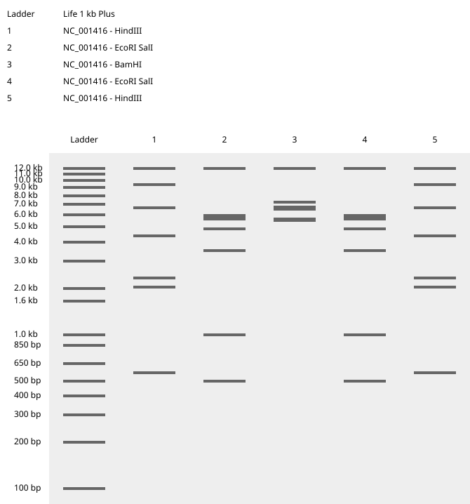
Part 01: Restriction Digestion Art Gel Image produced via Benchling to produced a rendition of the logo of a pop band called BTS. 3.1 Which protein have you chosen and why? Using one of the tools described in recitation (NCBI, UniProt, google), obtain the protein sequence for the protein you chose. Below is the protein sequence for ESR2 Human estrogen beta using UniProt. I have chosen this protein as it has been
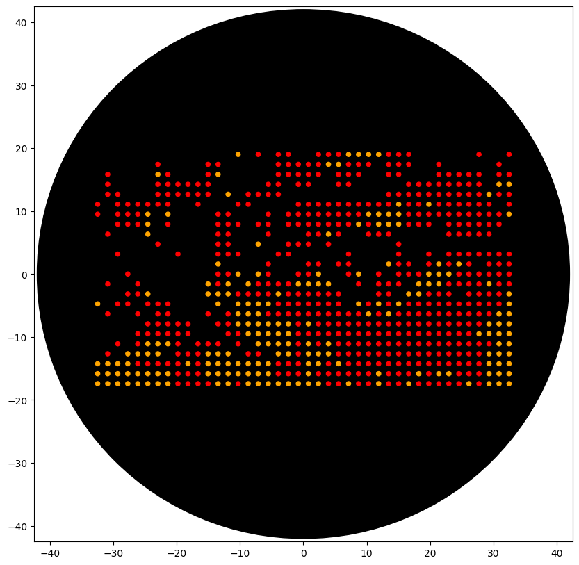
Part 01: Opentron python file to create a image. Here is my code for the image below alongwith the prerequisitve codes before and after the - I used Sonnet 4.6 for the task. from opentrons import types metadata = { ‘author’: ’tanishka’, ‘protocolName’: ‘recreate img of an eye’, ‘description’: ‘’, ‘source’: ‘HTGAA 2026 Opentrons Lab’, ‘apiLevel’: ‘2.20’ } ############################################################################## Robot deck setup constants - don’t change these ##############################################################################
Week 04 HW: Protein Design Part 1
Part A : How many molecules of amino acids do you take with a piece of 500 grams of meat? (on average an amino acid is ~100 Daltons) No. of AA molecules in 500gm of meat = Proteins consist of amino acids linked by peptide bonds, losing ~18 Da (water) per bond, so ~0.9–1 g protein yields ~1 mol amino acid residues (using your ~100 Da average residue weight). Protein in 500 g meat: ~120 g average (24% protein content). Moles of amino acids: 120 g / 100 g/mol = 1.2 mol. Molecules: 1.2 mol × 6.022 × 10²³ = ~7.2 × 10²³ (adjusts to ~2.3 × 10²⁴ if using precise 110 Da avg
Week 05 HW: Protein Design Part II
Part 1: Generate Binders with PepMLM The target is human SOD1 protein (UniProt P00441), focusing on A4V mutation, which is ALS. Mutant SOD1 Sequence (A4V): ATKVVCVLKGDGPVQGIINFEQKESNGPVKVWGSIKGLTEGLHGFHVHEFGDNTAGCTSAGPHFNPLSRKHGGPKDEERHVGDLGNVTADKDGVADVSIEDSVISLSGDHCIIGRTLVVHEKADDLGKGGNEESTKTGNAGSRLACGVIGIAQ Using PepMLM-650M, four peptides of 12 amino acids were generated and compared against known SOD1-binding peptide FLYRWLPSRRGG. PepMLM Confidence Scores Sequence Description Perplexity FLYRWLPSRRGG Real Binder — WHSPVVAVAHWE Sim 1 10.949699 WSVGWAAIAWWX Sim 2 16.027645 WRSYATAIALWK Sim 3 11.729657 WRYYATGAEWKE Sim 4 13.769973 Part 2: Evaluate Binders with AlphaFold3 Each peptide was modeled against the mutant SOD1 sequence using AlphaFold3 to assess structural docking and interface confidence (ipTM).
Week 06 HW: Genetic Circuits Part I
Part one 1: What are some components in the Phusion High-Fidelity PCR Master Mix, and what is their purpose? Phusion DNA Polymerase, Nucleotides, Optimised Phusion HF Reaction Buffer with MgCl2 - All at 2X concentration Generates long templates with high accuracy and speed, unattainable with a single enzyme, as per the Thermo Scientific protocol. It states that the error rate of Phusion DNA Polymerase is determined to be 4.4 × 10⁻⁷ in Phusion HF Buffer, which is approximately 50-fold lower than that of Thermus aquaticus DNA polymerase, and 6-fold lower than that of Pyrococcus furiosus DNA polymerase. Attached below are graphs that support these claims by comparing with traditional polymerase. The annealing temperature is at 60 degrees for all. https://documents.thermofisher.com/TFS-Assets/LSG/brochures/phusion-high-fidelity-dna-polymerases-flyer.pdf https://documents.thermofisher.com/TFS-Assets/LSG/manuals/MAN0012771_Phusion_HiFi_PCR_MasterMix_100rxn_UG.pdf
Week 07 HW: Genetic Circuits Part II
Part 1: Intracellular Artificial Neural Networks 1: What advantages do IANNs have over traditional genetic circuits, whose input/output behaviors are Boolean functions? IANNs process analog signals instead of only ON/OFF outputs. They handle noisy data better and perform complex decisions using fewer genetic parts. 2: Describe a useful application for an IANN; include a detailed description of input/output behavior, as well as any limitations an IANN might face to achieve your goal. An IANN can be used in cancer therapy cells. The cell detects tumor biomarkers and releases a killing protein only when signal levels match cancer conditions. A limitation is high energy use, which may slow growth or cause mutations.
Part One: General and Lecturer-Specific Questions 1. Cell-free protein synthesis gives better control over reaction conditions and allows direct addition of molecules like inhibitors or non-natural amino acids. It is useful for producing toxic proteins and for rapid protein prototyping. A cell-free system contains lysate with ribosomes and enzymes, a DNA/mRNA template, amino acids, and buffer salts. These components work together to produce proteins from genetic instructions. ATP regeneration is important because protein synthesis uses large amounts of energy. A creatine phosphate–creatine kinase system can recycle ADP into ATP and maintain continuous protein production.
Week 10 HW: Imaging and Measurement
Waters Part I — Molecular Weight 1: Based on the predicted amino acid sequence of eGFP and any known modifications, what is the calculated molecular weight? eGFP Sequence Analysis: The sequence provided includes the eGFP core, an LE linker, and a 6x-His purification tag. 2:Calculated Molecular Weight: 27,988.97 Da (Daltons). Calculate the molecular weight of the eGFP using the adjacent charge state approach. Using Figure 1, we select two adjacent peaks:
Week 11 HW: Bioproduction & Cloud Labs
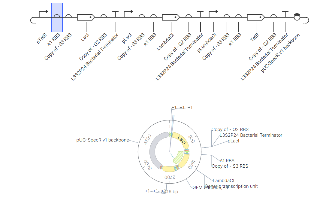
Part A: The 1,536 Pixel Artwork Canvas Contribution I designed a small section of the artwork using a few pixels to again create the BTS pop band logo, cos i am obsessed. Might not look like it, as the pixels were already too full. I liked how different ideas were combined into one large artwork. For next time, Divided sections for nodes could make collective teamwork visible. Part B: Cell-Free Protein Synthesis