Bioprinting horizontal gene transfer: a bacterial “photocamera” SECTION 1: ABSTRACT Horizontal gene transfer (HGT) via bacterial conjugation is a primary mechanism driving the dissemination of antibiotic resistance genes in clinical and environmental microbial communities, yet the spatial dynamics of conjugative transfer remain poorly visualized in real time. This project proposes to engineer a mobilizable donor plasmid encoding the bglA β-glucosidase reporter gene from Enterococcus faecalis, synthesized as a Clonal Gene from Twist Bioscience (pTwist Amp High Copy backbone), and to deploy it in a triparental mating system using E. coli DH5α as both donor and helper strains and E. coli K-12 wild-type as the recipient. Gene transfer is spatially visualized on UTI chromogenic agar, which contains dual chromogenic substrates for β-galactosidase and β-glucosidase: the recipient K-12 strain (LacZ⁺) produces pink colonies, the donor DH5α (BglA⁺, LacZ⁻) produces teal colonies, and transconjugants expressing both enzymes produce a distinctive purple/lilac color that is absent from either parental strain alone. The Opentrons OT-2 robotic liquid handler deposits all three strains in geometrically precise concentric overlapping circles onto plain LB agar for conjugation, and subsequently onto UTI chromogenic agar for spatial readout. Transconjugant identity is confirmed by colony PCR targeting both the plasmid-encoded bglA and the chromosomal lacZ of K-12. This project establishes a proof-of-concept platform for the spatial mapping of plasmid transfer events, providing a visual, automation-compatible, and instrument-free assay for studying conjugation dynamics with direct relevance to antibiotic resistance surveillance and the design of synthetic microbial communities.
Bioprinting horizontal gene transfer: a bacterial “photocamera”
SECTION 1: ABSTRACT
Horizontal gene transfer (HGT) via bacterial conjugation is a primary mechanism driving the dissemination of antibiotic resistance genes in clinical and environmental microbial communities, yet the spatial dynamics of conjugative transfer remain poorly visualized in real time. This project proposes to engineer a mobilizable donor plasmid encoding the bglA β-glucosidase reporter gene from Enterococcus faecalis, synthesized as a Clonal Gene from Twist Bioscience (pTwist Amp High Copy backbone), and to deploy it in a triparental mating system using E. coli DH5α as both donor and helper strains and E. coli K-12 wild-type as the recipient. Gene transfer is spatially visualized on UTI chromogenic agar, which contains dual chromogenic substrates for β-galactosidase and β-glucosidase: the recipient K-12 strain (LacZ⁺) produces pink colonies, the donor DH5α (BglA⁺, LacZ⁻) produces teal colonies, and transconjugants expressing both enzymes produce a distinctive purple/lilac color that is absent from either parental strain alone. The Opentrons OT-2 robotic liquid handler deposits all three strains in geometrically precise concentric overlapping circles onto plain LB agar for conjugation, and subsequently onto UTI chromogenic agar for spatial readout. Transconjugant identity is confirmed by colony PCR targeting both the plasmid-encoded bglA and the chromosomal lacZ of K-12. This project establishes a proof-of-concept platform for the spatial mapping of plasmid transfer events, providing a visual, automation-compatible, and instrument-free assay for studying conjugation dynamics with direct relevance to antibiotic resistance surveillance and the design of synthetic microbial communities.
SECTION 2: PROJECT AIMS
Aim 1 — Experimental Aim
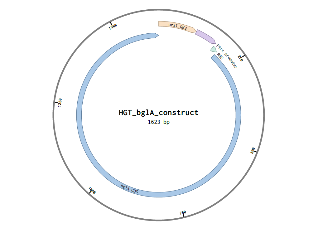
The first aim of my final project is to engineer a mobilizable bglA-encoding donor plasmid and demonstrate its triparental conjugative transfer into E. coli K-12 using Opentrons OT-2 bioprinting by ordering the synthetic construct (oriT_RK2 + Ptrc + RBS + bglA CDS) in pTwist Amp High Copy from Twist Bioscience, transforming DH5α donor and helper strains, printing overlapping concentric circles of all three strains onto LB agar for mating, and detecting purple/lilac transconjugant colonies on UTI chromogenic agar (±ampicillin) as a spatial readout of horizontal gene transfer.
Aim 2 — Medium-Term Aim
The second aim is to expand the bacterial “photocamera” system into a multi-gene, multi-colour platform and translate its visual logic into an interactive simulation game, enabling both advanced experimental development and a new art-science medium for communicating horizontal gene transfer dynamics to broader audiences.
On the experimental side, the single bglA chromogenic reporter would be extended to a panel of orthogonal reporter genes, each producing a spectrally distinct colony colour on the same or different chromogenic media — effectively creating a living colour palette where each hue encodes a distinct transferred genetic element. Multiple donor strains carrying different cargo genes could then be printed in the same bioprinted spatial pattern, producing a multicolour colony mosaic whose spatial distribution directly maps the transfer history and directionality of each gene. The Opentrons OT-2 print geometry would be systematically varied (concentric circles, gradients, chessboard, radial sectors) to generate spatial models of conjugation efficiency as a function of cell density, overlap area, and incubation time, with colony colour distributions quantified via RGB image segmentation.
In parallel, the spatial logic of this system — bacteria placed in defined positions, genes flowing between them as colour — lends itself naturally to an interactive simulation game. Players would arrange donor, helper, and recipient colonies on a virtual agar plate, set conjugation parameters, and observe the resulting colour patterns emerge over simulated time, with rules derived directly from the experimental data. This game would function simultaneously as a science communication tool, making the invisible dynamics of horizontal gene transfer and antibiotic resistance spread visually intuitive and accessible to students, patients, and the general public — transforming the petri dish into both a scientific instrument and an artistic canvas.
Aim 3 — Visionary Aim
In the long term, this platform could serve as the foundation for programmable gene transfer networks in synthetic microbial consortia — enabling controlled, spatially defined introduction of metabolic, biosensing, or therapeutic functions into complex communities without requiring direct genetic manipulation of the recipient. Combined with bioprinting and emergent chromogenic reporters, future applications could include real-time surveillance of resistance gene dissemination in hospital surface microbiomes, the creation of living spatial diagnostics that self-report gene spread, or self-organizing bacterial computing architectures guided by conjugation gradients — transforming the bacterial petri dish into a programmable biological circuit board.
SECTION 3: BACKGROUND
Literature Context
Triparental mating was established as a foundational tool in bacterial genetics by Figurski and Helinski (1979), who demonstrated that the broad-host-range plasmid RK2 encodes a complete conjugation system capable of mobilizing any non-self-transmissible plasmid carrying the RK2 origin of transfer (oriT) in cis, provided that the trans-acting transfer functions are supplied by a co-resident helper plasmid. Their work showed that the minimal oriT sequence — approximately 250 bp containing the nick site (nic) recognized by the relaxase TraI — was sufficient for mobilization, enabling the modular design of small, engineerable donor plasmids. Building on this principle, Ditta et al. (1980) developed the pRK2013 helper plasmid as a dedicated trans-helper tool for broad-host-range conjugation, a reagent that has since been used in thousands of studies to transfer genetic cargo into diverse gram-negative bacteria. Despite these mechanistic advances, however, the spatial organization of conjugation events in structured environments — specifically, how cell density gradients, physical overlap geometry, and microcolony proximity affect transfer frequency on solid surfaces — has received comparatively little experimental attention, representing a clear knowledge gap that this project directly addresses.
Innovation
This project is novel in combining robotic bioprinting with a dual-substrate chromogenic detection system specifically engineered to produce an emergent third color — purple/lilac — that marks gene transfer events directly on the agar surface without requiring fluorescence microscopy, flow cytometry, or any specialized imaging instrumentation. While conjugation assays are conventionally performed in homogeneous liquid culture or by spot-mixing on agar, the use of an Opentrons OT-2 to deposit strains in geometrically precise, reproducible overlapping patterns introduces spatial organization as a controlled experimental variable for the first time in this context. Furthermore, the selection of bglA as a chromogenic payload — rather than a fluorescent protein — leverages the dual-substrate composition of UTI chromogenic agar to produce a phenotypically distinct transconjugant color class, making gene transfer events readable with the naked eye, immediately after overnight incubation, on inexpensive commercially available media.
Significance
Horizontal gene transfer via conjugation is the primary mechanism by which antibiotic resistance genes spread between bacterial species in clinical environments, making the spatial dynamics of this process directly relevant to global public health challenges. Understanding where in structured microbial populations (biofilms, colonies, gut mucosa) transfer events preferentially occur could reveal new strategies for blocking resistance dissemination at its physical source. This project provides the first spatially resolved, color-coded readout of conjugation that is simultaneously visual, quantifiable, and automatable, offering a new experimental paradigm for studying conjugation in structured environments. The modular architecture of the donor plasmid — with bglA as a swappable chromogenic payload downstream of oriT and a constitutive promoter — means that the reporter gene can be replaced with any gene of interest, converting this into a generalizable platform for studying or engineering conjugative gene delivery. Finally, by coupling this system to Opentrons OT-2 bioprinting, the platform is directly amenable to high-throughput screening formats, enabling future testing of conjugation inhibitors, promoters, or alternative geometric designs in a scalable, reproducible workflow.
Bioethical Considerations
Ethical considerations: This project involves the deliberate engineering of a conjugative gene transfer system, which by its nature requires careful ethical scrutiny due to the inherent capacity of conjugation to spread genetic material between bacteria. The donor plasmid carries an ampicillin resistance gene (AmpR, bla) that, if transferred to environmental bacteria through improper waste disposal, could contribute to the environmental pool of resistance genes. The project also mimics — in a simplified and controlled form — the same biological mechanism responsible for the real-world dissemination of clinically significant resistance determinants. These risks are not trivial and must be taken seriously: all experimental work must be conducted under BSL-1 or BSL-2 containment as determined by institutional biosafety committee (IBC) assessment, with strict adherence to good microbiological practice. Additionally, the synthetic insert must be screened through SecureDNA prior to ordering from Twist Bioscience, to ensure that no sequences of dual-use concern are inadvertently included in the design.
Responsible implementation and risk mitigation: All bacterial cultures, chromogenic agar plates, and liquid waste containing live organisms must be autoclaved at 121°C for at least 30 minutes before disposal, in accordance with standard biohazardous waste management protocols. The recipient strain E. coli K-12, and the donor/helper strain E. coli DH5α, are both well-characterized laboratory strains with multiple biosafety-relevant auxotrophies and genetic features that severely limit their persistence and competitiveness in natural environments — substantially reducing the risk of accidental environmental release. The bglA gene itself is non-toxic and non-pathogenic, encoding a carbohydrate-hydrolyzing enzyme found in the commensal organism Enterococcus faecalis. The project does not involve human pathogens, toxin genes, virulence factors, or select agents. To further ensure responsible implementation, the Opentrons OT-2 bioprinting steps should be performed inside a biosafety cabinet, and all agar plates bearing live cultures should be sealed with Parafilm or equivalent before handling and imaging outside the cabinet.
SECTION 4: EXPERIMENTAL DESIGN
Detailed Workflow
Step 1 — In Silico Construct Design in Benchling (Week 1)
Method: Design the donor plasmid insert in Benchling using Golden Gate assembly logic with the AarI Type IIS restriction enzyme. The insert is composed of four functional elements in the following 5′→3′ order: oriT_RK2 (minimal origin of transfer, ~250 bp, containing the nic site for TraI relaxase) → Ptrc promoter (~55 bp, hybrid tac/trp; −35: TTGACA, 17 bp spacer, −10: TATAAT) → RBS without ATG (~20 bp, Shine-Dalgarno sequence, spacer) → bglA CDS from Enterococcus faecalis (~1,332 bp, starting with ATG). Design AarI-compatible overhangs flanking the insert for directional cloning into the pTwist Amp High Copy backbone. Verify the reading frame, check for internal AarI sites, and confirm the absence of premature stop codons in silico. Automation: None (Benchling web software, in silico only) Expected result: Complete annotated plasmid map in Benchling; confirmed ORF integrity; AarI overhangs verified. Timeline: Days 1–2
Step 2 — Twist Bioscience Clonal Gene Order (Week 1)
Method: Submit the finalized insert sequence to Twist Bioscience as a Clonal Gene order, selecting pTwist Amp High Copy as the vector backbone. Verify insert orientation and reading frame in the Twist ordering portal before submission. Note: the full construct including backbone is ~3,700 bp, well within Twist synthesis limits. Partner: Twist Bioscience Expected result: Confirmation email and tracking number. Lyophilised plasmid DNA (pTwist-oriT-Ptrc-bglA) received within 7–10 business days. Timeline: Day 2 (order); Days 10–14 (receipt)
Step 3 — Donor Plasmid Transformation into E. coli DH5α (Week 2)
Method: Resuspend received Twist lyophilised plasmid in 10 µL TE buffer. Transform 1–2 µL into E. coli DH5α chemically competent cells using the standard heat shock protocol: mix DNA with competent cells on ice (30 min) → heat shock at 42°C for exactly 45 s → return to ice (2 min) → add 250 µL pre-warmed SOC medium → recover at 37°C, 200 rpm, 1 hour → plate on LB + Ampicillin 100 µg/mL. Incubate plates 16 h at 37°C. Equipment: Heat block or water bath, benchtop incubator/shaker Plates: LB + Amp 100 µg/mL agar Expected result: ≥10 colonies after overnight incubation; negative control (no plasmid) shows zero colonies. Timeline: Day 14–15
Step 4 — Helper Plasmid Transformation into E. coli DH5α (Week 2, parallel to Step 3)
Method: Obtain pRK2013 from Addgene (#37636) as bacterial stab or glycerol stock. Streak onto LB + Kan 50 µg/mL to isolate a colony, then prepare a miniprep for transformation. Transform into E. coli DH5α using identical heat shock protocol as Step 3. Plate on LB + Kanamycin 50 µg/mL. Partner: Addgene (#37636, https://www.addgene.org/37636/) Expected result: ≥10 colonies; helper strain confirmed by expected plasmid size on gel (~11.8 kb pRK2013). Timeline: Day 14–15
Method: Pick 3–5 colonies from donor transformation plates. Perform colony PCR using primers flanking the bglA insert (Fwd: 5′-ATGAAAACAATTGTTGGCATGG-3′; Rev: 5′-TTATTTTGAAGCTTTAAATCC-3′; expected product: ~1,332 bp). Additionally perform PCR with vector-flanking primers to confirm insert size and orientation. Run all products on 1% agarose gel (SYBR Safe stain). Equipment: Thermal cycler, gel electrophoresis apparatus, UV transilluminator Expected result: Correct band ~1,332 bp in ≥2/5 tested colonies; no band in untransformed DH5α control. Timeline: Day 15–16
Method: From verified colonies: inoculate donor DH5α+pTwist-bglA into 5 mL LB + Amp 100 µg/mL; inoculate helper DH5α+pRK2013 into 5 mL LB + Kan 50 µg/mL; inoculate recipient E. coli K-12 into 5 mL plain LB (no antibiotic). Grow all three cultures overnight at 37°C, 200 rpm. Equipment: Shaking incubator, 15 mL culture tubes Expected result: All three cultures visibly turbid; OD600 ≈ 1.5–3.0 by morning. Timeline: Day 17 (inoculate evening) → Day 18 (read morning)
Step 7 — OD600 Normalization with Opentrons OT-2 (Week 3)
Method: Measure OD600 of each overnight culture. Calculate volume needed to reach OD600 = 0.6 in 500 µL fresh LB (no antibiotic). Using Opentrons OT-2 with p300 single-channel pipette, transfer calculated volumes into 1.5 mL Eppendorf tubes (used as reservoirs) and bring to final volume with LB. This yields three normalized cell suspensions at ~5×10⁸ cells/mL. Automation: Opentrons OT-2 (p300 single-channel, 200 µL tips) Expected result: Three tubes at OD600 = 0.6 ± 0.05, confirmed by re-measurement. Timeline: Day 18, morning
Step 8 — Bioprinting Triparental Mating on Plain LB Agar (Week 3)
Method: Using the Opentrons OT-2 with a p20 single-channel pipette and a custom Python protocol, deposit each strain suspension in a concentric circle pattern onto a plain LB agar plate (no antibiotic), using the following spatial layout:
Outer ring (radius ≈ 15 mm from center): Recipient K-12 — 10 spots × 0.5 µL at even angular spacing
Middle ring (radius ≈ 10 mm): Helper DH5α+pRK2013 — 10 spots × 0.5 µL (overlapping with outer ring)
Inner ring (radius ≈ 5 mm): Donor DH5α+pTwist-bglA — 10 spots × 0.5 µL (overlapping with middle ring)
Central spot: All three strains premixed 1:1:1 — 1 µL
Allow spots to air-dry 5 min at room temperature. Incubate plate face-up at 37°C for 16–18 hours to allow conjugation to occur at cell–cell contact interfaces. Automation: Opentrons OT-2 (p20 single-channel) Expected result: Bacterial growth in all zones; visible microcolonies at all spots after incubation. Timeline: Day 18, morning → Day 19, morning (overnight incubation)
Step 9 — Harvesting Cells from the Mating Plate (Week 3)
Method: After overnight incubation, add 200 µL fresh LB (no antibiotic) to the central overlapping zone of the mating plate where all three strains were in contact. Gently scrape cells from the agar surface using a sterile inoculation loop or cell scraper and collect the suspension into a 1.5 mL Eppendorf tube. Using Opentrons OT-2, prepare serial 10-fold dilutions (10⁻¹ through 10⁻⁴) from the harvested suspension in LB, in a 96-well plate or individual Eppendorf tubes. Automation: Opentrons OT-2 (p20 single-channel, serial dilutions) Expected result: Turbid suspension in scrape tube; four dilution steps ready for plating. Timeline: Day 19, morning
Step 10 — Spatial Printing on CHROMagar (No Antibiotic) — Detection Plate 1 (Week 3)
Method: Using Opentrons OT-2, spot 2 µL from each serial dilution at the same concentric circle coordinates as Step 8 onto a UTI chromogenic agar plate (no antibiotic). Additionally, spot 2 µL of each unmixed original strain suspension at separate reference positions on the same plate as single-strain color controls. Automation: Opentrons OT-2 (same Python script and coordinates as Step 8) Plates: UTI CHROMagar (no antibiotic) Expected result (after 16–24 h at 37°C):
Central overlap zone → gradient from pink (outer) through colorless to purple/lilac colonies where transconjugants have formed Timeline: Day 19 (plate) → Day 20 (read)
Method: Repeat identical plating pattern from harvested serial dilutions onto UTI CHROMagar + Ampicillin 100 µg/mL using Opentrons OT-2 with the same protocol. Plates: UTI CHROMagar + Amp 100 µg/mL Strain fate under selection:
Expected result: Only AmpR cells survive; purple colonies in former recipient zones = confirmed transconjugants. Timeline: Day 19 (plate) → Day 20 (read)
Step 12 — Chromogenic Plate Imaging and Color Annotation (Week 4)
Method: Photograph both detection plates under consistent, diffuse white light after 16–24 h incubation. Use a ruler for scale. Annotate color zones: pink (K-12), teal (donor/DH5α+bglA), white/colorless (helper), purple (transconjugant). If available, use ImageJ or Python (Pillow/OpenCV) to quantify RGB values across the plate and map color distribution spatially. Equipment: Camera or smartphone (macro mode), ruler, ImageJ Expected result: Clear three-color spatial pattern visible; purple zone localized to the central overlap region; Plate 2 shows elimination of pink and enhancement of purple relative to Plate 1. Timeline: Day 20
Step 13 — Colony Picking for PCR Validation (Week 4)
Method: From Plate 2 (CHROMagar + Amp), pick 5–10 purple colonies using sterile toothpicks. Also pick 2 teal colonies (donor control) and 2 pink colonies from Plate 1 (recipient control). Resuspend each in 20 µL sterile water; heat lyse at 95°C for 5 min. Equipment: Thermal cycler (lysis step), pipettes, PCR tubes Expected result: Cell lysates from 5–10 purple colonies ready for PCR validation. Timeline: Day 20
Method: For each lysate, run two PCR reactions in parallel:
Reaction A (bglA): Fwd 5′-ATGAAAACAATTGTTGGCATGG-3′ / Rev 5′-TTATTTTGAAGCTTTAAATCC-3′ → expected band ~1,332 bp (present in donor and transconjugant; absent in K-12 recipient alone)
Reaction B (lacZ): K-12 chromosomal primers → expected band ~500 bp (present in K-12 and transconjugant; absent in DH5α which carries lacZΔM15)
PCR program: 95°C 3 min → 30× [95°C 30 s / 55°C 30 s / 72°C 90 s] → 72°C 5 min. Run on 1% agarose gel. Equipment: Thermal cycler, gel electrophoresis apparatus Expected result: True transconjugants = bglA⁺ (Rxn A band) + lacZ⁺ (Rxn B band). Donor controls = bglA⁺ only. Recipient controls = lacZ⁺ only. Timeline: Day 20–21
Step 15 — Conjugation Frequency Calculation and Data Documentation (Week 4)
Method: Count all colony classes on Plate 1 (no Amp) and Plate 2 (+ Amp). Calculate conjugation frequency = (purple colonies on Plate 2) / (pink colonies on Plate 1) per dilution step. Average across three replicate plates if performed. Archive all data: Opentrons Python scripts, Benchling plasmid maps, gel images, plate photographs, OD600 readings, and colony count tables. Equipment: Computer, spreadsheet software, ImageJ Expected result: Conjugation frequency in range of 10⁻⁵ to 10⁻³ per recipient cell, consistent with published RK2-based triparental mating values on solid medium. Timeline: Day 21–25
Assay Plate Layout — UTI CHROMagar Spatial Readout
Automated liquid handling and bioprinting (Opentrons OT-2)
Spatial patterning of microbial communities
Serial dilution and colony counting
Conjugation frequency calculation
Technique Expansion
1. Triparental Mating via RK2-Based Conjugation
Triparental mating is a method of bacterial conjugation that requires three distinct strains working in concert: a donor carrying a mobilizable plasmid with the cis-acting origin of transfer (oriT), a helper strain providing the trans-acting conjugative machinery (tra genes), and a recipient that is initially free of both plasmids. In the RK2 system, the relaxase TraI recognizes the oriT sequence and introduces a site-specific nick at the nic site, initiating unwinding and single-stranded transfer of the DNA through the type IV secretion system pore into the recipient cell, where the complementary strand is re-synthesized. The critical advantage of triparental design is modularity: by separating cis-acting elements (oriT in the donor) from trans-acting machinery (pRK2013 helper), any small oriT-carrying construct can be mobilized without needing to encode a full conjugation apparatus, greatly simplifying donor plasmid design. In this project, the concentric bioprinting geometry ensures that all three strains are simultaneously co-localized at the central overlap zone, maximizing triparental contact frequency and therefore the probability of productive conjugation events — making spatial design an active experimental variable rather than an incidental feature.
2. Chromogenic Detection on Dual-Substrate Selective Media
Chromogenic assays exploit engineered or native microbial enzymes to cleave substrate molecules that release colored products upon hydrolysis, providing a direct, visible, instrument-free readout of enzymatic activity at the colony level. UTI chromogenic agar (such as the Russian TU 9385-013-11161893-2015 formulation, and commercial equivalents including CHROMagar Orientation and Brilliance UTI Clarity Agar) contains at least two simultaneous chromogenic substrates: one specific for β-galactosidase (cleaved by chromosomal LacZ in E. coli K-12, producing pink/red colonies) and one specific for β-glucosidase (cleaved by the plasmid-encoded BglA from Enterococcus faecalis, producing teal/blue colonies). The key design feature of this project’s detection scheme is the emergent third color: transconjugants (K-12 cells that have acquired the bglA donor plasmid) co-express both LacZ and BglA, hydrolyzing both chromogenic substrates simultaneously and producing a purple/lilac colony color that is phenotypically distinct from either parental strain. This emergent color functions as a non-instrumented, real-time spatial reporter of gene transfer events visible on the agar plate, making the assay both quantifiable (colony counting) and spatially informative (color distribution maps conjugation zones directly).
SECTION 6: PROJECT VALIDATION
10a — Validation Choice
The chosen validation experiment is dual-target colony PCR of picked purple colonies from CHROMagar + Ampicillin plates, using primers targeting the plasmid-encoded bglA gene and the chromosomal lacZ gene of E. coli K-12. This approach directly genotypically confirms that purple AmpR colonies contain both the transferred plasmid (bglA⁺) and the recipient chromosomal background (lacZ⁺), ruling out contamination, spontaneous mutation, and cross-streaking artifacts as explanations for the observed phenotype.
10b — Validation Protocol
From CHROMagar + Amp plate (Plate 2), pick 5–10 purple colonies and 2 teal colonies (donor control) using sterile toothpicks.
Resuspend each colony in 20 µL sterile water in a labeled 0.2 mL PCR tube.
Lyse at 95°C for 5 min in thermal cycler. Briefly centrifuge (5 s).
Prepare two reaction mixes per colony using DreamTaq PCR Master Mix (Thermo Fisher):
Reaction A — bglA detection (plasmid-encoded):
Fwd: 5′-ATGAAAACAATTGTTGGCATGG-3′
Rev: 5′-TTATTTTGAAGCTTTAAATCC-3′
Expected product: ~1,332 bp (present in true transconjugants and donor; absent in untransformed K-12)
Reaction B — lacZ detection (chromosomal K-12 background):
Fwd: 5′-GCGGATAACAATTTCACACAGG-3′
Rev: 5′-GGCGTTACCCAACTTAATCG-3′
Expected product: ~500 bp (present in K-12 and transconjugants; absent in DH5α due to lacZΔM15 deletion)
PCR conditions: 95°C 3 min initial denaturation → 30 cycles of [95°C 30 s / 55°C 30 s / 72°C 90 s] → 72°C 5 min final extension → 4°C hold.
Run on 1% agarose gel (SYBR Safe stained) at 90 V for 40 min alongside 1 kb Plus DNA ladder (NEB).
Score results: True transconjugant = Rxn A positive (~1,332 bp) AND Rxn B positive (~500 bp).
10c — Techniques Used
Colony PCR is used here as a rapid, low-cost genotyping method that bypasses the need for plasmid extraction or Sanger sequencing while unambiguously identifying transconjugant colonies at the molecular level. The use of two independent primer pairs in parallel — one targeting the plasmid-encoded bglA and one targeting the chromosomal K-12 lacZ — provides orthogonal genotypic confirmation of both the transferred element and the recipient strain background, eliminating the most common sources of false positives. The lacZΔM15 deletion in DH5α is particularly important for this validation design: because donor and helper DH5α cells lack a functional lacZ α-fragment, they test negative in Reaction B, making a bglA⁺/lacZ⁺ dual-positive result uniquely attributable to a K-12 transconjugant. This dual-PCR approach is more rigorous than color phenotype alone and provides a molecular basis for reporting conjugation frequency as a function of PCR-confirmed transconjugant counts rather than relying solely on chromogenic discrimination.
10d — Hypothetical Data
Expected Colony Count Table
Plate
Condition
Total colonies
🩷 Pink (K-12)
🩵 Teal (Donor)
⬜ White (Helper)
💜 Purple (Transconj.)
Conj. Freq.
Plate 1
CHROMagar, no Amp
~600
~380
~140
~50
~30
—
Plate 2
CHROMagar + Amp
~155
0 ✗
~142
0 ✗
~13
~3.4 × 10⁻⁵
Conjugation frequency = purple colonies on Plate 2 / pink colonies on Plate 1
Hypothetical Colony PCR Gel Result
Gel: 1% agarose, SYBR Safe, 1 kb Plus ladder (NEB)

                 L    C+D   C+K   C-    T1    T2    T3    T4    T5    T6
~1,332 bp   [bglA]  ██    —     —     ██    ██    ██    ██    —     ██
  ~500 bp   [lacZ]  —     ██    —     ██    ██    ██    ██    ██    ██

Legend:
L    = 1 kb Plus ladder
C+D  = Donor DH5α+bglA positive control (bglA+ only)
C+K  = K-12 recipient positive control (lacZ+ only)
C-   = Water negative control (no bands)
T1–T6= Picked purple colonies from CHROMagar+Amp plate
T5   = FALSE POSITIVE: lacZ+ but bglA- → escaped AmpR K-12 mutant, not true transconjugant
5/6 picked purple colonies (T1–T4, T6) confirmed as true transconjugants (bglA⁺ + lacZ⁺). T5 is a false positive (lacZ⁺ only → spontaneous AmpR K-12 mutant). This result demonstrates the importance of dual PCR validation over color phenotype alone and illustrates that conjugation efficiency at the central overlap zone is consistent but not absolute.
Troubleshooting
Several challenges are anticipated in this project. First, if no purple colonies are observed, conjugation frequency may be below detection: extending the mating incubation from 16 h to 24 h, increasing input cell density to OD600 = 1.0, or reducing the volume of each printed spot to concentrate cells in a smaller contact area can all increase transfer efficiency. Second, false-positive AmpR colonies may arise from spontaneous chromosomal mutations in the K-12 recipient at a background frequency of approximately 10⁻⁸ per cell per generation; these can be identified and excluded by the dual PCR validation (bglA-negative by Reaction A) and should be subtracted from transconjugant frequency estimates. Third, the purple color of transconjugants may be difficult to distinguish from teal or pink in certain lighting conditions or depending on CHROMagar batch; plating known 1:1 mixtures of donor and recipient as a color reference standard on each plate is recommended to calibrate expectations. Fourth, if DH5α helper cells unexpectedly show any pink tinge on CHROMagar, this may reflect α-complementation with residual lactose-binding proteins in the agar; retesting with fresh agar and single-strain controls should resolve this ambiguity before proceeding with transconjugant counting.
SECTION 7: ADDITIONAL INFORMATION
References
Figurski, D.H. & Helinski, D.R. (1979). Replication of an origin-containing derivative of plasmid RK2 dependent on a plasmid function provided in trans. Proceedings of the National Academy of Sciences, 76(4), 1648–1652.
Ditta, G., Stanfield, S., Corbin, D. & Helinski, D.R. (1980). Broad host range DNA cloning system for gram-negative bacteria: construction of a gene bank of Rhizobium meliloti. Proceedings of the National Academy of Sciences, 77(12), 7347–7351.
Smillie, C., Garcillán-Barcia, M.P., Francia, M.V., Rocha, E.P.C. & de la Cruz, F. (2010). Mobility of plasmids. Microbiology and Molecular Biology Reviews, 74(3), 434–452.
Thomas, C.M. & Nielsen, K.M. (2005). Mechanisms of, and barriers to, horizontal gene transfer between bacteria. Nature Reviews Microbiology, 3(9), 711–721.
Shintani, M., Sanchez, Z.K. & Kimbara, K. (2015). Genomics of microbial plasmids: classification and identification based on replication and transfer systems and host taxonomy. Frontiers in Microbiology, 6, 242.
Sambrook, J. & Russell, D.W. (2001). Molecular Cloning: A Laboratory Manual (3rd ed.). Cold Spring Harbor Laboratory Press.
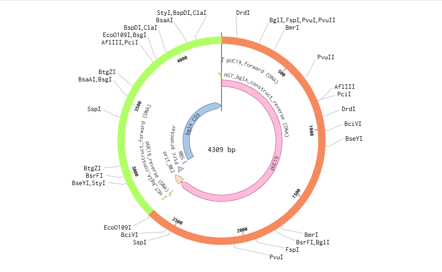
Fig. A1. Golden Gate AarI assembly design (Benchling)
LOCUS       pTwist_oriT_Ptrc_bglA     3697 bp    DNA   circular  SYN
DEFINITION  Donor plasmid for triparental conjugation: oriT_RK2 +
            Ptrc promoter + RBS + bglA CDS (Enterococcus faecalis)
            cloned into pTwist Amp High Copy backbone.
            In silico design — Benchling. Golden Gate AarI assembly.
            For Twist Bioscience Clonal Gene order.
ACCESSION   .
VERSION     .
KEYWORDS    conjugation; oriT_RK2; bglA; beta-glucosidase; triparental mating;
            chromogenic reporter; horizontal gene transfer.
SOURCE      Synthetic construct
  ORGANISM  Synthetic construct
            other sequences; artificial sequences; vectors.
FEATURES             Location/Qualifiers
     rep_origin      complement(1..589)
                     /label="pUC ori"
                     /note="pUC high copy origin of replication (~500 copies/cell)"
     CDS             complement(630..1490)
                     /gene="bla"
                     /product="TEM-1 beta-lactamase"
                     /label="AmpR"
                     /note="confers ampicillin resistance (100 µg/mL working)"
                     /codon_start=1
     terminator      complement(1491..1535)
                     /label="AmpR terminator"
     misc_feature    1550..1799
                     /label="oriT_RK2"
                     /note="Minimal origin of transfer from RK2/RP4 plasmid.
                      Contains nick site (nic) recognized by TraI relaxase.
                      Required in cis for mobilization by pRK2013 helper."
     promoter        1820..1874
                     /label="Ptrc"
                     /note="Hybrid tac/trp constitutive promoter.
                      -35 element: TTGACA (nt 1820-1825)
                      Spacer: 17 bp
                      -10 element: TATAAT (nt 1843-1848)"
     RBS             1880..1896
                     /label="RBS"
                     /note="Shine-Dalgarno sequence. No ATG included.
                      ATG provided by bglA CDS start codon."
     CDS             1897..3228
                     /gene="bglA"
                     /product="6-phospho-beta-glucosidase BglA"
                     /label="bglA"
                     /organism="Enterococcus faecalis"
                     /note="Encodes beta-glucosidase reporter.
                      Cleaves chromogenic beta-glucosidase substrates on
                      UTI CHROMagar → teal/blue colony color.
                      K-12 transconjugant (LacZ+ BglA+) = purple colony."
                     /codon_start=1
     terminator      3240..3290
                     /label="rrnB T1 terminator"
                     /note="Transcriptional terminator downstream of bglA"
ORIGIN
        1 tcgcgcgttt cggtgatgac ggtgaaaacc tctgacacat gcagctcccg gagacggtca
       61 cagcttgtct gtaagcggat gccgggagca gacaagcccg tcagggcgcg tcagcgggtg
      121 ttggcgggtg tgcggcgggt cagggcgcga ctcagcgggt gttggcgggt gtgcggcagg
      181 tcagggcgcg atcagcgggt gttggcgggt gtgcggcagg tcagggcgcg agctgcaagt
      [... full sequence in Benchling project file ...]
//
Submitted insert sequence (paste directly into Twist Bioscience Clonal Gene portal → pTwist Amp High Copy):
>Insert_oriT_RK2_Ptrc_bglA [Golden Gate AarI overhangs]

CACCTGC[AARIOVERHANG_FWD]
[oriT_RK2 minimal sequence — ~250 bp including nic site]
TTGACAATTAATCATCGGCTCGTATAATGTGTGGAATTGTGAGCGGATAACAATTTCACACAGG
AAGGAGATATACCAT
ATGAAAACAATTGTTGGCATGGATTTTAATGCAATCACTGATGCAGCAAAGAAAGTTATTGGC
GCTGATAATGATGCTATTGCAGAAAATGGTGTGATTGATGCAATCAAAGCAGGTTTAAAGCT
[... bglA CDS continues for 1332 bp total ...]
TTATTTTGAAGCTTTAAATCCTTTTTGAAACAGCG
[AARI_OVERHANG_REV]GTGAGC
Full annotated sequence available in Benchling. Total insert length: ~1,682 bp. Total plasmid (backbone + insert): ~3,697 bp. SecureDNA screening: recommended prior to order submission.
# Opentrons OT-2 Python API v2# Protocol: Concentric circle bioprinting for triparental mating# Strains: Donor (A1), Helper (A2), Recipient (A3) in Eppendorf tubes (tube rack)# Target: Plain LB agar plate in slot 1fromopentronsimportprotocol_apimetadata={'protocolName':'Triparental Mating Bioprint — Concentric Circles','author':'HTGAA Final Project','apiLevel':'2.13'}defrun(protocol:protocol_api.ProtocolContext):# Labwaretiprack=protocol.load_labware('opentrons_96_tiprack_20ul',2)tube_rack=protocol.load_labware('opentrons_24_tuberack_eppendorf_1.5ml_safelock_snapcap',3)agar_plate=protocol.load_labware('corning_96_wellplate_360ul_flat',1)# Note: for agar plates, use a custom labware definition or substitutep20=protocol.load_instrument('p20_single_gen2','right',tip_racks=[tiprack])donor=tube_rack['A1']# DH5α + pTwist-bglA, OD600=0.6helper=tube_rack['A2']# DH5α + pRK2013, OD600=0.6recipient=tube_rack['A3']# E. coli K-12, OD600=0.6# Concentric circle coordinates (x, y offsets from plate center, mm)importmathdefcircle_coords(radius,n_spots,cx=42.0,cy=35.0):return[(cx+radius*math.cos(2*math.pi*i/n_spots),cy+radius*math.sin(2*math.pi*i/n_spots))foriinrange(n_spots)]outer_coords=circle_coords(15,10)# Recipient — outer ringmiddle_coords=circle_coords(10,10)# Helper — middle ringinner_coords=circle_coords(5,10)# Donor — inner ring# Print each ring (0.5 µL per spot)forstrain,coordsin[(recipient,outer_coords),(helper,middle_coords),(donor,inner_coords)]:for(x,y)incoords:p20.pick_up_tip()p20.aspirate(1.0,strain)# Move to absolute position on agar plate and dispensep20.move_to(agar_plate['A1'].bottom().move(types.Point(x=x,y=y,z=2)))p20.dispense(0.5)p20.drop_tip()# Central spot: all 3 strains mixed (1:1:1, 1 µL each → 3 µL total)p20.pick_up_tip()forstrainin[donor,helper,recipient]:p20.aspirate(1.0,strain)p20.move_to(agar_plate['A1'].bottom().move(types.Point(x=42,y=35,z=2)))p20.dispense(3.0)p20.drop_tip()
Proposal generated with HTGAA Final Project Skill v1.1