Week 2 HW: DNA Read, Write, & Edit
Part 1: Benchling & In-silico Gel Art
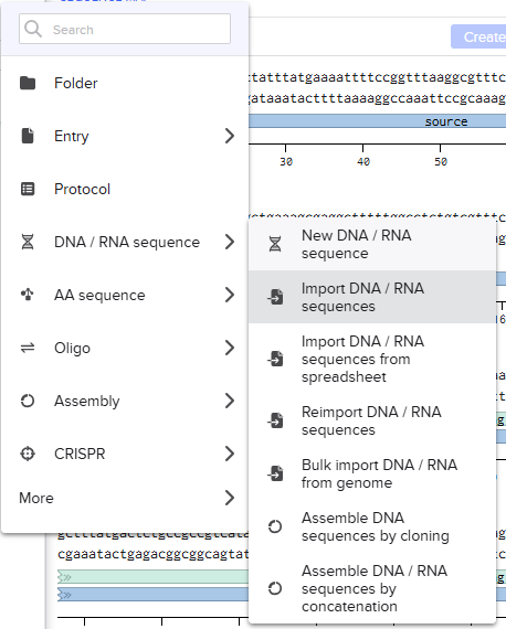
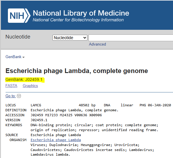
After locating the E. coli phage Lambda genome in the NCBI database, I loaded the sequence into my Benchling project by searching for the GenBank accession number (GenBank: J02459.1):
I then went to the DNA Gel Art Interface (https://rcdonovan.com/gel-art) and went through the gallery for inspiration and to observe what bands looked like for the digestion enzymes listed both individually and in combination with one another, taking screenshots of these images to determine which of these I could use as “building blocks” for my image.
I took inspiration from the following design on the gel art website, thinking I could modify it to make “boxes” using three lanes that could look like eyes that could then be used for a “smiley face” design:
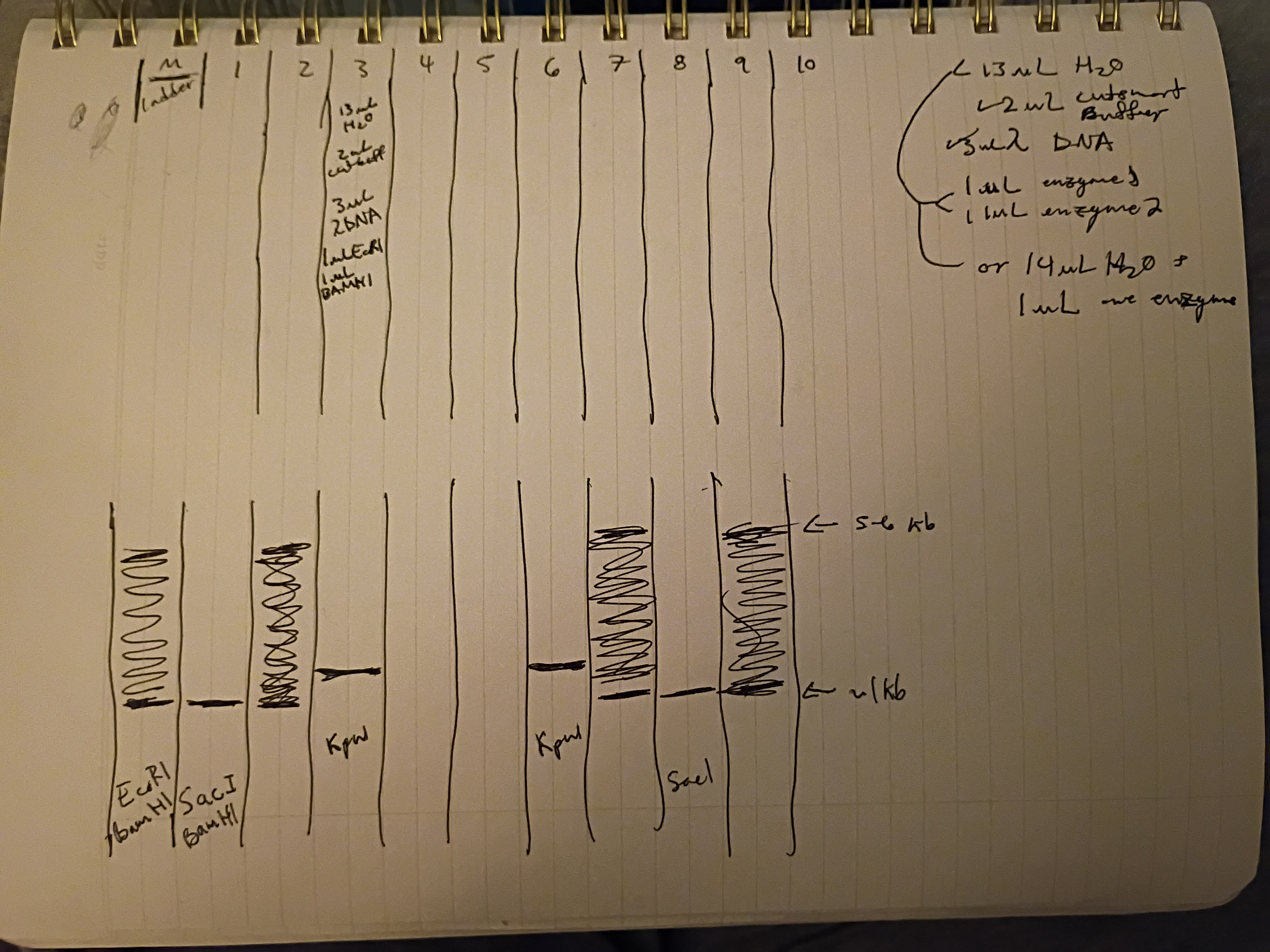
I used my notes/screenshots to determine what other bands could be used for the top parts of these boxes and for the parts of the smile, planning them out on paper first:
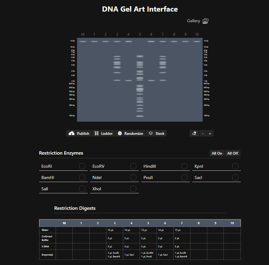
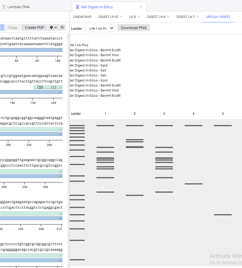
I then tested this design using the in-silico digest in Benchling:
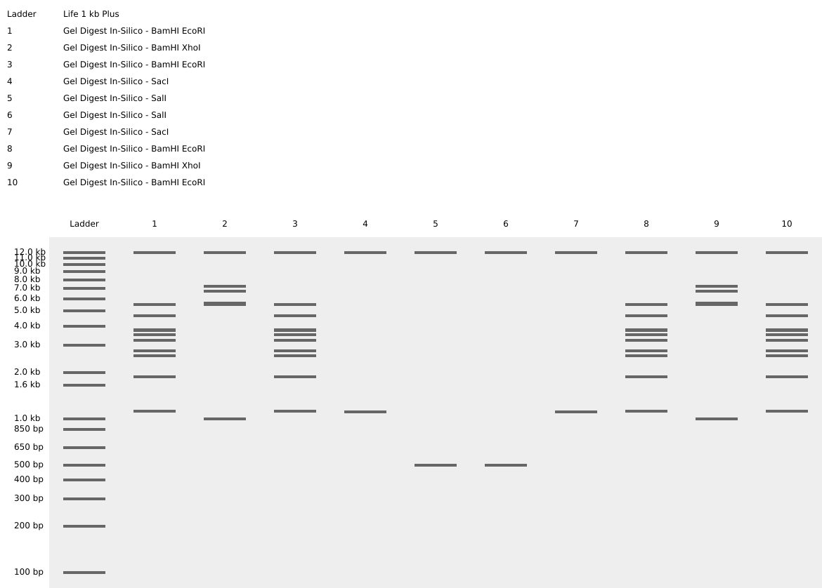
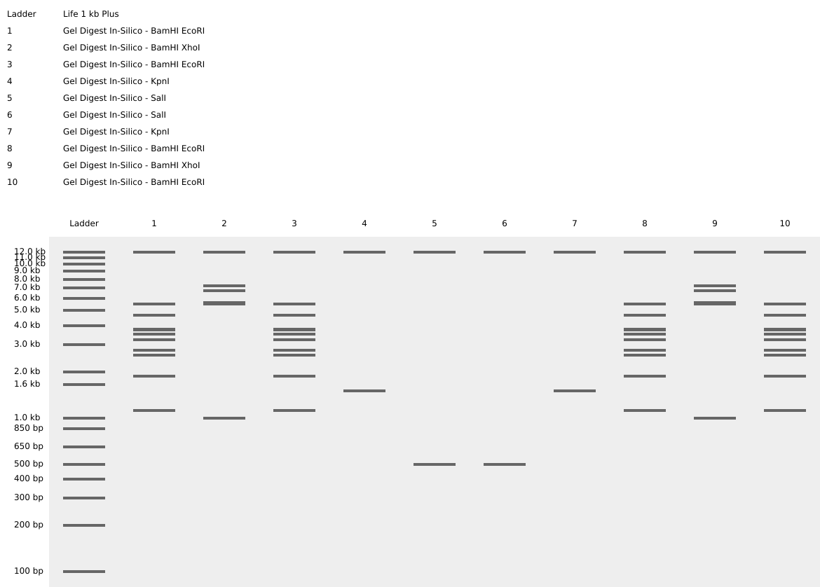
I did not like that the bands meant to represent the edges of the smile were level with the bottoms of the eyes, so I adjusted the design to make the smile start higher up in the lanes:
Final In-Silico Gel Design: https://benchling.com/s/seq-FfTcfmv79fwJMljaZij3?m=slm-Gjs2Z4pczSrUZzB76zUr
Part 2: Gel Art - Restriction Digests and Gel Electrophoresis Wet Lab
Note: I will not be able to make most wet labs due to EMT school causing scheduling conflicts but was able to attend this first in-person node lab and will participate in-person when possible.
Protocol Part 1a: Preparing a 1% agarose electrophoresis gel
*Chemicals:
-Agarose powder
-1x TBE buffer
-SmartGlow Safe Green Pre Stain (https://www.labproservices.com/buy-smartglow-safe-green-pre-stain-1-0ml-ea-1-e4500-ps-ea.html?gad_source=1&gad_campaignid=20923765122&gbraid=0AAAAAqHTWoNkTzYIzDQi_alxD1vIk_2bP&gclid=CjwKCAiAncvMBhBEEiwA9GU_fvIAPFHPYWljiQUeFtU-1_CGZxCC4XhO-unP2Wn_t-_yl_bK6QYBLBoCFlwQAvD_BwE)
*Equipment
-Pipette Set
-Gel tray
-9 or 13-well comb
-Microwave
-Precision scale
*Consumables
-Pipette tips
-250 mL beaker
-Graduated cylinder (100mL)
TBE Buffer Preparation The TBE buffer provided in our lab was already diluted to the proper 1X working solution.
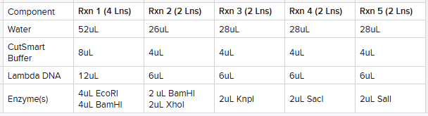
Gel Preparation To prepare an agarose gel for electrophoresis: Added 0.4 g of agarose powder (exact weight came out to 0.3999g) and 40mL of 1x TBE buffer to a microwavable beaker (1% w/v) Placed the flask in a microwave, heated in short pulses of 15–20 seconds, swirling gently between pulses, until the agarose was completely dissolved, watching to prevent any boil-overs. Allowed the solution to cool until the beaker was warm but comfortable to touch. Once cooled, added 2μL of SmartGlow DNA pre-stain to the solution and mixed gently. Slowly poured the agarose into the tray to avoid forming bubbles, placed the comb into the gel tray to create wells. 9 or 13 well-combs were available; I selected the 13-well comb and adapted my design to this. Allowed the gel to solidify for about 30 minutes at room temperature. Once set, carefully removed the comb, and the gel was ready for use.
Protocol Part 1b: Restriction Digest
*Chemicals
-1X Lambda DNA
-The following enzymes: EcoRI-HF, BamHI-HF, Xhol, KpnI-HF, SacI-HF, SalI-HF (see calculated quantities below)
-Nuclease-free water
*Equipment and Consumables
-Thermocycler
-PCR tube rack
I planned out an adjusted design from what I had in-silico due to the availability of 13 lanes:
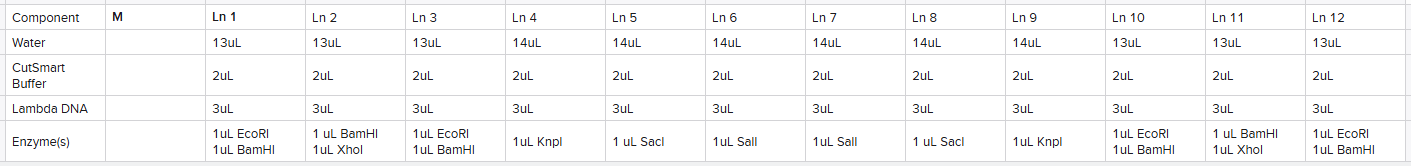
Based upon this plan, I made 5 reaction tubes as follows calculated based upon the number of lanes I planned on using the reaction for:
I then placed the rxn tubes in the thermocycler set to 37ºC and incubated for 30 minutes (while the agarose gel solidified).
Protocol Part 2: Gel Run
*Chemicals
-6x loading dye
-1X TBE
-SmartGlow Safe Green Pre Stain
*Equipment
-Gel Box
-Power supply (ours did not require the addition of leads, everything was built into the gel box with just a power supply connected to an outlet, voltage already adjusted to what was required)
Gel Run Comb was removed once the gel was set. Gel box was filled with approximately 30-40mL 1x TBE (enough to ensure gel was covered entirely). 0.72uL of pre-stain was added to the 1X TBE. For each rxn (per lane), I mixed 7uL of my digest from the rxn tube with 2uL of 6X loading dye on a sheet of parafilm prior to loading into wells, following the schema described above for all lanes. Made sure the top was place on the gel box the correct way (negative to negative, positive to positive, gel positioned with loaded lanes at the negative end). Ran the gel at 80V - 115V for around 45 minutes (originally checked after 30 minutes, let run about another 15 to allow for further separation of bands). Checked that there were bubbles in the buffer and that lanes were moving after about 10 minutes.
Protocol Part 3: Imaging Your Results
*Equipment
-Blue light transilluminator (for our lab, this was actually built into the gel electrophoresis chamber/gel box)
-Phone camera
*Consumables and Safety
-Gloves
To view my results, I simply turned on the blue light built into our gel boxes, removing the need to transfer the gel to a separate transilluminator. Put a light-blocking device around the gel box to block out ambient room light and allow for better photos to be taken. Took a picture using my phone’s camera through an imaging hole on the top of the light-blocking device. Disposed of the gel in the solid waste bin (burn box).
Final Results
Here is a picture of the final results:
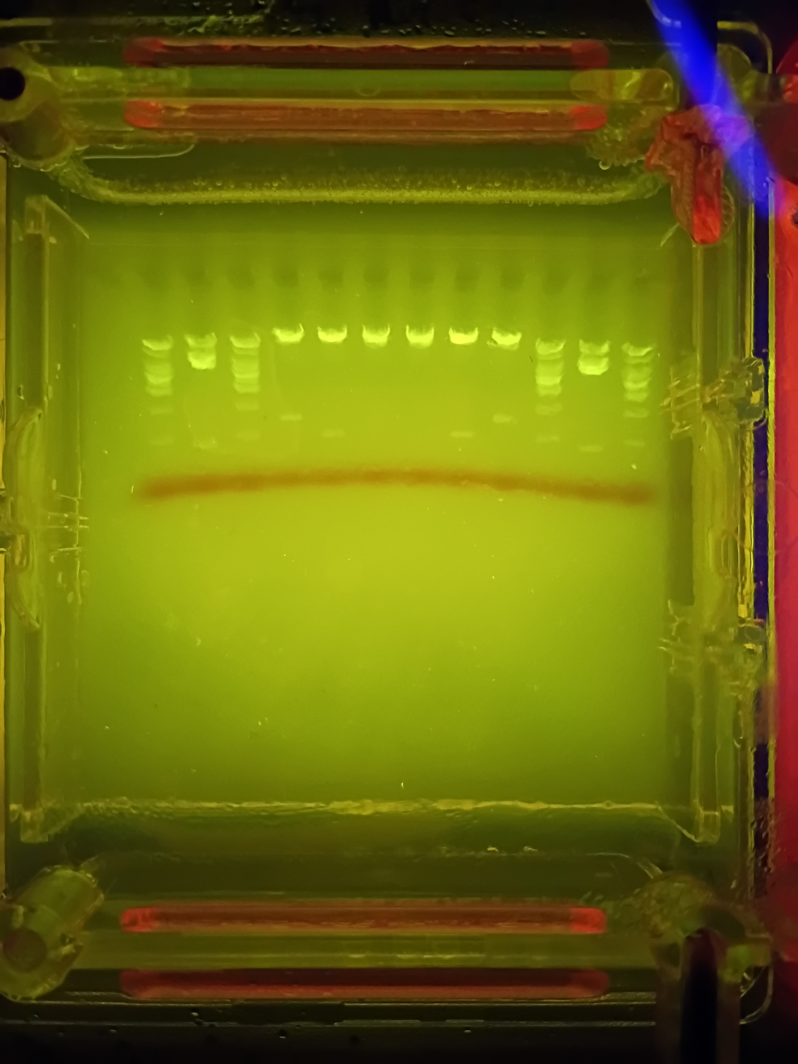
I forgot to account for the fact that some of the single bands created were rather small sections base-pair wise and thus would not be very visible. I also was hoping for better separation from my deisgn from the uncut sections at the top, making my smiley face a bit hard to make out (especially since the bottom of the smile is not actually visible). Someone commented that they think the design looked like a neat bridge, so if anyone asks…it was definitely a bridge I was trying to make here!
Part 3: DNA Design Challenge
3.1. Choose your protein.
The protein I have chosen is Chitin deacetylase 2 (CDA2), as I am interested in the usage of chitosan in hemostatic dressings to treat traumatic injury by stopping hemorrhage. Most chitosan produced for the purpose of hemostatic agents is produced chemically, but there is newer research being performed looking into using enzymes such as CDA2 for a “greener” method for the production of chitosan by removing N-acetyl groups from chitin. Chitin is naturally found in the exoskeleons of insects and crustaceans as well as fungi and certain algae. Chitosan produced from shellfish was used more heavily in the past and can still be found today in some hemostatic bandages, but this chitosan had to be irrigated out of wounds and posed danger to those with shellfish allergies.
The specific protein I found in UniProt was found in Saccharomyces cerevisiae (strain ATCC 204508 / S288c):
sp|Q06703|CDA2_YEAST Chitin deacetylase 2 OS=Saccharomyces cerevisiae (strain ATCC 204508 / S288c) OX=559292 GN=CDA2 PE=1 SV=1 MRIQLNTIDLQCIIALSCLGQFVHAEANREDLKQIDFQFPVLERAATKTPFPDWLSAFTG LKEWPGLDPPYIPLDFIDFSQIPDYKEYDQNHCDSVPRDSCSFDCHHCTEHDDVYTCSKL SQTFDDGPSASTTKLLDRLKHNSTFFNLGVNIVQHPDIYQRMQKEGHLIGSHTWSHVYLP NVSNEKIIAQIEWSIWAMNATGNHTPKWFRPPYGGIDNRVRAITRQFGLQAVLWDHDTFD WSLLLNDSVITEQEILQNVINWNKSGTGLILEHDSTEKTVDLAIKINKLIGDDQSTVSHC VGGIDYIKEFLS
3.2. Reverse Translate: Protein (amino acid) sequence to DNA (nucleotide) sequence.
CDA2_YEAST Chitin deacetylase 2 OS=Saccharomyces cerevisiae (strain ATCC 204508 / S288c) AA sequence to nucleotides using the CUSABIO Protein to DNA Sequence Converter tool (including a start and stop codon):
ATGATGCGTA TTCAATTAAA TACTATTGAT TTACAATGTA TTATTGCTTT ATCTTGTTTA GGTCAATTTG TTCATGCTGA AGCTAATCGT GAAGATTTAA AACAAATTGA TTTTCAATTT CCTGTTTTAG AACGTGCTGC TACTAAAACT CCTTTTCCTG ATTGGTTATC TGCTTTTACT GGTTTAAAAG AATGGCCTGG TTTAGATCCT CCTTATATTC CTTTAGATTT TATTGATTTT TCTCAAATTC CTGATTATAA AGAATATGAT CAAAATCATT GTGATTCTGT TCCTCGTGAT TCTTGTTCTT TTGATTGTCA TCATTGTACT GAACATGATG ATGTTTATAC TTGTTCTAAA TTATCTCAAA CTTTTGATGA TGGTCCTTCT GCTTCTACTA CTAAATTATT AGATCGTTTA AAACATAATT CTACTTTTTT TAATTTAGGT GTTAATATTG TTCAACATCC TGATATTTAT CAACGTATGC AAAAAGAAGG TCATTTAATT GGTTCTCATA CTTGGTCTCA TGTTTATTTA CCTAATGTTT CTAATGAAAA AATTATTGCT CAAATTGAAT GGTCTATTTG GGCTATGAAT GCTACTGGTA ATCATACTCC TAAATGGTTT CGTCCTCCTT ATGGTGGTAT TGATAATCGT GTTCGTGCTA TTACTCGTCA ATTTGGTTTA CAAGCTGTTT TATGGGATCA TGATACTTTT GATTGGTCTT TATTATTAAA TGATTCTGTT ATTACTGAAC AAGAAATTTT ACAAAATGTT ATTAATTGGA ATAAATCTGG TACTGGTTTA ATTTTAGAAC ATGATTCTAC TGAAAAAACT GTTGATTTAG CTATTAAAAT TAATAAATTA ATTGGTGATG ATCAATCTAC TGTTTCTCAT TGTGTTGGTG GTATTGATTA TATTAAAGAA TTTTTATCTT AA
3.3. Codon optimization.
Codon optimization for the host organism that is intended to express the sequence is necessary as each organism may have different tRNAs available in different quantities, impacting the speed at which translation may take place or even be possible. I used Benchling’s back translate with codon optimization tool to optimize this sequence for the host organism E.coli K12 and avoid the BglI digest enzyme cut-site because it is non-pathogenic and would allow for selection of only transformed bacteria in an experiment if I use a plasmid vector that includes a gene that confers antibiotic resistance to an antibiotic that kills E. coli K12 wild type.
Signal Peptide: ATGCGCATTCAGCTTAATACCATCGATTTACAATGCATCATTGCGTTGAGCTGTCTGGGCCAATTTGTTCATGCCGAAGCAAACCGTGAGGACCTGAAACAGATTGATTTCCAGTTTCCGGTGCTGGAACGC
Chain CDA2: GCAGCGACCAAGACGCCGTTCCCGGATTGGCTGTCGGCTTTTACGGGCCTTAAAGAGTGGCCGGGCCTTGACCCGCCGTATATTCCCCTGGATTTTATCGATTTCAGCCAAATTCCAGATTACAAAGAATATGACCAGAACCATTGTGATAGTGTGCCACGCGATAGCTGCTCGTTCGACTGCCACCATTGCACCGAGCACGATGACGTCTACACTTGTTCCAAACTGAGTCAAACCTTTGACGATGGTCCTAGCGCCTCTACGACGAAACTGTTAGATCGCTTAAAACATAACTCAACCTTTTTTAATCTGGGCGTCAACATTGTTCAGCACCCCGACATTTATCAGCGTATGCAGAAAGAAGGCCATCTGATTGGAAGCCATACCTGGTCTCATGTATATCTGCCTAATGTGAGCAATGAGAAGATCATTGCGCAGATTGAATGGAGCATCTGGGCGATGAACGCCACCGGCAATCATACACCGAAATGGTTTCGTCCGCCGTACGGGGGCATTGACAATCGCGTTCGGGCCATTACCCGTCAATTCGGTCTGCAAGCGGTGTTGTGGGATCACGATACTTTCGATTGGTCGCTGTTGCTTAACGATTCCGTGATAACCGAACAGGAAATCCTGCAGAACGTAATTAATTGGAACAAAAGTGGCACAGGTTTGATTCTAGAACACGACAGCACGGAAAAGACTGTTGACTTAGCAATCAAGATTAATAAACTCATCGGTGATGATCAGTCGACCGTGTCACACTGTGTCGGAGGGATAGATTACATCAAAGAATTTCTCTCA
Together: ATGCGCATTCAGCTTAATACCATCGATTTACAATGCATCATTGCGTTGAGCTGTCTGGGCCAATTTGTTCATGCCGAAGCAAACCGTGAGGACCTGAAACAGATTGATTTCCAGTTTCCGGTGCTGGAACGCGCAGCGACCAAGACGCCGTTCCCGGATTGGCTGTCGGCTTTTACGGGCCTTAAAGAGTGGCCGGGCCTTGACCCGCCGTATATTCCCCTGGATTTTATCGATTTCAGCCAAATTCCAGATTACAAAGAATATGACCAGAACCATTGTGATAGTGTGCCACGCGATAGCTGCTCGTTCGACTGCCACCATTGCACCGAGCACGATGACGTCTACACTTGTTCCAAACTGAGTCAAACCTTTGACGATGGTCCTAGCGCCTCTACGACGAAACTGTTAGATCGCTTAAAACATAACTCAACCTTTTTTAATCTGGGCGTCAACATTGTTCAGCACCCCGACATTTATCAGCGTATGCAGAAAGAAGGCCATCTGATTGGAAGCCATACCTGGTCTCATGTATATCTGCCTAATGTGAGCAATGAGAAGATCATTGCGCAGATTGAATGGAGCATCTGGGCGATGAACGCCACCGGCAATCATACACCGAAATGGTTTCGTCCGCCGTACGGGGGCATTGACAATCGCGTTCGGGCCATTACCCGTCAATTCGGTCTGCAAGCGGTGTTGTGGGATCACGATACTTTCGATTGGTCGCTGTTGCTTAACGATTCCGTGATAACCGAACAGGAAATCCTGCAGAACGTAATTAATTGGAACAAAAGTGGCACAGGTTTGATTCTAGAACACGACAGCACGGAAAAGACTGTTGACTTAGCAATCAAGATTAATAAACTCATCGGTGATGATCAGTCGACCGTGTCACACTGTGTCGGAGGGATAGATTACATCAAAGAATTTCTCTCA
3.4. You have a sequence! Now what? My selected codon optimized sequence could be expressed in the host organism if this sequence were placed into a plasmid (preferably with an antibiotic resistance gene for an antibiotic that the E. coli K12 wild type is susceptible to) and the plasmid were uptaken by the bacteria, generally induced with heat-cold shock or electroporation. The presence of an antibiotic resistance gene in this plasmid that is lacking in the wild type would allow for me to select only for transformed bacteria by growing the bacteria in antibiotic-containing media.
Part 4: Prepare a Twist DNA Synthesis Order Alas, I’m going to have to come back to this section and complete at another time. I have been attempting to make a Twist account for the past several days and have received an error message each time after attempting to access from different browsers:
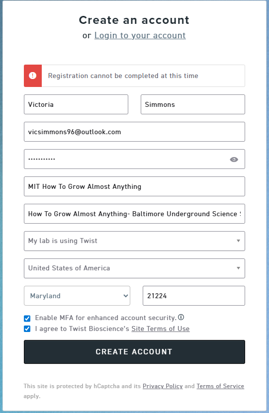
I attempted to communicate with Twist through the chat system, but it is manned by actual people that are not working this long weekend. They should get back to me 17 February 2026, so I’ll come back to this for practice after this is resolved and I can make my account. In the meantime:
5.1 DNA Read (i) What DNA would you want to sequence (e.g., read) and why? This could be DNA related to human health (e.g. genes related to disease research), environmental monitoring (e.g., sewage waste water, biodiversity analysis), and beyond (e.g. DNA data storage, biobank). (ii) In lecture, a variety of sequencing technologies were mentioned. What technology or technologies would you use to perform sequencing on your DNA and why? Also answer the following questions: Is your method first-, second- or third-generation or other? How so? What is your input? How do you prepare your input (e.g. fragmentation, adapter ligation, PCR)? List the essential steps. What are the essential steps of your chosen sequencing technology, how does it decode the bases of your DNA sample (base calling)? What is the output of your chosen sequencing technology?
Maybe this is a bit of a trick answer, but I work in biosurveillance/biodefense, so I would be interested in looking for sequences of novel pathogens in order to identify possible disease outbreaks before they spread into pandemics. The question then becomes, how do you look for a sequence that you don’t know?
- For biosurveillance of novel, potential pandemic-causing pathogens in a population, the preferred sample type would either be waste water (collected either from the municipal supplies or from specific large-gathering places such as airports) or nasal or oropharyngeal swabs in transport media taken from patients that, for example, shows respiratory illness symptoms but test negative on respiratory panels looking for common flu or cold-caueing viruses and bacteria. Swabs could also be taken of asymptomatic passengers at airports that decide to opt into such proposed programs.
- Nucleic acid from the samples of choice would then be extracted, typically using kits (e.g. Qiagen) for the specific sample type or other high-throughput methods (e.g. KingFisher or liquid handler-based extraction methods). This nucleic acid would then be quantified and checked for any contaminants, such as proteins, via methods such as NanoDrop or electrophoresis. QC: Quantify DNA concentration (e.g., Qubit) and check for fragmentation or contamination (e.g., NanoDrop, electrophoresis).
- The extracted DNA would then need to be sheared into smaller, randomized fragments (sequencing fragmentation). A library prep step would then be performed for end repair, A-tailing, and ligation of adapters/indices for multiplexing, allowing multiple samples to be sequenced together.The sample would then be sequenced on a NextGenn platform such as Illumina (e.g., NovaSeq, NextSeq) to produce millions of short reads. Short read sequencing via sequencing by synthesis (SBS) is the most common method on the market, though long read methods like NanoPore are also used.
- Bioinformatics analysis, which can be read-centric or assembly-centric, then takes place and includes steps to: filter out low-quality reads and trim adapters (preprocessing); remove host reference genomes (e.g., human) as applicable;identify microbial species and their relative abundances (e.g., using Kraken2, MetaPhlAn) (taxonomic profiling); use assemblers (e.g., MEGAHIT, SPAdes) to stitch reads into longer contiguous sequences (contigs);predict genes (e.g., Prodigal) and annotate functions (e.g., Prokka, DIAMOND) to understand the metabolic potential; and cluster contigs into Metagenome-Assembled Genomes (MAGs) to identify novel organisms (e.g., MetaBAT2) (binning). Data can also be verified via methods such as qPCR and visualized by performing alpha/beta diversity analysis, and metabolic pathway mapping using tools like R, Python, or QIIME 2.
5.2 DNA Write (i) What DNA would you want to synthesize (e.g., write) and why? These could be individual genes, clusters of genes or genetic circuits, whole genomes, and beyond. As described in class thus far, applications could range from therapeutics and drug discovery (e.g., mRNA vaccines and therapies) to novel biomaterials (e.g. structural proteins), to sensors (e.g., genetic circuits for sensing and responding to inflammation, environmental stimuli, etc.), to art (DNA origamis). If possible, include the specific genetic sequence(s) of what you would like to synthesize! You will have the opportunity to actually have Twist synthesize these DNA constructs! :)
(ii) What technology or technologies would you use to perform this DNA synthesis and why? Also answer the following questions: What are the essential steps of your chosen sequencing methods? What are the limitations of your sequencing method (if any) in terms of speed, accuracy, scalability?
I would be interested in on-demand production of mRNA vaccines, an endeavor that is already being explored by the 100 days mission and by BARDA. One way that this could be done would be by sequencing a pathogen of interest and then selecting for a protein expressed on the outside of the bacteria or virus (one that would be interacting with the immune system directly) and construct an mRNA with the instructions for how your own ribosomes could produce the protein in order to train the immune system against the specific protein found on the pathogen (building up an immunity or resistance against the pathogen without using a killed or live-attenuated version of the pathogen). Using the SARS-CoV-2 mRNA vaccine as a reference, the genetic sequence for the SARS-CoV-2 spike protein is synthesized (e.g. through solid-phase phosphoramidite chemistry) and then inserted into a plasmid. From there, they separate the two strands of plasmid DNA. RNA polymerase, the molecule that transcribes RNA from DNA, then uses the spike protein gene to create a single mRNA molecule. Other molecules break down the rest of the plasmid to ensure that only the mRNA is packaged as a vaccine. This single-stranded mRNA is then encapsulated in a lipid bilayer to protect the fragile molecule. This method is limited by both the limitations previously discussed for phosphoramidite chemistry (losing efficiency and accuracy after a certain length, necessitating synthesis of smaller pieces to be stitched together) and by the fragility of the mRNA molecule (which requires protection via a lipid bilayer and, at the present, a continuous cold chain from production to end user). mRNA vaccines being made on demand should decrease the amount of time between novel pathogen identification/outbreak and a vaccine, but may of limited use in areas where a maintained (very) cold chain is unfeasible, such as areas in the global south. The hope is that these methods may be more accessible as the technology progresses, allowing for on-demand production at the point of the end user (reducing the need for the cold chain).
5.3 DNA Edit (i) What DNA would you want to edit and why? In class, George shared a variety of ways to edit the genes and genomes of humans and other organisms. Such DNA editing technologies have profound implications for human health, development, and even human longevity and human augmentation. DNA editing is also already commonly leveraged for flora and fauna, for example in nature conservation efforts, (animal/plant restoration, de-extinction), or in agriculture (e.g. plant breeding, nitrogen fixation). What kinds of edits might you want to make to DNA (e.g., human genomes and beyond) and why?
(ii) What technology or technologies would you use to perform these DNA edits and why? Also answer the following questions: How does your technology of choice edit DNA? What are the essential steps? What preparation do you need to do (e.g. design steps) and what is the input (e.g. DNA template, enzymes, plasmids, primers, guides, cells) for the editing? What are the limitations of your editing methods (if any) in terms of efficiency or precision?
I think a good candidate for gene editing would be using the CRISPR/Cas9 technology to replace harmful/pathogenic variant BRCA1 and BRCA2 inherited gene mutations with functional BRCA1 and BRCA2 genes in individuals that have these harmful mutations. Many of these individuals know that they have these gene mutations due to a family history (generally confirmed during genetic testing/counseling) and have no recourse other than preventative mastectomies and other surgeries, certain medications, and increased cancer-screenings. While this gene does not run in my family, I think it would be a good candidate for clinical gene editing due to public awareness of this gene and the fact that many of the pathogenic variants of the BRCA gene are due to frame shift mutations caused by small insertions or deletions (indels). CRISPR/Cas9 gene-editing technology involves using a guide RNA (in our case, a synthetic single guide RNA) to match a desired target gene, and Cas9 (CRISPR-associated protein) endonuclease which causes a double-stranded DNA break, allowing modifications to the genome. In order to cut, a specific sequence of DNA of between 2 and 5 nucleotides (the exact sequence depends upon the bacteria which produces the Cas9) must lie at the 3’ end of the guide RNA, called the protospacer adjacent motif (PAM). Repair after the DNA cut may occur via two pathways: non-homologous end joining, typically leading to a random insertion/deletion of DNA; or homology directed repair where a homologous piece of DNA is used as a repair template (e.g. a syntheaized non-pathogenic gene variant), allowing for precise genome editing. The homologous section of DNA with the required sequence change may be delivered with the Cas9 nuclease and sgRNA, theoretically allowing changes as precise as a single base-pair. Based upon my limited knowledge of CRISPR/Cas9 technology, I would think targeted delivery would be one of the largest technical challenges in using this technology to gene edit in a way that produces non-pathogenic BRCA gene variants. If this method were to be used to treat children or adults, then we would be looking to make alterations in somatic cells where we would expect the gene variant to cause disease; this can differ between individuals, but generally impacts more than the breast tissue (individuals with BRCA mutations also have higher incidence of uterine, ovarian, and other reproductive cancers, as well as elevated risk of pancreatic cancers). Ensuring that all of the cells that could become cancerous are effectively treated would be extremely difficult (and I am doubtful there would be an effective method to screen or otherwise check this, clinically speaking). Even if all of the cells that could become cancerous due to this gene mutation were treated in an individual, this individual would still be able to pass on their pathogenic gene variant via germline, unless this were also treated. Treating the patient’s germline via CRISPR/Cas9 would ensure that offspring would inherit the “fixed” gene variant, but leads to ethical considerations regarding eugenics and “designer babies”.