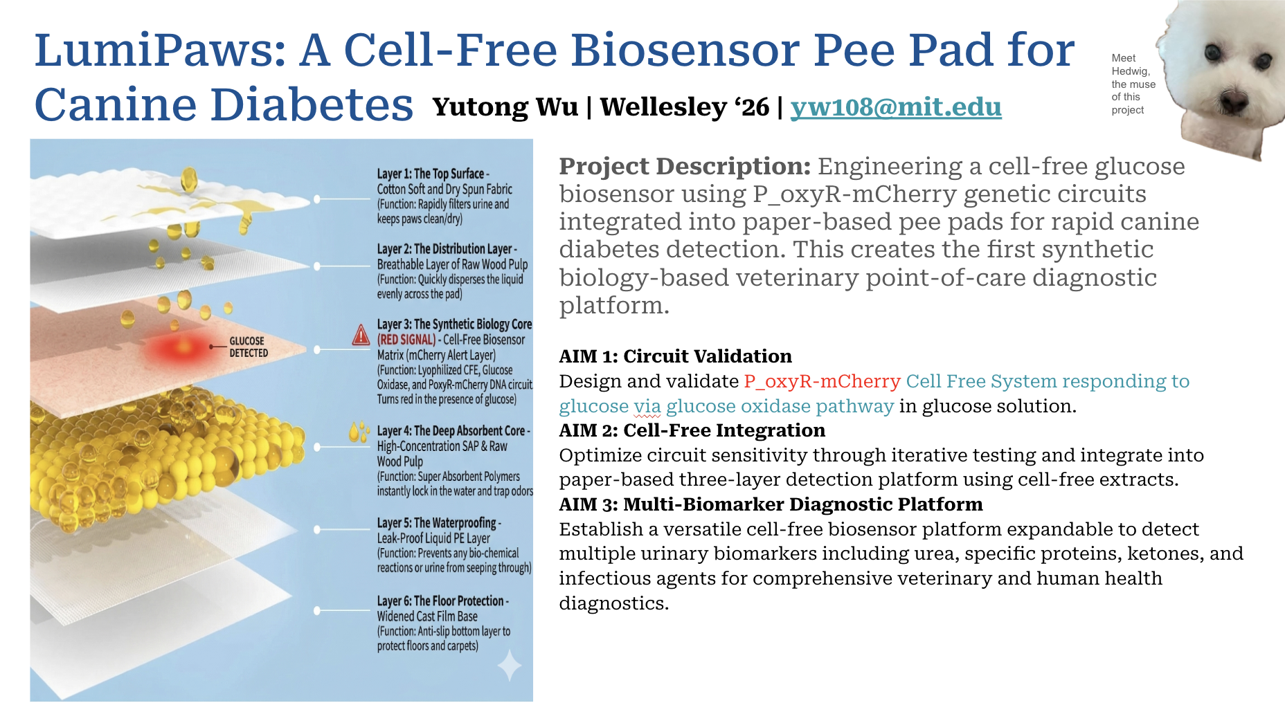
LumiPaws
LumiPaws engineers an OxyR-responsive genetic circuit into a freeze-dried paper substrate that detects urinary glucose through visual fluorescence. Urine testing is the foundation of preventive care for pets as a way to shift from waiting until the animal is already sick to spotting trouble before it starts.
Yutong Wu · Wellesley College ‘26 · yw108@mit.edu
01 · Overview
Canine diabetes affects approximately 1 in 300 dogs, and most diagnoses arrive late: after weight loss, excessive thirst, or ketoacidosis bring the family to a clinic. There is no accessible at-home screening tool. Veterinary blood glucose tests require a visit, and pet owners have no early warning system.
LumiPaws turns the everyday pee pad into a diagnostic. A freeze-dried cell-free reaction sits in the absorbent core of a six-layer pad; when a diabetic dog urinates on it, glucose is enzymatically converted to hydrogen peroxide, which activates an engineered OxyR transcription factor and drives mCherry expression, producing a red signal visible to the owner via .
Why red? Urine is naturally yellow, which is a problem for any color-based test, since most signals blend right into the background. LumiPaws uses mCherry, a red fluorescent protein, precisely because red cuts cleanly through yellow rather than getting lost in it. The signal is readable under a focused light at 587 nm, mCherry’s excitation peak, and every kit ships with a small handheld torch tuned to that exact wavelength.
| Metric | Value | Context |
|---|---|---|
| 1 : 300 | Dogs with diabetes | Rising rapidly over the last decade |
| > 100 mg/dL | Clinical glucosuria threshold | Renal threshold ~180–220 mg/dL serum |
| ~ $0 | Equipment to use | Visual fluorescence; no hardware |
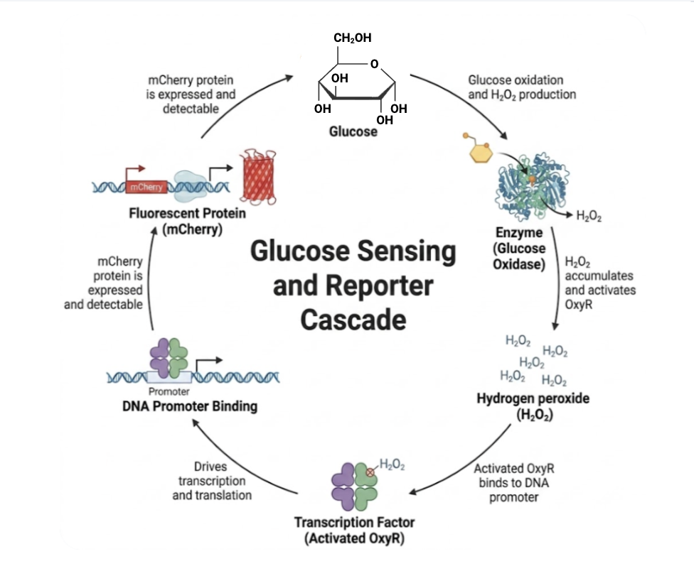 Insert: Glucose → GOx → H₂O₂ → OxyR → mCherry signal flow
02 · Project Aims
Aim I — Circuit Validation
Design and validate a P_oxyR-mCherry cell-free system responsive to glucose via the glucose oxidase pathway. Quantify dose-response across H₂O₂ and glucose concentrations in tube and on paper.
Aim II — Cell-Free Integration
Optimize circuit sensitivity through iterative promoter design and freeze-dry the validated reaction onto a paper-based three-layer detection platform suitable for urine-triggered activation.
Aim III — Multi-Biomarker Platform
Establish a versatile cell-free biosensor platform expandable to detect urinary biomarkers including urea, ketones, proteins, and infectious agents for comprehensive veterinary and human diagnostics.
03 · Circuit Design
Two rounds of iterative engineering.
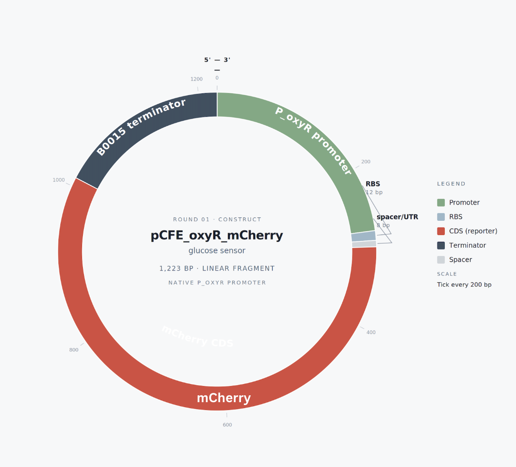
Round 01 — Native P_oxyR (minimum viable circuit)
Concept. First iteration used the native E. coli ahpC promoter with OxyR’s endogenous binding site driving mCherry directly. A minimal architecture intended to test whether OxyR-dependent activation could be detected in a cell-free extract.
Architecture. EcoRI → P_ahpC → RBS B0034 → mCherry → rrnB T1 → HindIII. Total length ~1.2 kb. Linear gene fragment ordered from Twist.
Outcome. Worked in vivo but produced negligible signal in cell-free — the native σ⁷⁰-dependent promoter is poorly transcribed by the T7-rich CFPS systems. This motivated Round 2.
Round 02 — T7–OxyR hybrid promoter (current)
Concept. Engineered a hybrid promoter fusing the T7 core sequence with the OxyR operator from ahpC. T7 RNAP provides high-yield transcription; OxyR gates that activity in an H₂O₂-dependent manner. Two cassettes on one template: constitutive OxyR expression upstream, T7–OxyR gated mCherry downstream.
Architecture. EcoRI → T7 → RBS → OxyR → rrnB T1 → insulator → T7-OxyR hybrid promoter → RBS → mCherry → Tphi → HindIII. Two terminator variants used to eliminate sequence repeats. Total length 1,903 bp.
Variants ordered. Two gene fragments: v1 (5 bp spacer) and v2 (3 bp spacer) between T7 core and OxyR operator — to identify the optimal spatial geometry for transcriptional regulation.
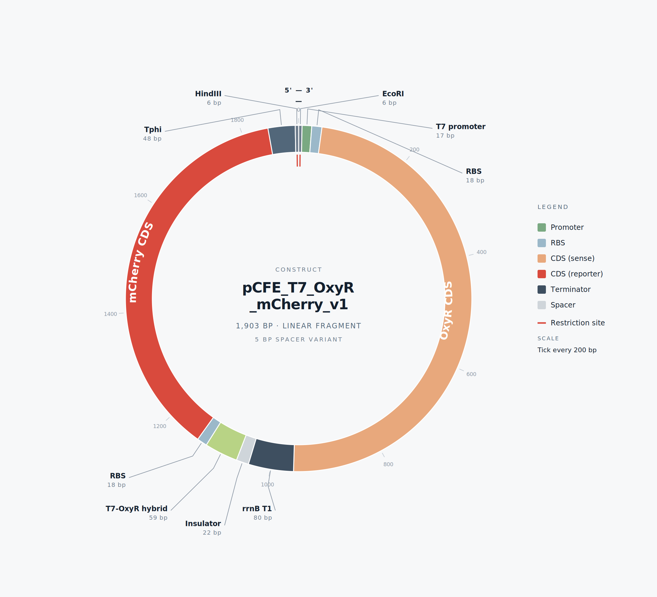 Insert: T7–OxyR hybrid construct map
04 · Parts & Components
Final assembled gene fragment, 5’ to 3’. Synthesized as Gene Fragment (Adapters OFF) for direct use in cell-free reactions with GamS protein.
| Element | Source | Function | Size |
|---|---|---|---|
| EcoRI flank | Synthetic | Cloning site / restriction handle | 6 bp |
| T7 promoter | T7 bacteriophage | Drives OxyR expression in CFPS | 17 bp |
| RBS B0034 | iGEM Registry | Strong ribosome binding site | 18 bp |
| OxyR CDS | E. coli K-12 MG1655 | H₂O₂-sensing transcription factor | 918 bp |
| rrnB T1 terminator | BBa_B0010 | Terminates Chapter 1 transcription | 80 bp |
| Insulator spacer | Synthetic neutral | Prevents read-through | 22 bp |
| T7–OxyR hybrid | Engineered (this work) | Gated transcriptional element | ~59 bp |
| RBS B0034 | iGEM Registry | RBS for mCherry | 18 bp |
| mCherry CDS | Codon-optimized (IDT) | Red fluorescent reporter | 708 bp |
| Tphi terminator | T7 bacteriophage | Terminates Chapter 2 transcription | 48 bp |
| HindIII flank | Synthetic | Cloning site / restriction handle | 6 bp |
05 · Methods
📄 → View the full experiment protocols (Google Doc)
01 — Construct Design. DNA design and annotation in Benchling. Parts sourced from NCBI (OxyR Gene ID 948462), iGEM Registry (RBS, terminators), and IDT CodonOpt for E. coli-optimized mCherry.
02 — Gene Synthesis. Twist Bioscience Gene Fragments (Adapters OFF), 1,903 bp linear DNA. Two spacer variants synthesized in parallel to triangulate optimal T7–OxyR geometry.
03 — Cell-Free Expression. Ginkgo CFPS Economy Kit (primary) and NEB PURExpress (backup). GamS protein added to protect linear DNA from exonuclease activity in crude extracts.
04 — Enzymatic Conversion. Glucose oxidase from Aspergillus niger (Sigma G7141) co-lyophilized with the cell-free reaction. Converts urinary glucose into H₂O₂ via molecular oxygen.
05 — Fluorescence Readout. mCherry signal quantified on a plate reader (ex 569 nm, em 610 nm) with kinetic measurements every 10 minutes over 4–6 hours at 30 °C. Dose-response curves fit to extract EC50 and dynamic range.
06 — Paper Integration. Optimized reactions freeze-dried onto Whatman #1 filter paper discs and integrated into a six-layer pad architecture: spunbond top → distribution → biology core → SAP → PE waterproofing → base.
06 · Results & Expectations
What success looks like.
 Insert: mCherry fluorescence vs. H₂O₂ concentration across both variants
Quantitative targets
Validation of the T7–OxyR hybrid promoter in cell-free reactions across an H₂O₂ titration. Success at this stage unlocks the full glucose cascade and paper integration.
| Key | Description | Target |
|---|---|---|
| LOD | Limit of detection | < 50 µM H₂O₂ |
| EC50 | Half-maximal activation | 100 – 500 µM |
| Range | Dynamic range, fold induction | 5 – 15 × |
| T½ | Time to detectable signal | 1 – 2 hr |
| CV | Replicate variability | < 25 % |
07 · Project Timeline
From design to deployment.
- ✅ Phase 01 — Construct design in Benchling. Two T7–OxyR hybrid promoter variants assembled with non-repeating terminators. Sequence verified, EcoRI/HindIII flanks added.
- ✅ Phase 02 — Gene fragment ordered from Twist. Both variants submitted as Gene Fragment (Adapters OFF), 1,903 bp linear DNA each. Co-shipped via MIT/Harvard institutional batch.
- ✅ Phase 03 — Cell-free reagents acquired. Ginkgo CFPS Economy Kit, GamS protein, glucose oxidase, and H₂O₂ standards in hand. Plate reader access confirmed.
- 🔵 Phase 04 — Pilot run + H₂O₂ dose-response (Active). Friday: pilot with mScarlet positive control and three H₂O₂ conditions. Saturday: 24-well replicate across full H₂O₂ titration with both variants.
- ⚪ Phase 05 — Full glucose cascade. Add glucose oxidase + glucose titration (0–1000 mg/dL). Confirm end-to-end glucose → H₂O₂ → mCherry signal in tube.
- ⚪ Phase 06 — Paper prototype. Freeze-dry optimized reaction onto Whatman discs. Test rehydration with synthetic urine + glucose. Iterate on stability and signal time.
08 · Future Vision
Your pet can’t talk, but their urine can.
LumiPaws is a platform, not a single product. The cell-free system at the heart of the pad can be re-engineered to detect a growing range of urinary biomarkers — and the format can expand from dog pee pads to cat litter, creating a unified at-home diagnostic ecosystem for companion animals.
🔵 Kidney Function — Urea + Creatinine
Cell-free expression of urease and creatininase, paired with engineered ammonia- or pH-responsive transcription factors, can flag early kidney disease — one of the most common causes of mortality in older dogs and cats.
🟡 Diabetes — Glucose + Ketones
Adding β-hydroxybutyrate dehydrogenase to the same cell-free chassis extends the diabetes panel from glucose alone to diabetic ketoacidosis, a life-threatening complication that requires immediate veterinary care.
🩷 Pathogen DNA Detection
Toehold switches and CRISPR-Cas12a-based reporters can be freeze-dried alongside the OxyR circuit to detect bacterial or viral DNA in urine — screening for urinary tract infections, leptospirosis, and other pathogens without ever culturing a sample.
🟢 From Pads to Litter
Cats use litter, not pads. The same cell-free chemistry can be embedded into color-changing litter substrates, giving cat owners the same early-warning system. One platform, two delivery formats, every household.
The platform thesis
Every cell-free reaction we engineer becomes a reusable module. Swap the enzyme, swap the promoter operator, swap the reporter color — and the same freeze-dried paper substrate becomes a different test. LumiPaws starts with glucose because the chemistry is well-characterized, but the roadmap is multiplexed urinalysis at home: kidney, liver, pancreas, infection, hydration — read from a single sample, every day, by the people who already pay closest attention to their pets.
Acknowledgments
HTGAA 2026 · Twist Bioscience · Ginkgo Bioworks · NEB · Sigma-Aldrich
© 2026 Yutong Wu · HTGAA Final Project · Wellesley College ‘26