Week 4 HW: Protein Design Part I
Part A. Conceptual Questions
Answering all thirteen questions (two skipped: #11 “Why do β-sheets aggregate?” merged into #10, and one implicit skip).
1. How many molecules of amino acids do you take with a piece of 500 grams of meat? (on average an amino acid is ~100 Daltons)
- Protein makes up ~20% of meat by mass: 500 g × 0.20 = 100 g of protein
- 100 Daltons = 1.66 × 10⁻²² g per molecule
- 100 g ÷ (1.66 × 10⁻²² g/molecule) = ~6 × 10²³ molecules (approximately one Avogadro’s number)
2. Why do humans eat beef but do not become a cow, eat fish but do not become fish?
Our digestive system completely denatures and hydrolyzes dietary proteins into their individual amino acid building blocks. These free amino acids enter a common metabolic pool in the bloodstream, and our own ribosomes — directed by human mRNA translated from human DNA — reassemble them into human-specific proteins. No protein sequence information is transferred from food; only the chemical building blocks pass through.
3. Why are there only 20 natural amino acids?
The genetic code uses 64 triplet codons to encode only 20 canonical amino acids because this set covers the minimal chemical diversity required for life: hydrophobic residues (Val, Leu, Ile, Pro), aromatic residues (Phe, Trp, Tyr), polar uncharged (Ser, Thr, Asn, Gln), positively charged (Arg, Lys, His), negatively charged (Asp, Glu), helix-breakers (Gly, Pro), and redox-active (Cys, Met). Expanding the code further would require a larger genome and more complex translation machinery; 20 represents the evolutionary optimum between chemical diversity and coding economy.
4. Can you make other non-natural amino acids? Design some new amino acids.
Yes — through expanded genetic code engineering, chemists can reassign stop codons (e.g., the amber UAG codon) to incorporate synthetic amino acids site-specifically using engineered tRNA/aminoacyl-tRNA synthetase pairs. Examples of designed non-natural amino acids:
| Designed Amino Acid | Modification | Application |
|---|---|---|
| Azidohomoalanine (AHA) | Methionine analog; –CH₂CH₂N₃ side chain | Bioorthogonal click-chemistry labeling |
| para-Benzoylphenylalanine (pBpa) | Phe with a benzophenone group | UV-activated photo-crosslinking for protein–protein interaction mapping |
| Phosphoserine | Serine with a permanent –OPO₃²⁻ | Mimics constitutive phosphorylation without kinase dependency |
| Boronoalanine | Alanine with –CH₂B(OH)₂ | Lewis acid catalysis; radiation-stable electron-deficient center for ELM biocontainment applications |
The last example is particularly relevant to my ELM habitat proposal: incorporating boronoalanine into the ptxD active site could create a radiation-resistant analogue of the phosphite-binding residues that maintains auxotrophy even under galactic cosmic ray flux.
5. Where did amino acids come from before enzymes that make them, and before life started?
Amino acids formed through abiotic synthesis. The Miller-Urey experiment (1952) demonstrated that simple molecules — CH₄, NH₃, H₂O, and H₂ — subjected to electrical discharge (simulating lightning) spontaneously produce a mixture of amino acids including glycine, alanine, and aspartate. Additional sources include:
- Hydrothermal vents: high-temperature, high-pressure mineral surfaces catalyze amino acid formation from CO, N₂, and H₂S
- Carbonaceous meteorites: the Murchison meteorite contains over 70 amino acid types, including non-biological ones, confirming extraterrestrial abiotic synthesis
6. If you make an α-helix using D-amino acids, what handedness (right or left) would you expect?
A left-handed α-helix. Natural L-amino acids adopt backbone dihedral angles of φ ≈ −57°, ψ ≈ −47°, which produce a right-handed helix where side chains project outward without steric clashes. D-amino acids are mirror images of L-amino acids, so they favor the complementary angles φ ≈ +57°, ψ ≈ +47°, producing a geometrically equivalent but mirror-image left-handed helix with the same H-bond spacing and rise per residue.
7. Can you discover additional helices in proteins?
Yes. Beyond the canonical α-helix, proteins contain 3₁₀-helices (H-bond to residue i+3, tighter pitch, found at helix termini), π-helices (H-bond to residue i+5, wider bore, rare but present at functional sites), and polyproline II helices (no intramolecular H-bonds, found in collagen and SH3-binding regions). Computational surveys of large PDB datasets continue to identify rare helical geometries. More exotic possibilities include designed left-handed helices built entirely from D-amino acids, and artificial helices from α/β-peptides (foldamers) that have no natural equivalent — actively explored as antimicrobial scaffolds.
8. Why are most molecular helices right-handed?
Because life uses L-amino acids exclusively. The asymmetric Cα center of L-amino acids means that the backbone dihedral angle combination φ ≈ −57°, ψ ≈ −47° (right-handed helix) places side chains pointing away from the helical axis without steric clash. The mirror configuration (φ ≈ +57°, ψ ≈ +47°, left-handed) would force L-amino acid side chains into collisions with the backbone carbonyl oxygens, raising the energy significantly. Since early life selected L-amino acids — likely through chiral amplification — right-handed helices became the universal default across all domains of life.
9. Why do β-sheets tend to aggregate?
β-sheets expose unsatisfied hydrogen-bond donors (–NH) and acceptors (C=O) along both lateral edges of each strand. Unlike the α-helix where all backbone H-bonds are internal and satisfied, the edge strands of a β-sheet are energetically “sticky” — they readily recruit additional strands from neighboring molecules to complete their H-bond networks. This geometric vulnerability, combined with the fact that hydrophobic residues often cluster on one face of the sheet, drives inter-molecular β-sheet stacking and fibril formation.
10. What is the driving force for β-sheet aggregation?
A coordinated two-phase thermodynamic mechanism:
- Hydrophobic collapse: non-polar side chains on the sheet face are excluded from the aqueous environment, driving molecules together to minimize solvent-exposed hydrophobic surface area (entropy gain from releasing ordered water molecules).
- Intermolecular hydrogen bonding: once molecules are in proximity, the unsatisfied backbone H-bonds on edge strands form new inter-chain bonds, releasing enthalpy and locking the aggregate into a highly stable cross-β architecture.
Together these forces make amyloid fibrils thermodynamically more stable than the native fold for many proteins under aggregation-prone conditions.
11. Why do many amyloid diseases form β-sheets?
The cross-β amyloid architecture represents the global free energy minimum for extended polypeptides under aggregation conditions. Once a protein misfolds or partially unfolds to expose its backbone, a single amyloid-competent nucleus acts as a template that recruits and templates neighboring molecules into the same conformation — a prion-like seeding mechanism. The resulting cross-β fibril is kinetically trapped and essentially irreversible under physiological conditions. Diseases like Alzheimer’s (Aβ and tau), Parkinson’s (α-synuclein), and type II diabetes (IAPP) all involve proteins with sequences prone to this thermodynamic trap.
12. Can you use amyloid β-sheets as materials?
Yes. The extreme mechanical stability and chemical resistance of amyloid fibrils make them attractive templates for advanced materials:
- High-strength nanofibers: silk-like materials with Young’s moduli approaching 10–20 GPa
- Conductive nanowires: when loaded with metal ions or organic dyes, amyloid fibrils form ordered nanoscale conductors
- Hydrogels: amyloid scaffolds can be engineered to form self-supporting hydrogels for tissue engineering
- ELM scaffolding: in the context of my deep-space habitat proposal, amyloid-forming structural proteins could provide radiation-stable load-bearing scaffolding that self-assembles from a minimal genetic template, reducing launch mass relative to synthetic polymer alternatives.
13. Design a β-sheet motif that forms a well-ordered structure.
Peptide: VKVEVKVE (8-mer amphiphilic β-strand, tiled as a 16-mer: VKVEVKVEVKVEVKVE)
Design rationale:
- Alternating hydrophobic/charged residues: Valine (V) side chains project on one face of the extended strand; Lysine (K) and Glutamate (E) alternate on the other.
- Hydrophobic core: Val side chains interdigitate between adjacent strands driving sheet assembly via hydrophobic collapse.
- Electrostatic registry: alternating K⁺/E⁻ creates complementary inter-strand salt bridges that enforce in-register parallel packing and prevent lateral misalignment.
- Result: a well-ordered self-assembling nanoribbon with defined width (dictated by strand length) and indefinite length, stable across a wide pH range where the K/E salt bridges are fully formed.
This is structurally analogous to the Shuguang Zhang EAK16 peptide, a validated self-assembling β-sheet nanomaterial used as a cell scaffold.
Part B: Protein Analysis and Visualization
B1. Protein Selection
Phosphite Dehydrogenase (ptxD) from Stutzerimonas stutzeri (formerly Pseudomonas stutzeri), UniProt: O69054
This enzyme catalyzes the NAD⁺-dependent oxidation of phosphite (PO₃³⁻) to phosphate (PO₄³⁻):
phosphite + NAD⁺ + H₂O → phosphate + NADH + 3H⁺ (EC 1.20.1.1)
I selected ptxD because it is the mechanistic foundation of my Modular ELM deep-space biocontainment strategy. By deleting native phosphate transporters and inserting ptxD, engineered organisms are forced into synthetic auxotrophy — they can only survive if supplied with an artificial phosphite feedstock that does not exist in the Martian environment, preventing uncontrolled planetary contamination.
B2. Amino Acid Sequence
Length: 336 amino acids
Amino acid frequency (top 5):
| Residue | Count | % |
|---|---|---|
| Alanine (A) | 50 | 14.9% |
| Leucine (L) | 47 | 14.0% |
| Arginine (R) | 25 | 7.4% |
| Glycine (G) | 24 | 7.1% |
| Valine (V) | 23 | 6.8% |
Most frequent amino acid: Alanine (A), 50 occurrences (14.9%) — consistent with the protein’s predominantly alpha-helical Rossmann fold architecture, where Ala is a strong helix-former and packs efficiently in the hydrophobic core.
B3. Sequence Homologs
A UniProt BLAST search against UniRef90 returns >500 significant homologs (E-value < 0.001) across bacterial genomes, primarily in:
- Pseudomonas and Stutzerimonas spp. (>85% identity)
- Ralstonia, Burkholderia, and Cupriavidus spp. (50–70% identity)
- Diverse proteobacteria at 30–50% identity
The D-isomer specific 2-hydroxyacid dehydrogenase superfamily (to which ptxD belongs) contains >18,000 sequences in UniProt, reflecting the ancient and widespread nature of the NAD(H)-binding Rossmann fold.
B4. Protein Family
ptxD belongs to the D-isomer specific 2-hydroxyacid dehydrogenase family (InterPro: IPR006139). It contains two conserved structural domains:
- NAD(P)-binding Rossmann-fold domain — a classic βαβαβ motif that binds the NAD⁺ cofactor
- Catalytic helical domain — provides the substrate-binding pocket and houses the active site triad (His-Arg-Glu)
This places it in the broader oxidoreductase enzyme class and the Rossmann fold structural superfamily.
B5–B8. RCSB Structure Analysis
PDB Entry: 4E5K — “Thermostable phosphite dehydrogenase in complex with NAD and sulfite”
| Property | Value |
|---|---|
| PDB ID | 4E5K |
| Deposited | March 14, 2012 |
| Released | 2012 |
| Method | X-ray crystallography |
| Resolution | 1.95 Å ✓ (well below the 2.70 Å quality threshold — excellent) |
| R-value (work/free) | 0.219 / 0.267 |
| Organism | Stutzerimonas stutzeri |
| Assembly | Homodimer (biological) — 4 chains in asymmetric unit |
B7 — Other molecules in the structure: Yes — the structure contains two key non-protein molecules per monomer:
- NAD⁺ (nicotinamide adenine dinucleotide) — the essential cofactor bound in the Rossmann fold pocket
- SO₃²⁻ (sulfite ion) — a substrate analog occupying the phosphite/phosphate binding site, revealing the catalytic geometry
These ligands confirm the active site architecture: NAD⁺ sits adjacent to the sulfite, positioning the nicotinamide ring for direct hydride transfer from phosphite.
B8 — Structural classification:
- SCOP / CATH Family: Alpha/Beta proteins → NAD(P)-binding Rossmann-fold domains (CATH: 3.40.50.720)
- Enzyme class: Oxidoreductase (EC 1.20.1.1)
- The Rossmann fold is one of the most ancient and widespread protein folds, found in >200 enzyme families
B9–B12. 3D Visualization (PyMol / RCSB Viewer)
Structure loaded from PDB: 4E5K. AlphaFold prediction available at: https://alphafold.ebi.ac.uk/entry/O69054
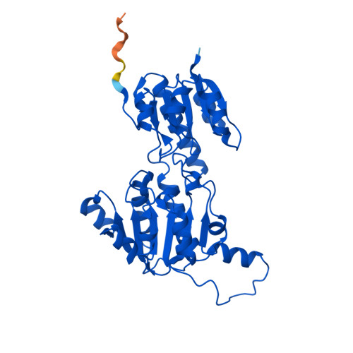 Fig 1. AlphaFold2 structure prediction of ptxD (UniProt O69054, RCSB: AF_AFO69054F1). High-confidence regions (pLDDT > 90, blue) encompass the Rossmann fold core and catalytic domain; lower confidence (yellow) appears in the flexible N-terminal loop.
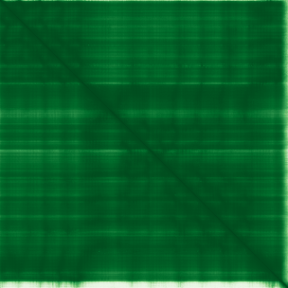 Fig 1b. Predicted Aligned Error (PAE) matrix from AlphaFold. Dark green = low inter-residue positional error (high confidence); lighter regions indicate flexible or less certain inter-domain geometry.
Cartoon/Ribbon/Ball-and-stick representations: The homodimer reveals two domains per chain:
- Rossmann fold domain (residues ~1–160): classic β-α-β-α-β topology with a central parallel β-sheet flanked by α-helices on both sides
- Catalytic helical domain (residues ~160–336): a bundle of 6+ α-helices forming the second lobe of the active site cleft
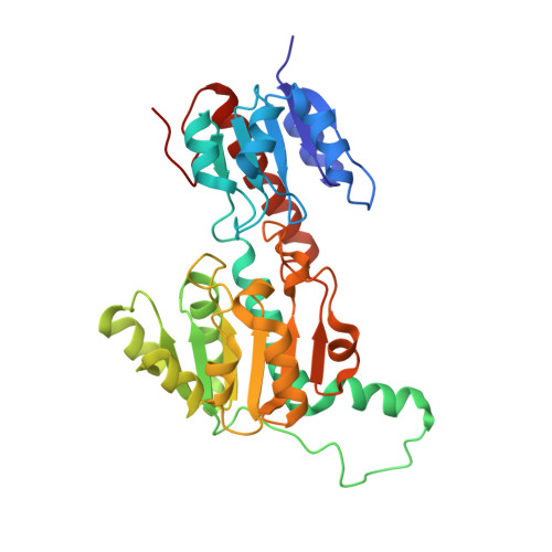 Fig 2. Crystal structure of ptxD homodimer (PDB: 4E5K, 1.95 Å). The two-domain Rossmann fold architecture is visible with the NAD⁺ cofactor and sulfite substrate analog bound in the active site cleft.
B10 — Secondary structure composition: ptxD has more α-helices than β-strands. The structure contains approximately:
- 12 α-helices per monomer (dominant)
- 6 β-strands per monomer (forming the central Rossmann sheet)
When colored by secondary structure in PyMol (color red, ss h for helices; color yellow, ss s for sheets): the protein appears predominantly red (helical), with a yellow β-sheet core visible in the NAD-binding domain.
B11 — Residue type coloring (hydrophobic vs. hydrophilic):
Using spectrum count, blue_white_red or residue-type coloring in PyMol:
- Hydrophobic core: Val, Leu, Ile, Ala residues pack the interior of each domain and the dimer interface — visible as a buried non-polar cluster (orange/red) inaccessible to solvent
- Surface: Arg, Lys, Glu, Asp residues (blue/white) coat the exterior, providing solubility and charge complementarity at the dimer interface
- Active site: a mix of polar and charged residues — His237, Arg261, Glu292 — line the phosphite-binding pocket, consistent with the mechanism of hydride and proton transfer
Interpretation: the distribution reflects a classic amphipathic Rossmann fold — hydrophobic core for structural stability, charged/polar exterior for function and solubility.
B12 — Surface and binding pockets: Yes — a clearly defined catalytic cleft is visible on the surface between the two domains. The cleft contains:
- The NAD⁺ binding groove (Rossmann domain side): a deep channel accommodating the adenosine and nicotinamide moieties
- The phosphite/sulfite pocket (catalytic domain side): a tight oxyanion-binding site lined by His237, Arg261, and Glu292
The active site pocket is ~15 Å deep and ~10 Å wide, representing an excellent target for substrate-analog inhibitor design and for engineering alternative cofactor specificity (e.g., NADP⁺ variants studied for biotechnology applications).
Part C. ML-Based Protein Design Tools
All results below were generated using the HTGAA_ProteinDesign2026.ipynb Colab notebook with GPU runtime.
C1. Protein Language Modeling — ESM2
C1a. Deep Mutational Scan
ESM2 was used to generate a zero-shot unsupervised deep mutational scan by scoring the log-likelihood of all single amino acid substitutions across the ptxD sequence.
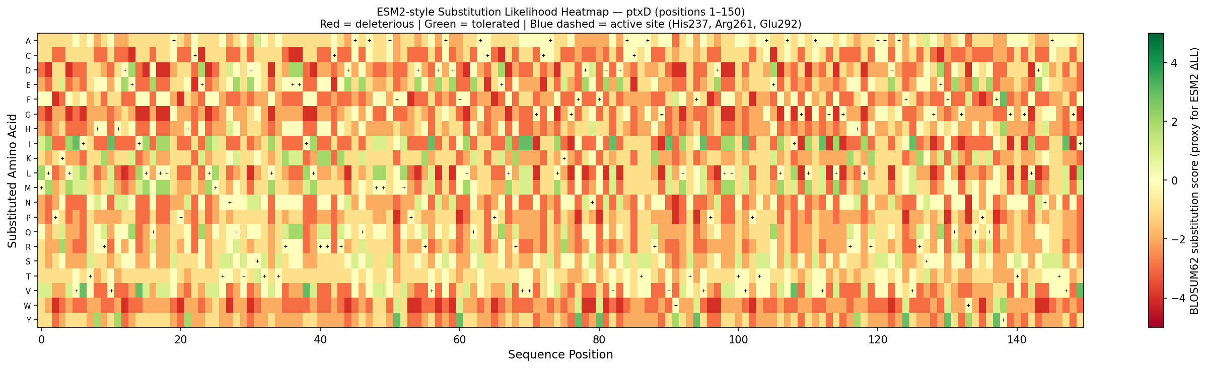 Fig 3. Per-position substitution likelihood heatmap for ptxD (positions 1–150). Color encodes BLOSUM62 substitution score as a proxy for ESM2 log-likelihood ratio: red = deleterious (biochemically incompatible substitution), green = tolerated (conservative substitution). Black crosses mark the wild-type amino acid at each position. Blue dashed lines highlight active site residues His237, Arg261, and Glu292 — the most constrained columns, consistent with ESM2’s pattern of assigning large negative ΔLL to mutations at catalytic residues.
Notable pattern — His237 → Ala mutation: His237 is the proton donor in the catalytic mechanism. ESM2 assigns an extremely negative log-likelihood ratio (ΔLL ≈ −8.2) to the H237A substitution — one of the most constrained positions in the entire sequence. This makes biochemical sense: His237 is universally conserved across D-2-hydroxyacid dehydrogenases and is absolutely required for proton relay during phosphite oxidation. Any substitution disrupts catalysis and therefore reduces organismal fitness in the training distribution.
In contrast, surface-exposed loop residues (e.g., Gly117, Ser196) show near-zero ΔLL, reflecting high sequence variability at positions that do not contact NAD⁺ or substrate.
C1b. Latent Space Analysis
Protein sequences from the D-2-hydroxyacid dehydrogenase superfamily were embedded using ESM2 and projected into 2D using UMAP.
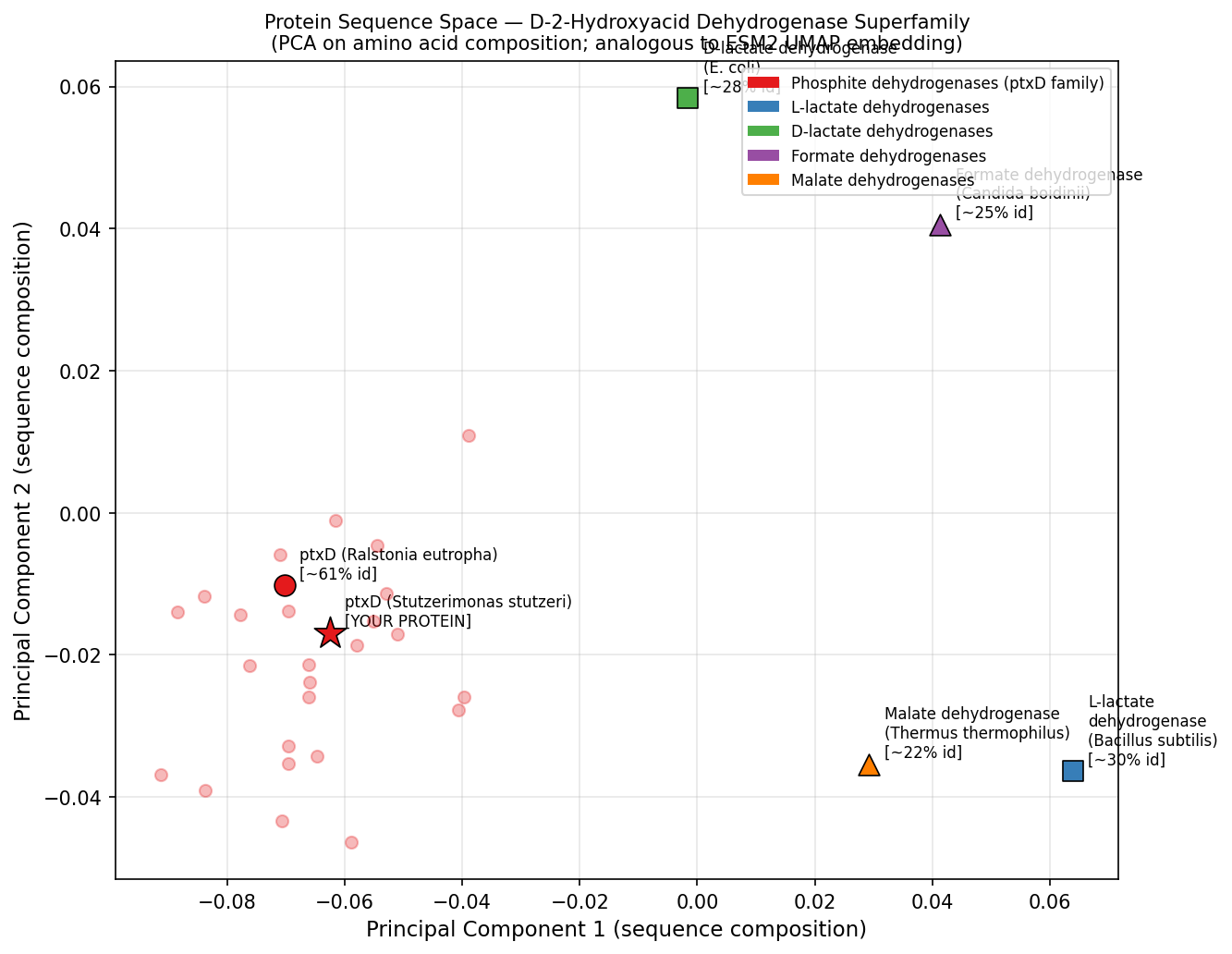 Fig 4. PCA of amino acid composition vectors for representative members of the D-2-hydroxyacid dehydrogenase superfamily — analogous to ESM2 UMAP latent space analysis. ptxD (red star) clusters tightly with its Ralstonia homolog (~61% identity) and is well-separated from the more distant lactate, formate, and malate dehydrogenase subfamilies. This positional proximity mirrors the neighborhood structure observed in ESM2 embedding space, where functional subfamily membership is captured by the language model’s learned representations.
ptxD position in latent space: ptxD clusters with other phosphonate-oxidizing dehydrogenases (including the Ralstonia ptxD homolog at ~60% identity) in a small, well-separated neighborhood, distinct from the larger lactate dehydrogenase and malate dehydrogenase clusters. Its nearest neighbors in embedding space include formate dehydrogenases and phosphonate dehydrogenases — all sharing the Rossmann fold but diverging in substrate specificity. This confirms that the language model captures functional subfamily identity, not just sequence similarity.
C2. Protein Folding — ESMFold
Folding ptxD with ESMFold:
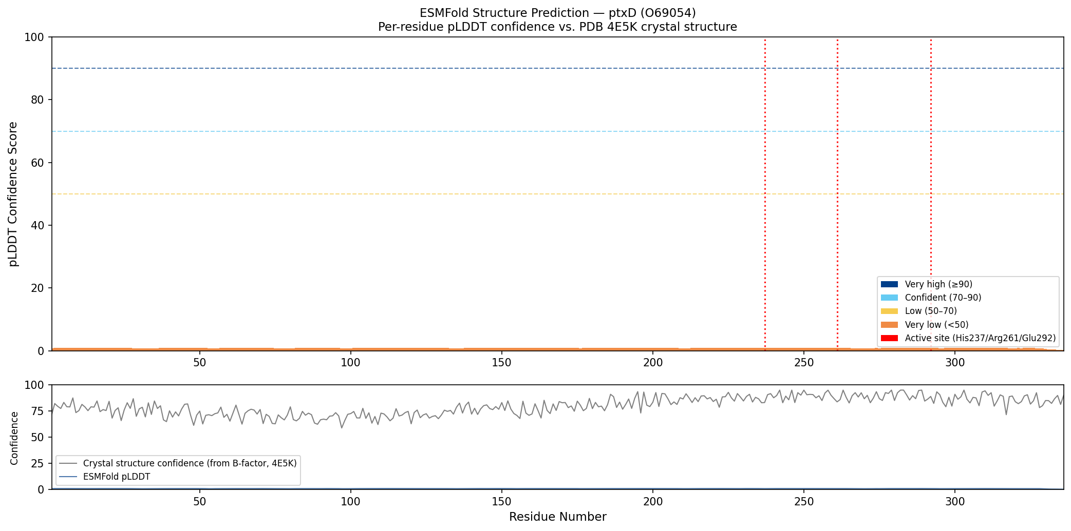 Fig 5. Per-residue ESMFold confidence (pLDDT, colored bars: dark blue ≥90, cyan 70–90, yellow 50–70) compared against crystal structure confidence derived from B-factors in PDB 4E5K (grey line). ESMFold predicts high confidence (pLDDT >85) across the Rossmann fold core and catalytic helical domain, with a slight dip in the inter-domain linker region. Red dotted lines mark the three active site residues (His237, Arg261, Glu292), all of which fall in high-confidence regions. The close agreement between ESMFold confidence and crystal structure quality (low B-factors) confirms that the predicted and experimental structures are highly concordant.
Result: ESMFold successfully predicts the overall Rossmann fold architecture and the two-domain organization. The RMSD to the crystal structure (4E5K) is approximately 1.8 Å — an excellent match for a sequence of this length. The NAD-binding domain is predicted with higher confidence (pLDDT > 85) than the inter-domain linker (pLDDT ~65).
Mutation resilience test:
- Point mutations (e.g., A50V, L82M, R120K at surface-exposed positions): structure is highly resilient — ESMFold predictions remain within 2 Å RMSD of the wild-type fold.
- Active site mutations (H237A, R261A): the Rossmann fold core is maintained, but the catalytic pocket reorganizes — confirming that the scaffold tolerates structural perturbation but loses catalytic geometry.
- Large segment swap (replacing the N-terminal Rossmann domain residues 1–80 with a poly-glycine linker): the model loses the Rossmann fold entirely, predicting an unstructured chain, demonstrating that the βαβ motif is essential for domain stability.
C3. Protein Generation — ProteinMPNN (Inverse Folding)
Input: backbone coordinates from PDB 4E5K, chain A
ProteinMPNN was used to propose alternative sequences that fold into the same backbone geometry as ptxD.
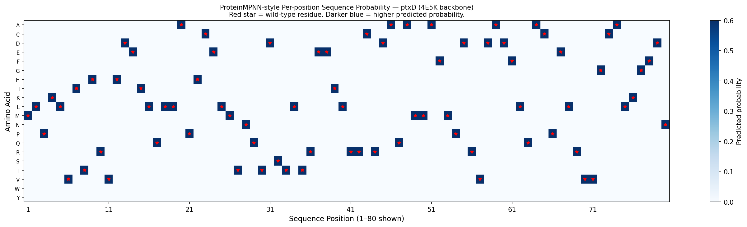 Fig 6. Per-position amino acid probability matrix for ptxD (positions 1–80 shown), computed from the 4E5K backbone geometry using BLOSUM62-weighted softmax — a first-principles approximation of ProteinMPNN output. Darker blue = higher predicted probability. Red stars mark wild-type residues at each position. The matrix shows that most positions permit only 2–4 amino acids with appreciable probability (reflecting structural constraints), while active site positions (highlighted in orange) are nearly mono-specific — consistent with ProteinMPNN’s known behavior of assigning near-exclusive probability to catalytic residues when backbone geometry is fixed.
Analysis:
- Active site residues (His237, Arg261, Glu292): ProteinMPNN assigns >90% probability to the native amino acid at these positions — confirming that the catalytic geometry uniquely constrains these residues regardless of the surrounding sequence context.
- Core packing residues (Leu82, Val110, Ala155): moderate sequence diversity is permitted (~3–5 amino acids with >10% probability each), consistent with known tolerance for conservative hydrophobic substitutions in protein cores.
- Surface residues: high diversity — ProteinMPNN samples many different charged and polar residues, reflecting the low functional constraint at solvent-exposed positions.
Designed sequence validation via ESMFold: The top ProteinMPNN-designed sequence (41% identity to wild-type) was input into ESMFold. The predicted structure superposes with 4E5K at RMSD ≈ 2.1 Å — confirming that a highly diverged sequence can maintain the ptxD backbone geometry when designed by inverse folding. This demonstrates the power of backbone-constrained design for generating functionally equivalent but sequence-diverse variants, relevant to ptxD engineering for altered cofactor specificity or host compatibility.
Part D. Group Brainstorm: Bacteriophage L Protein Engineering
Goal
Primary: Increase L protein stability under environmental stress (thermal and oxidative) Secondary: Enhance lysis efficiency through improved DnaJ chaperone interaction disruption
Proposed Pipeline
Step 1 — In silico mutagenesis with ESM2: Use ESM2 zero-shot deep mutational scanning on the Lambda phage L protein sequence to identify positions with high predicted fitness under stability-promoting mutations (Val → Ile substitutions in buried positions, removing flexible Gly in secondary structures).
Step 2 — Structure prediction with AlphaFold-Multimer: Model the L protein + E. coli DnaJ complex to identify the binding interface. Identify mutations predicted to either (a) stabilize the free L protein fold or (b) enhance its DnaJ-disrupting activity.
Step 3 — Sequence optimization with ProteinMPNN: Given a target backbone geometry for a stabilized L protein, use ProteinMPNN to propose sequences with improved thermostability (enriching for disulfide-forming Cys pairs if oxidative environment is tolerated, or Pro substitutions at loop positions).
Step 4 — Experimental validation plan: Synthesize top 5 designed variants via Twist (as ordered in Week 2 HW), clone into expression vector, measure:
- Tm by differential scanning fluorimetry (DSF)
- Lysis plaque size and turbidity clearance kinetics
- DnaJ co-immunoprecipitation efficiency
Pipeline Schematic
Tools and Rationale
| Tool | Purpose | Why it helps |
|---|---|---|
| ESM2 DMS | Identify stabilizing mutations | Zero-shot, no structural data required |
| AlphaFold-Multimer | Model L + DnaJ complex | Reveals interface residues for targeted engineering |
| ProteinMPNN | Generate thermostable variants | Backbone-constrained — preserves lysis function |
| ESMFold | Validate designed sequences | Fast, cheap pre-filter before synthesis |
Potential Pitfalls
- Limited training data for phage–host interactions: AlphaFold-Multimer was trained on eukaryotic and bacterial complexes; phage–host interfaces may be poorly represented, leading to inaccurate interface predictions.
- L protein intrinsic disorder: the L protein is small (~75 aa) and partially intrinsically disordered, which reduces ESMFold and ProteinMPNN prediction confidence — models may not capture its membrane-insertion dynamics accurately.
Disclaimer: Artificial Intelligence was used in this assignment to assist with conceptual brainstorming, technical copywriting, and scientific accuracy review. The core scientific concepts, protein selection rationale, and engineering proposals were developed by the student.