Week 6: Gibson Assembly
Using PCR-based mutagenesis, Gibson Assembly, and bacterial transformation, we intended to introduce targeted mutations into the amilCP gene to produce magenta and blue color variants in E. coli.
Part 1: PCR
We ran two separate PCR reactions from the mUAV plasmid template using Phusion HF PCR Master Mix:
Backbone Fragment (Backbone Fwd + Backbone Rev primers): amplifies the origin of replication, chloramphenicol resistance gene, promoter, and RBS.
Color Insert Fragment (Color Fwd + Color Rev primers): amplifies the region from 24 bp before the chromophore to the terminator. The Forward primer contains a mutation at the CP site to introduce the desired color mutation(we chose magenta and blue variants).
Each reaction was run in a 25 µL volume with 0.8 µL template (38.5 ng/µL), 2.5 µL of each primer (5 µM stock), and 12.5 µL Phusion HF PCR Mix.
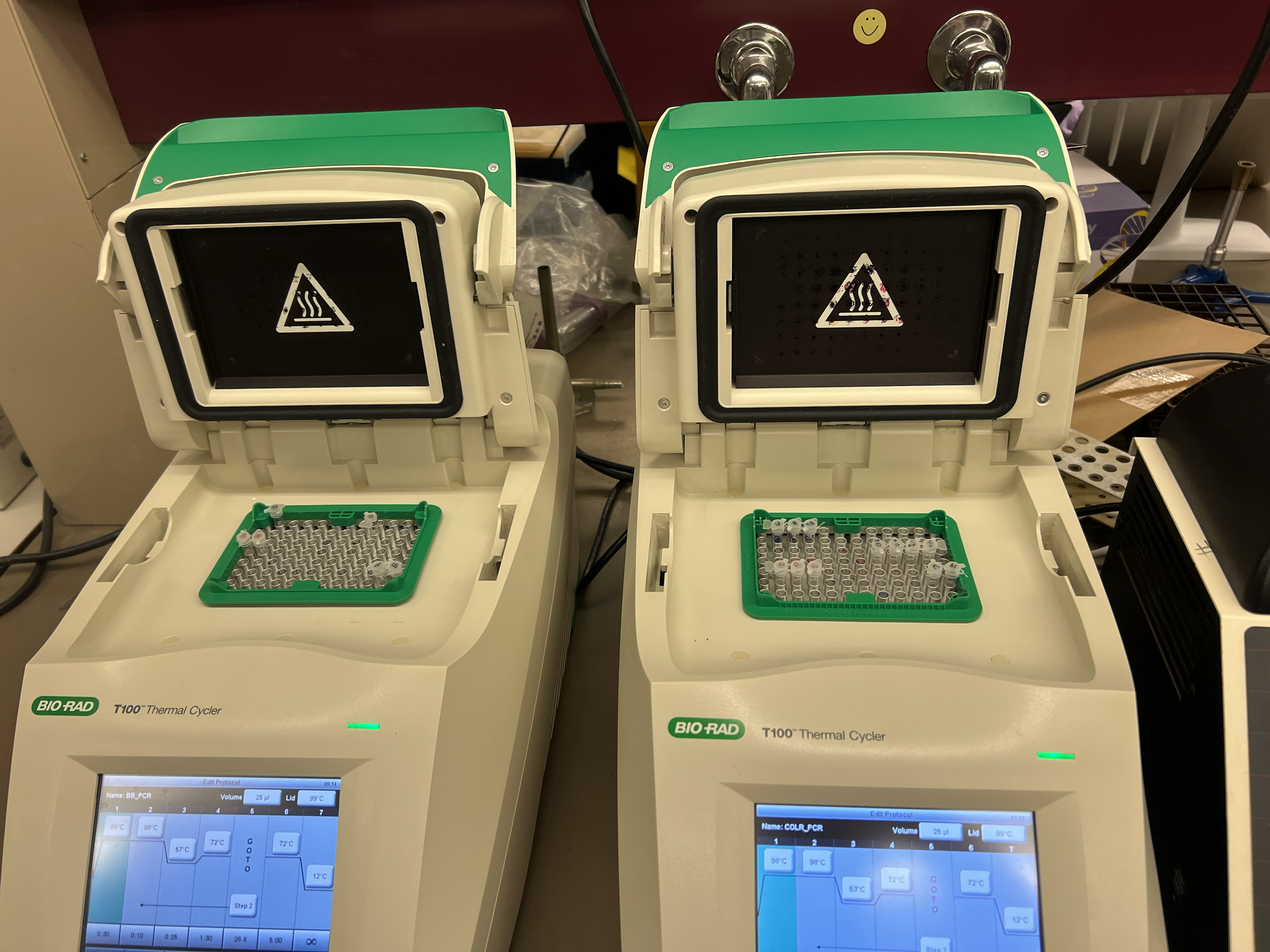
Thermocycler conditions:
Backbone PCR: 98°C 30s → 26 cycles of (98°C 10s / 57°C 25s / 72°C 1.5 min) → 72°C 5 min → 12°C hold
Color PCR: 98°C 15s → 26 cycles of (98°C 10s / 53°C 20s / 72°C 15s) → 72°C 5 min → 12°C hold
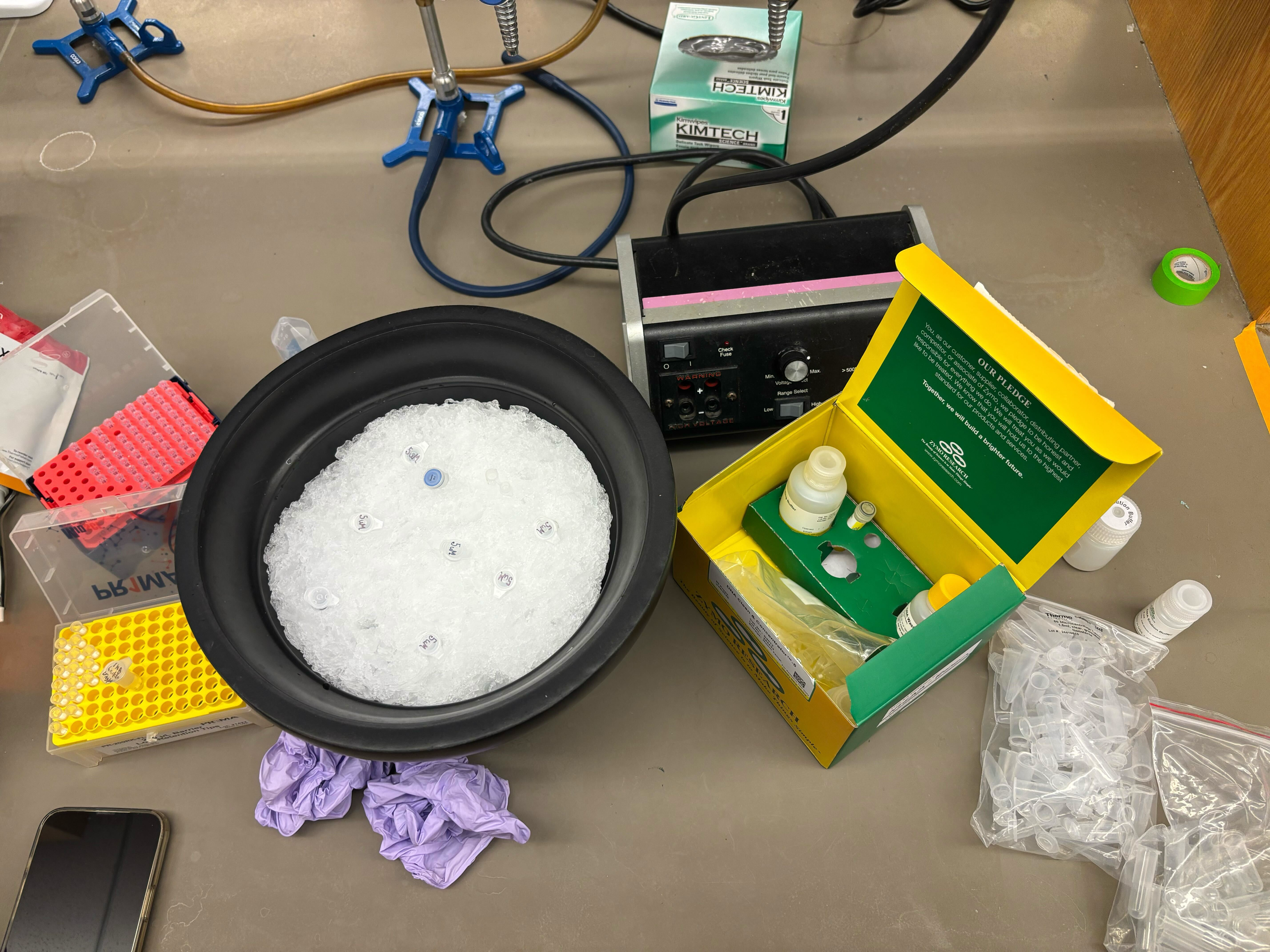
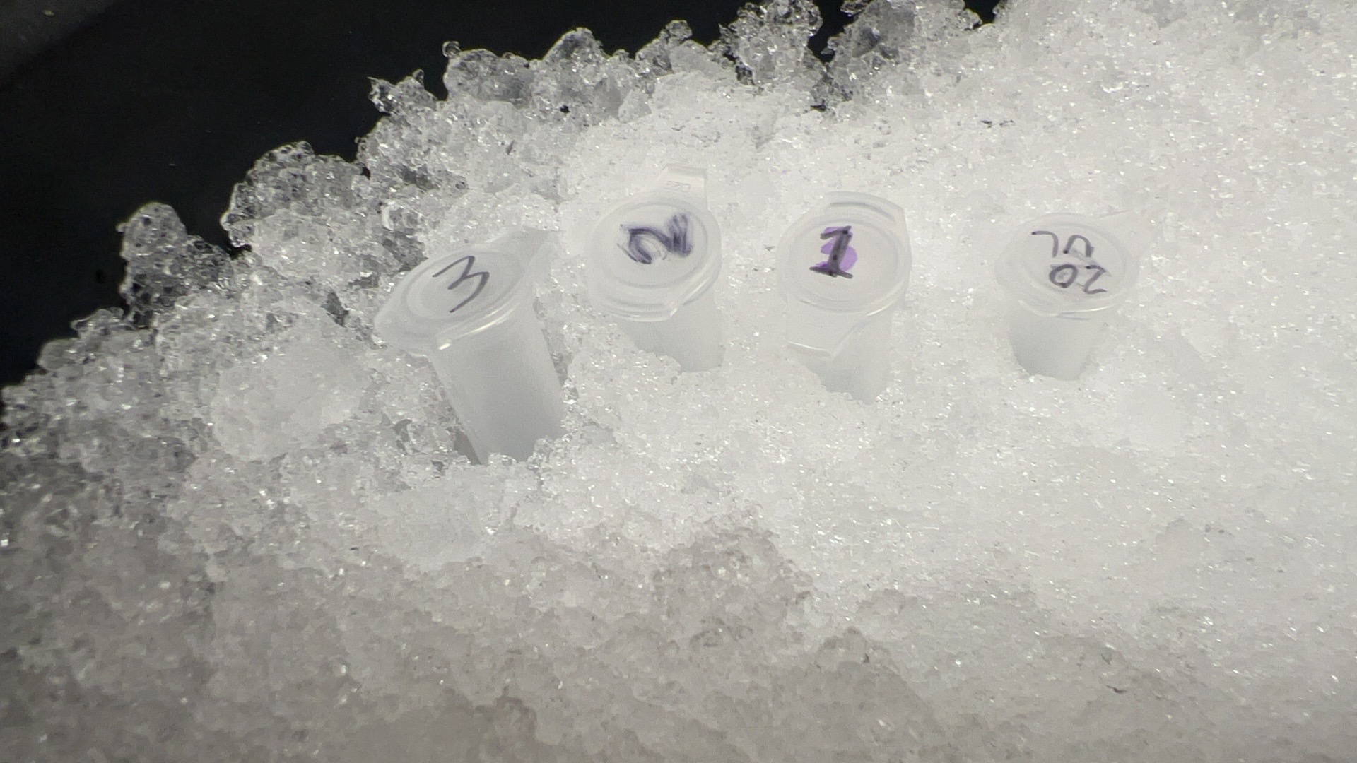
Part 1a: DNA Purification
PCR products were purified using the Zymo DNA Clean & Concentrator kit. Each 20µL sample was mixed with 100 µL DNA Binding Bufferinto micro centrifuge tube and loaded onto a ZymoSpin column, and centrifuged. After two washes with 200 µL DNA Wash Buffer, DNA was eluted in 6 µL nuclease-free water.
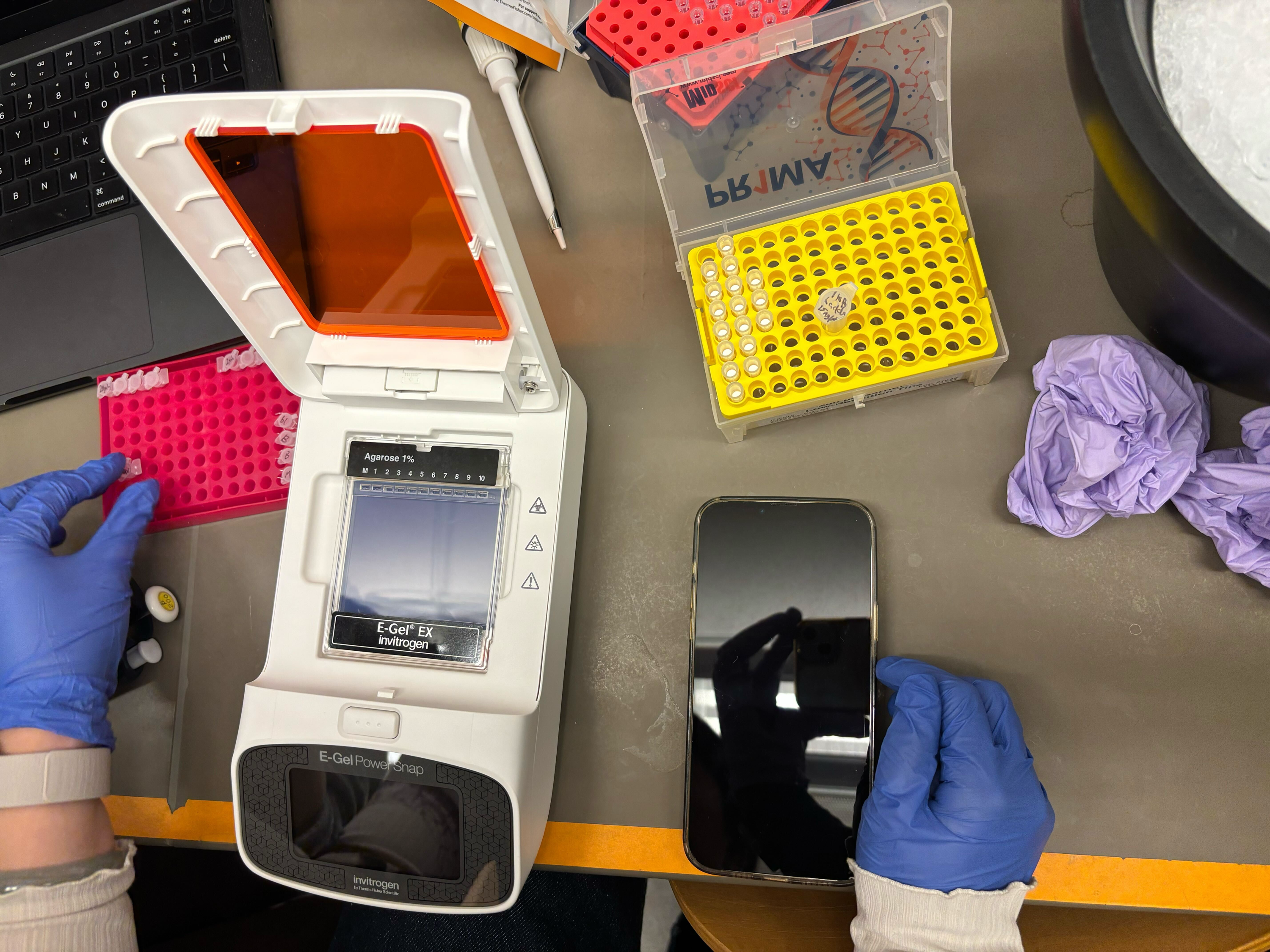
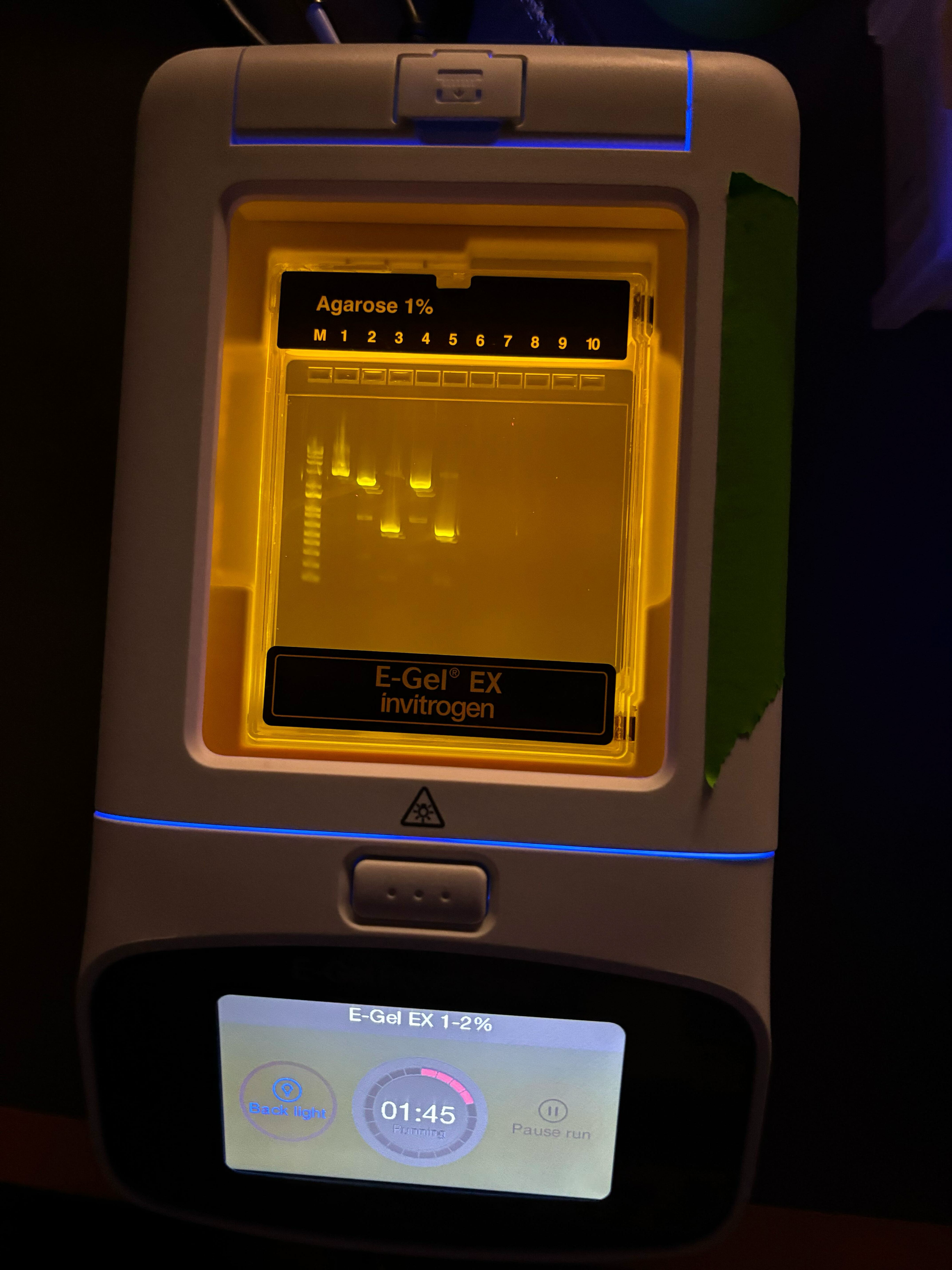
Part 1b: Diagnostic Gel Electrophoresis
Purified samples were run on through gel electrophoresis to confirm correct fragment sizes. 2 µL of each sample was mixed with 18 µL of water and loaded into the gel wells along with 20µL of pre-diluted ladder and original mUAV plasmid as references.
Part 2a: Gibson Assembly
Purified backbone and color fragments were assembled using Gibson Assembly Master Mix. Reactions were set up in 10 µL total volume and incubated at 50°C for 15 minutes. After incubation, 100 µL of nuclease-free water was added to dilute the reaction before transformation.
Gibson Assembly works by: (1) exonuclease chewing back 5’ ends to expose complementary single-stranded overhangs, (2) annealing of complementary overhangs between backbone and insert, (3) polymerase filling in gaps, and (4) ligase sealing nicks to form a complete circular plasmid.
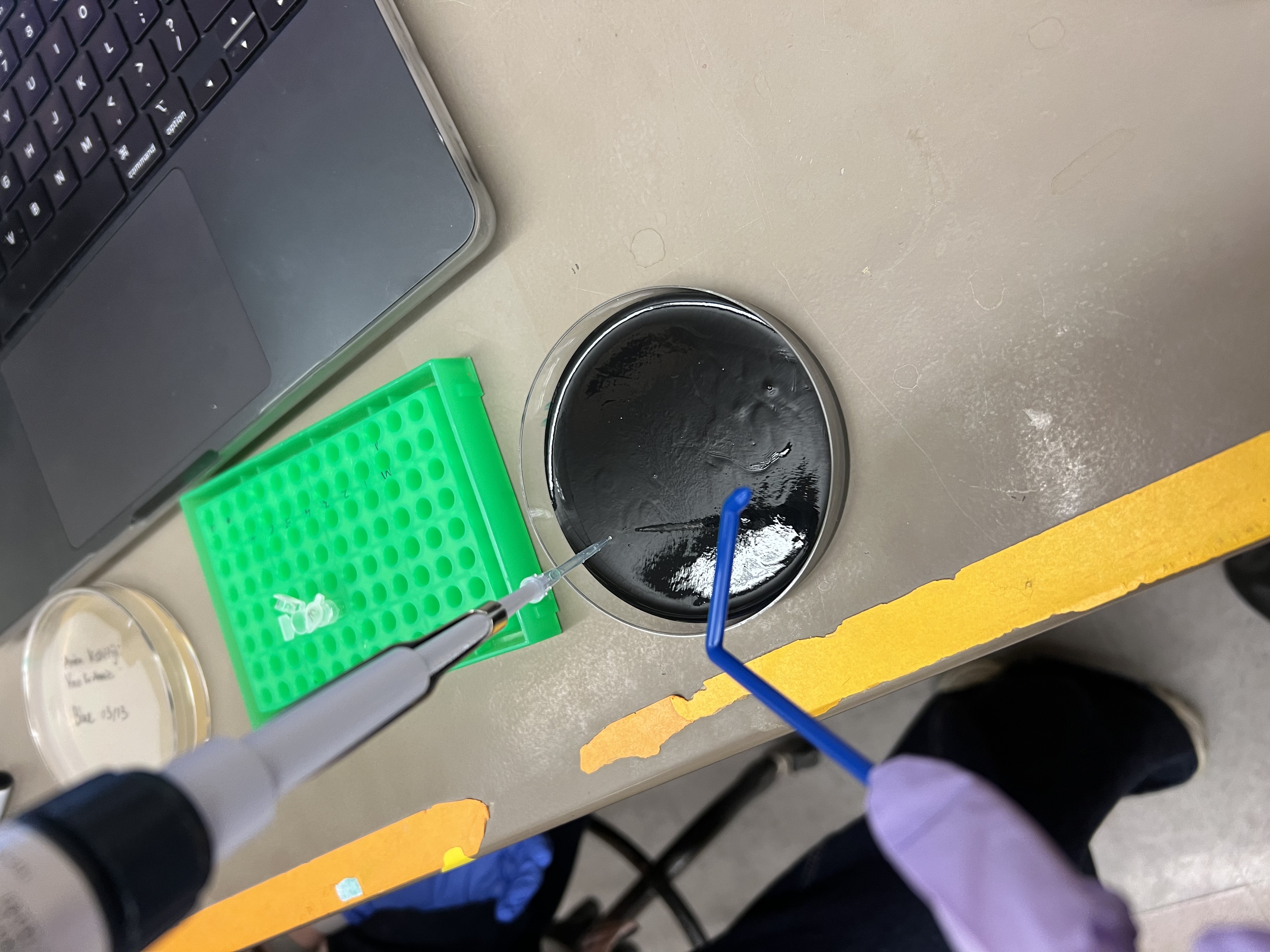
Part 2b: Transformation
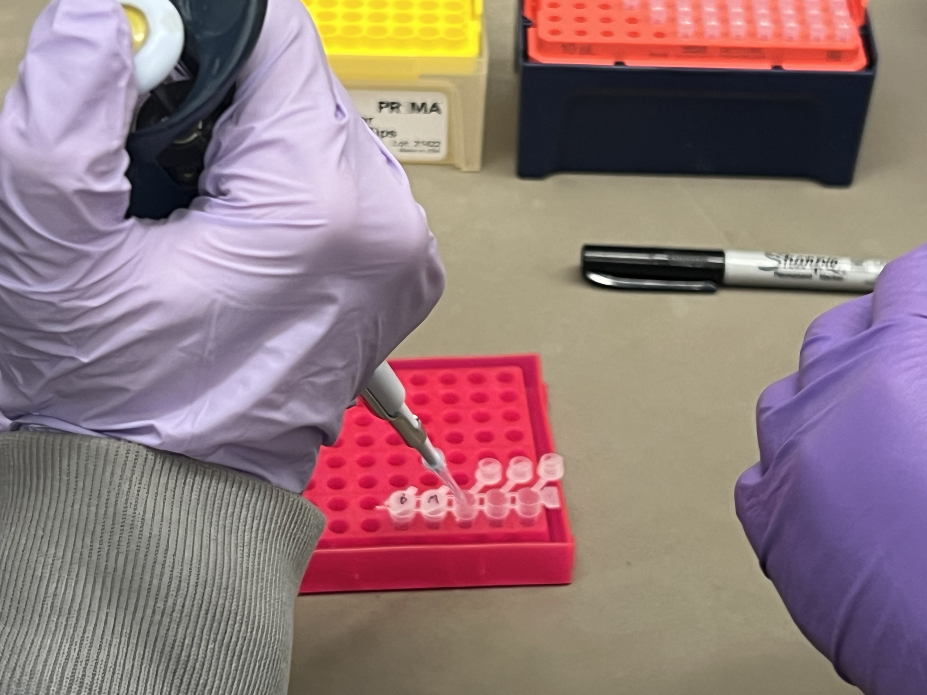
Gibson Assembly products were transformed into DH5α chemically competent E. coli cells using heat shock:
- Thawed competent cells on ice for 10 minutes
- Added 4 µL of diluted Gibson Assembly product to 20 µL of competent cells
- Incubated on ice for 30 minutes
- Heat shocked at 42°C for exactly 45 seconds, then returned to ice for 5 minutes
- Added 100 µL SOC media and incubated with shaking for 60 minutes
- Plated 100 µL onto agar plates
- Incubated plates at 37°C for 72 hours
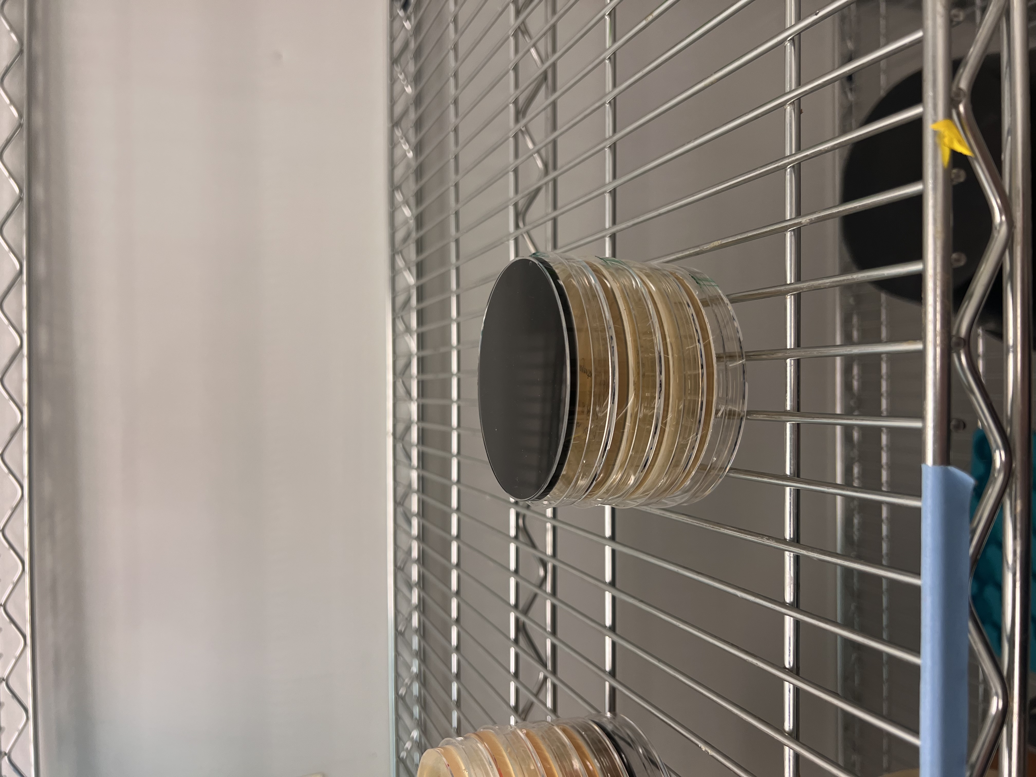
Results
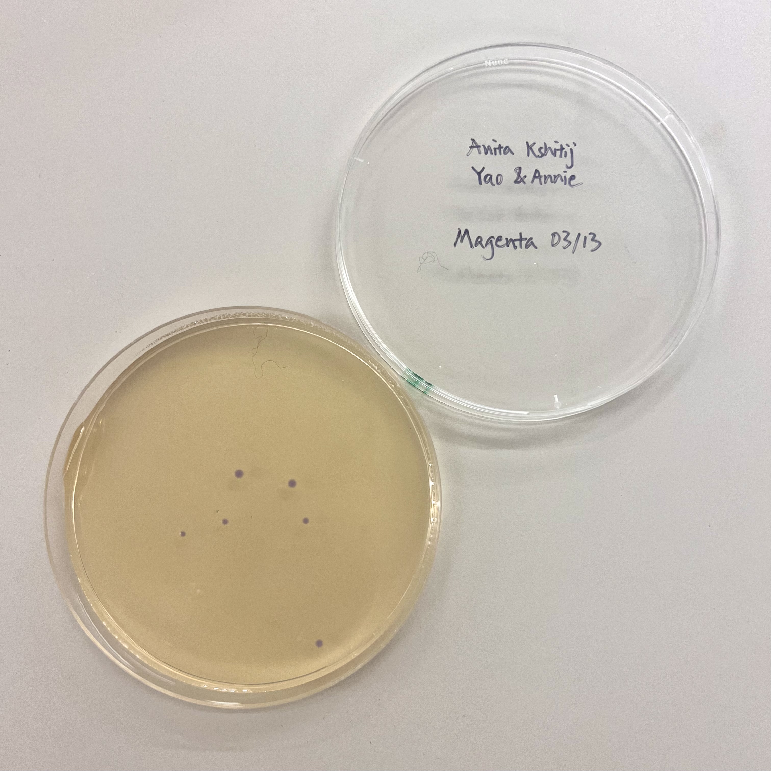
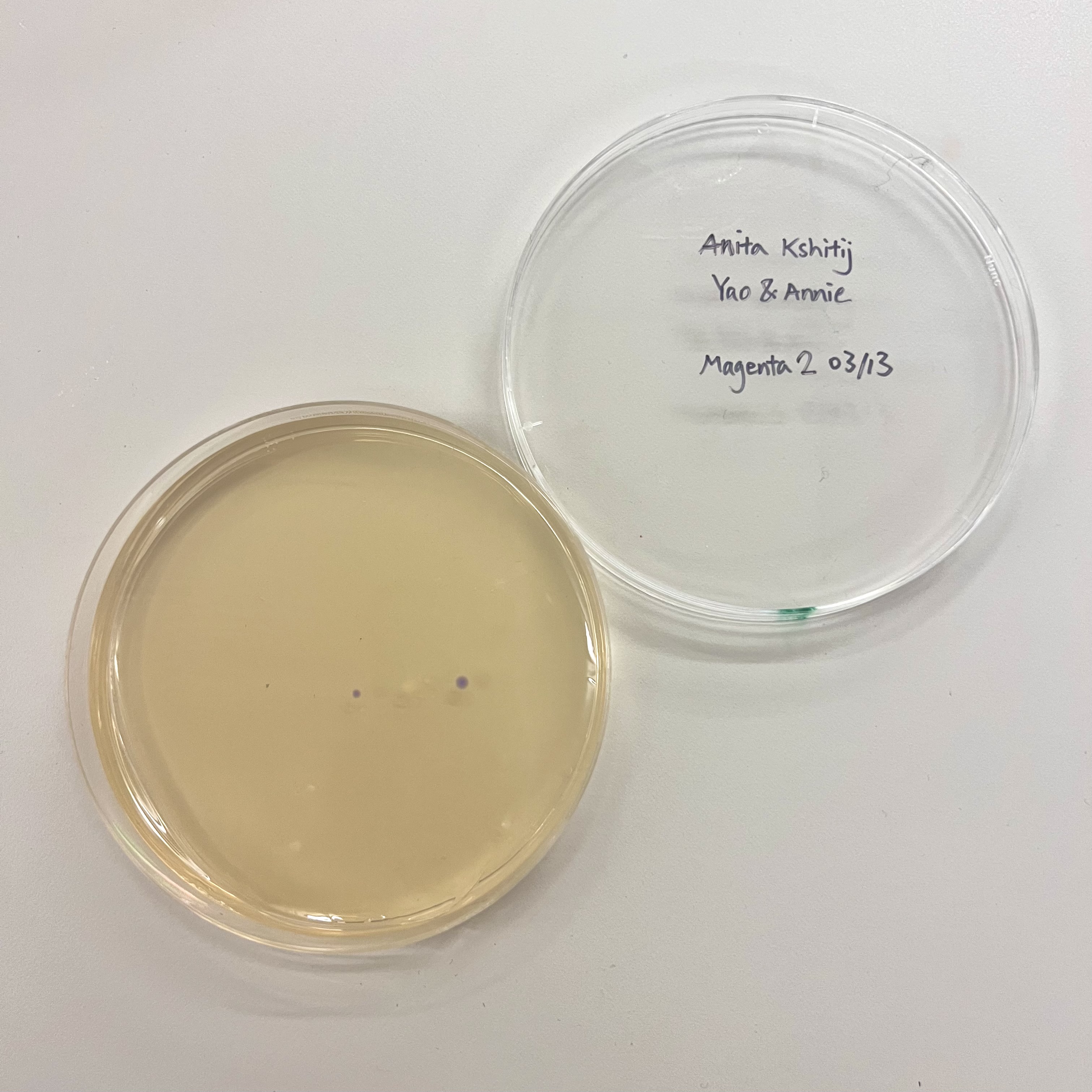
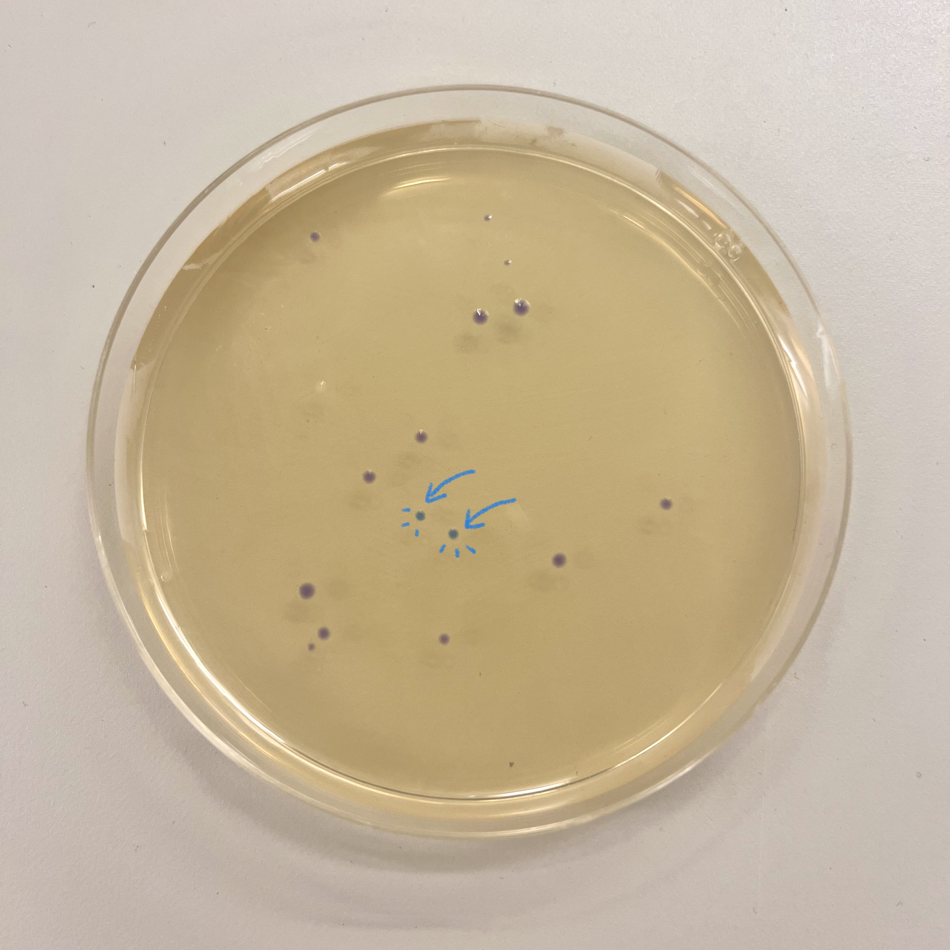
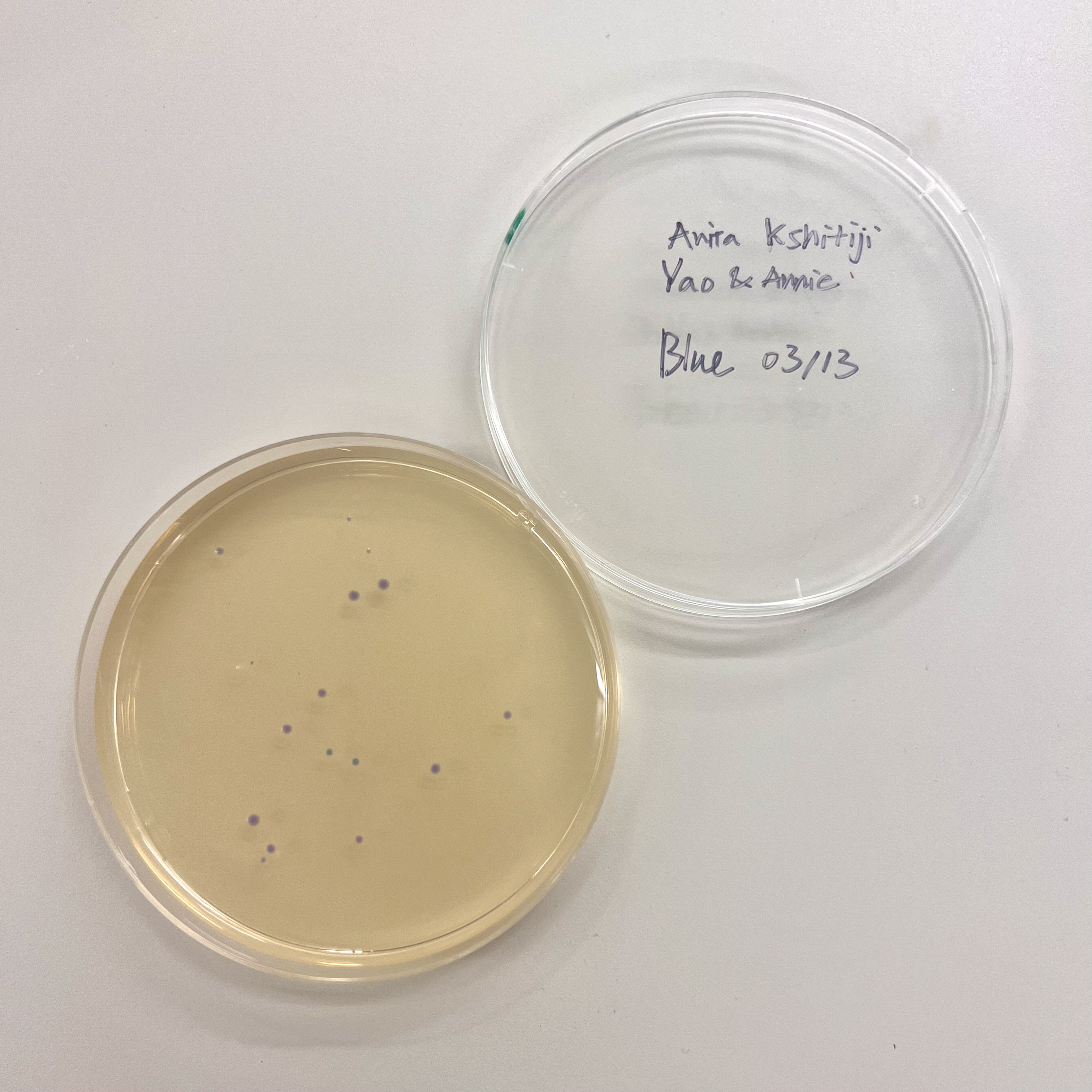 All plates had very low colony count overall. The blue plate had 2 visibly blue-colored colonies, small partial success. Both magenta plates showed no magenta coloration, so the colonies that did grow were likely untransformed or reverted to original amilCP color of purple.