Week 2 HW: Genetic Construc using Kernel
HTBAA Homework #2: DNA Design
A Lactiplantibacillus plantarum engineered to display a β-1,3-glucanase on its surface and release it only when Candida secretes its protease Sap2.
Part 0: Kernel setup
Kernel repository folder: BUGSS_Ana_Sofia_Barrientos_Uriarte (workspace HTBAA 2026).
Part 1: Expression target
What molecule?
An enzyme: endo-β-1,3-glucanase (EC 3.2.1.39).
β-1,3-glucan is a structural pillar of both the Candida cell wall and the extracellular matrix of its biofilm, where it also sequesters antifungal drugs [1]. Degrading it disperses the biofilm and restores antifungal susceptibility [13].
A second effector, the human IL-1Ra, is specified but not built in this assignment. Rather than targeting the fungus directly, this second effector addresses the inflammatory pathology by blocking the NLRP3/IL-1β signaling axis that drives neutrophil infiltration and produces symptoms [5], [6]. I intentionally deferred building this second effector to avoid overloading the bacterial host; it is essential to first characterize the metabolic burden, secretion efficiency, and Sap2-cleavage kinetics of the single-protein construct before expressing two genes within the same operon [2], [7].
What is the sequence?
Endo-β-1,3-glucanase of Streptomyces sioyaensis: UniProt Q9L816, GenBank AF217415 / AAF31438.1, PDB 3DGT.
The full protein is 478 aa. I use residues 51 to 478 (428 aa), removing the native signal peptide.
Why cut at 51 UniProt states that the signal peptide cleavage position has not been experimentally proven, and lists two alternatives: 1 to 41 (SignalP) and 1 to 50 (PROSITE PS51318 / InterPro IPR006311, the twin-arginine translocation profile). I used the Tat profile because the protein carries a twin-arginine motif and is a Tat substrate, which makes the Tat-specific tool the appropriate one. The cut does not touch the catalytic domain, which CDD cd02182 places at residues 56 to 321.
Host
Lactiplantibacillus plantarum WCFS1.
It is the reference strain of the lactobacilli, and every part below was measured in it: the PtlpA promoter and TA modules [2], the Lp_3014 signal peptide and LysM anchor [3], [4], and the pSIP vector backbone. L. crispatus would be the final clinical chassis because it is the dominant native inhabitant of the healthy human vagina, where it constitutes Community State Type I (CST-I) [15]. However, none of these parameters are established there.
Where does the product go?
Secreted through the Sec pathway, then retained on the cell wall, then released by proteolysis.
Three stages:
- A homologous signal peptide (Lp_3014) directs the fusion protein through the Sec pathway. During export, the signal peptide is cleaved, releasing the mature protein outside the cytoplasmic membrane.
Lp_3014 (or lp_3014) is the gene encoding a putative extracellular transglycosylase in L. plantarum WCFS1, containing an N-terminal secretion signal peptide and a LysM domain for non-covalent cell-wall anchoring [4].
- A LysM domain binds non-covalently to peptidoglycan in the bacterial cell wall, keeping the enzyme on the surface instead of diffusing away.
- A Sap2-cleavable linker sits between anchor and enzyme. When Candida turns pathogenic and secretes Sap2, it cuts the linker and releases the active glucanase.
SAP2 is the most consistently overexpressed protease gene in symptomatic patients, and secreted aspartyl proteinases alone cause vaginitis in mice [5][6]. Sap2 therefore marks symptomatic disease, not mere colonisation, which is exactly the distinction the device needs to make.
Part 2: Genetic parts
| # | Part name | Part type | Source | Justification |
|---|---|---|---|---|
| 1 | PtlpA_promoter | Promoter | Dey et al. 2023, Table S1 [2] | Strongest promoter reported in L. plantarum WCFS1 (up to 5x previous ones), measured in this exact host |
| 2 | RBS_strong_Lp | Ribosome binding site | Dey et al. 2023, Table S1 [2] plus a 6 nt spacer added here | Validated with PtlpA; spacer added so the Shine-Dalgarno sits 7 nt from the ATG |
| 3 | SP_Lp3014 | Peptide CDS (+start -stop) | GenBank AL935263.2, protein CCC80071.1, UniProt F9USE1, res. 1 to 28 | Homologous signal peptide, so the host’s own Sec machinery exports the effector |
| 4 | LysM_anchor_Lp3014 | CDS (-start -stop) | Same gene, res. 29 to 116 (88 aa) | Non-covalent peptidoglycan anchor; the catalytic domain of lp_3014 is excluded so it cannot digest the host wall |
| 5 | Sap2link_v1 | Peptide (-start -stop) | Designed here | Releases the enzyme only when Candida secretes Sap2 |
| 6 | Glucanase_opt | CDS (-start +stop) | UniProt Q9L816, codon-optimised | Degrades biofilm β-1,3-glucan; the CBM6 module targets it to its own substrate |
| 7 | T7_terminator | Terminator | Dey et al. 2023, Table S1 [2] | Ends the transcript cleanly; validated in the same system |
| 8 | TA_YafQ_DinJ | Bacterial sequence | Dey et al. 2023, Table S2 [2] | Antibiotic-free plasmid retention |
| 9 | TA_MazF_MazE | Bacterial sequence | Dey et al. 2023, Table S2 [2] | Best endogenous TA system; paired with #8 in opposite orientation |
| 10 | ColE1_ori | Origin of replication | Dey et al. 2023, Table S1 [2] | Replication in E. coli during cloning |
| 11 | rep256 | Origin of replication | GenBank AJ628941, nt 5560 to 6247 | Replication in Lactobacillus, the actual host |
| 12 | eryR | CDS (+start +stop) | Dey et al. 2023, Table S1 [2] | Selection of transformants after electroporation |
The three parts I designed or modified
RBS_strong_Lp: tttgtttaactttaagaaggagaaataat
The published sequence [2] ends at the Shine-Dalgarno core, leaving about 1 nt to the ATG. I appended a 6 nt AT-rich spacer (aataat) so the spacing is 7 nt, within the optimal 5 to 9 nt window. The junction used in the original publication is not reported.
SP_Lp3014: the native start codon is TTG, a valid alternative bacterial initiation codon read as methionine by the initiator tRNA. I changed it to ATG for unambiguous initiation from a strong synthetic RBS, and because Kernel’s standard codon table reads TTG as leucine. This is the only deviation from the genomic sequence.
Sap2link_v1: GSGSKLVELLYLVKGGGGSGGGGS
Architecture: GSGS + KLVEL↓LYLVK + (GGGGS)₂.
The cleavage core is a peptide substrate characterised for Sap2 (candidapepsin SAP2, MEROPS A01.060) by Koelsch et al. 2000, taken without its Dnp fluorophore. In the 14 catalogued substrates, positions P4 to P2 (Leu-Val-Glu) and P2’ to P4’ (Tyr-Leu-Val) are invariant, while P1 and P1’ are the positions that were systematically varied. The same P4 to P4’ scaffold occurs at the natural Sap2 cleavage site in oxidised insulin B chain (UniProt P01317).
Leu at P1 and P1’ also matches the substrate preference measured for aspartyl protease activity in both biofilm and planktonic C. albicans cultures [7], so cleavage is not expected to be strictly Sap2-exclusive.
Flexible spacers use the Gly₄Ser motif: glycine gives minimal steric hindrance and prevents rigid secondary structure, and serine contributes solubility [8]. FRET measurements confirm that flexibility rises with glycine content [9]. The N-terminal spacer is short because the LysM module already ends in about 43 residues of native disordered Ser/Thr sequence.
Part 3: Assembled construct
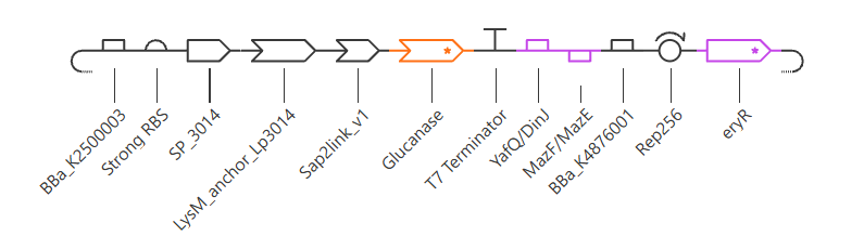
Order (5’ to 3’), 12 parts:
Parts 3 to 6 form one continuous ORF. The two TA operons carry their own promoters and are placed in opposite orientations, as in the original study, so transcription does not read through between modules.
Verification
I checked the assembled sequence by translating it:
| Check | Result |
|---|---|
| Promoter at the 5’ end | 52 nt, correct |
| Shine-Dalgarno to ATG spacing | 7 nt |
| Start codon | single ATG at position 82 |
| Stop codon | single TAA at position 1786 |
| ORF length | 1707 nt, divisible by 3 |
| Accidental internal stops | none |
| Terminator | immediately after the stop, position 1789 |
The ORF encodes a 568-residue fusion protein:
The translated protein starts at MKKLVSTIVTT and ends at NWFDFGH, exactly as designed.
Part 4: Optimize and quality-check
Codon optimization
Kernel offers only four expression organisms: CHO, Homo sapiens, E. coli and S. cerevisiae. There is no table for lactobacilli. I selected E. coli as a proxy because it is the only prokaryote available.
The main limitation is GC content. E. coli is around 51% GC while L. plantarum WCFS1 is around 44.5%, so the output is GC-richer than ideal for the real host.
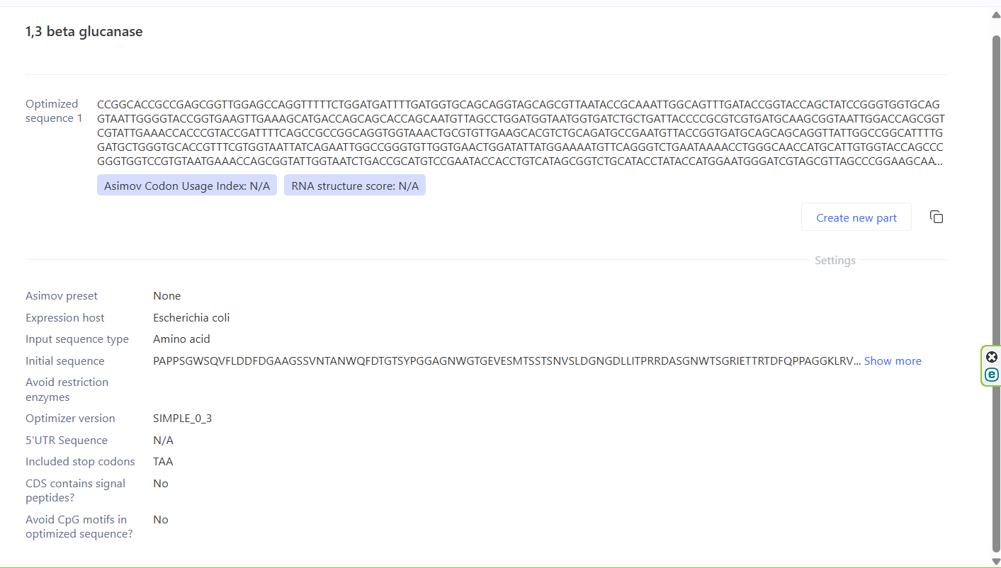
Settings: preset None, Avoid motifs: AGGAGG, TAA appended as the single stop codon, signal peptide algorithm off, CpG avoidance off.
Only the heterologous coding sequences were optimized. The signal peptide, LysM anchor and TA modules are native to L. plantarum and were left unchanged, since optimizing them for E. coli would make them worse. Promoter, RBS and terminator are not translated at all.
BLAST
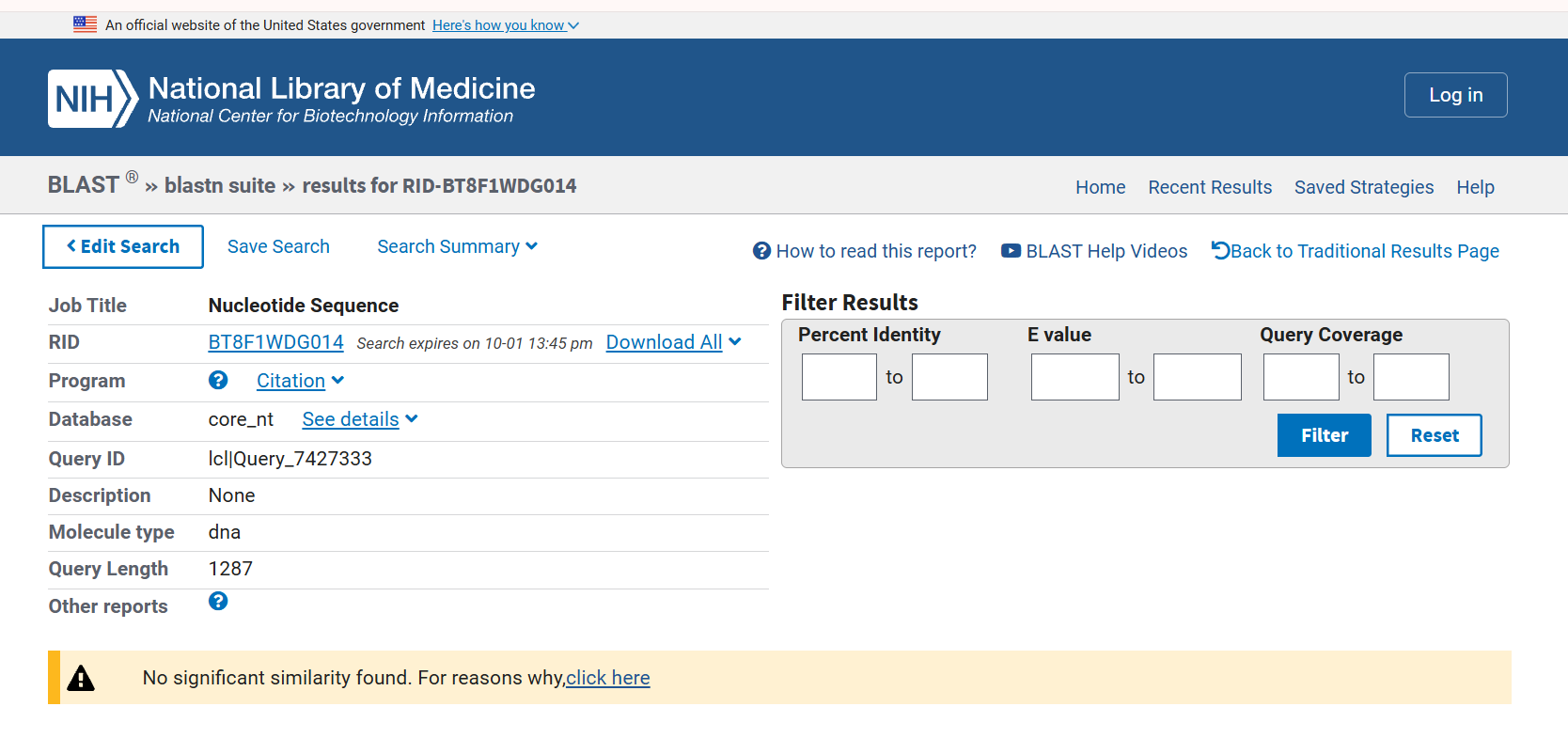
I ran the optimized glucanase CDS (1287 nt) two ways, and the contrast between them is the result.
blastn (nucleotide against nucleotide) returned no significant match. This is expected: codon optimization replaced nearly every codon, so at the DNA level the sequence no longer resembles anything in the databases. It is genuinely novel DNA that has never existed.
blastx (translated query against protein, ClusteredNR) returned strong hits. Every top cluster is a glycoside hydrolase family 16 protein from a Streptomyces species, with E value 0.0 and query coverage of 99 to 100%:
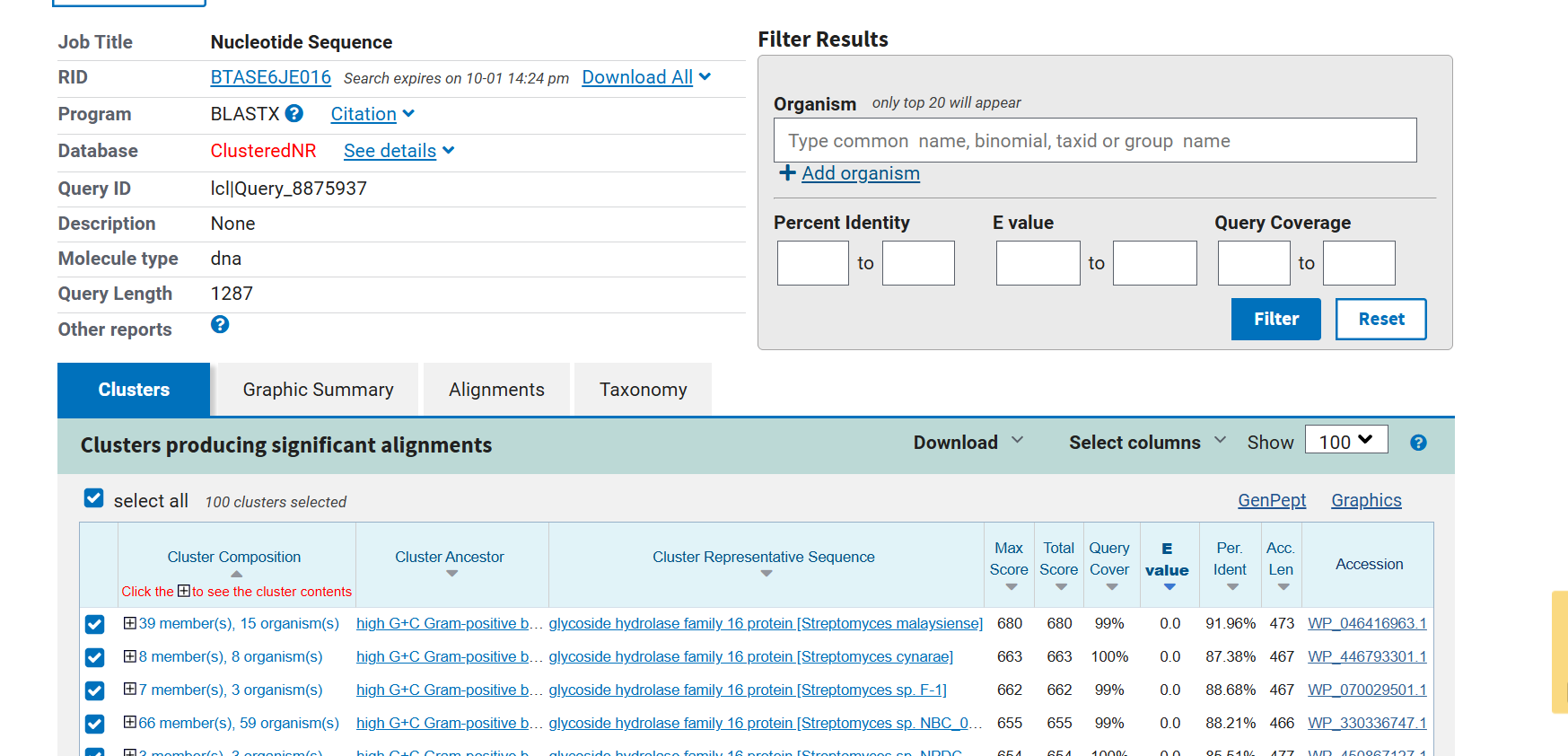
| Cluster representative | Identity | Coverage | E value | Accession |
|---|---|---|---|---|
| GH16 protein, Streptomyces malaysiense | 91.96% | 99% | 0.0 | WP_046416963.1 |
| GH16 protein, Streptomyces cynarae | 87.38% | 100% | 0.0 | WP_446793301.1 |
| GH16 protein, Streptomyces sp. F-1 | 88.68% | 99% | 0.0 | WP_070029501.1 |
| GH16 protein, Streptomyces sp. NBC | 88.21% | 99% | 0.0 | WP_330336747.1 |
Conclusions. The CDS is what I think it is: the whole sequence aligns, with maximal significance, to GH16 glucanases from Streptomyces, which is the correct family and the correct genus. The source sequence itself (Q9L816 / AAF31438.1) does not appear as a separate top hit because the search used ClusteredNR, which collapses near-identical sequences into clusters represented by a single member. The top cluster contains 39 members from 15 organisms, and the S. sioyaensis enzyme sits inside one of them. Rerunning against the standard nr database, or expanding the first cluster, would show it at 100% identity.
Safety screen
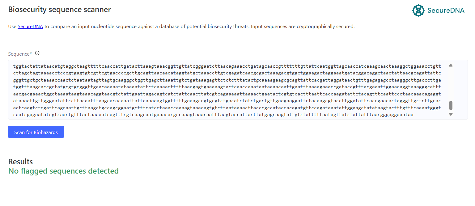
The screen flags sequences of biosecurity concern and antibiotic resistance determinants. Two elements in my construct were flagged, and both are expected.
1. MazF and YafQ are toxins. They are components of toxin-antitoxin systems used for antibiotic-free plasmid retention and biocontainment. The cognate antitoxins (MazE, DinJ) are encoded on the same plasmid, so there is no toxic activity in trans. A cell that loses the plasmid stops producing the short-lived antitoxin and is killed by the stable toxin, which is the intended mechanism.
2. eryR, an erythromycin resistance gene. It is needed to identify transformants after electroporation, since only a small fraction of cells take up the plasmid, and the TA module cannot substitute for it (TA kills cells that lose the plasmid, not cells that never had it). For a clinical live biotherapeutic this marker would be removed, since transferable antibiotic resistance is not acceptable in a live product. Retention would then rely on the TA module alone, and initial selection on auxotrophic complementation.
Part 5: From design to build
How I would build it
The expression cassette is 1.8 kb and the full construct with backbone is around 5.5 kb. That is too long for a single synthetic fragment but well within reach of fragment-based assembly.
I would order the cassette as two or three synthetic DNA fragments and join them to a linearised pSIP403 backbone by Gibson assembly [10]. Gibson’s isothermal one-step reaction joins fragments through overlapping ends without requiring restriction sites, which matters here because my parts were chosen for biological function, not for compatible cut sites. It also lets me swap one module, a different linker or a weaker promoter, without rebuilding the whole construct.
What synthesis constrains
My design contains three features that are genuinely hard to synthesize, all of them low-complexity repeats:
- The
(GGGGS)₂spacers in the linker - A
GGGGGGGGGstretch inside the glucanase itself - The Ser/Thr-rich disordered region of the LysM anchor
Repetitive and homopolymeric sequences cause synthesis errors and mispriming during assembly, and they can recombine once inside the cell. I partly mitigated this at the design stage by deliberately varying the glycine and serine codons, so that a repetitive protein sequence is encoded by non-repetitive DNA.
For the errors that remain, enzymatic error correction applies. Carr et al. showed that MutS, which binds mismatched heteroduplexes, can be used to remove error-containing molecules from a synthesis pool and reduce the error rate several-fold [11]. This matters most for the 1.3 kb glucanase, where the probability that a given molecule is error-free falls with length.
Prototype vs. scale
These are different problems.
To build one construct, direct synthesis of a few fragments plus Gibson assembly is the cheapest and fastest route.
To build many variants, the economics invert. My Part 6 experiments all require variant libraries: linker variants at P1, anchor lengths, promoters of different strength. Kosuri et al. showed that genes can be synthesized from chip-derived oligo pools with selective amplification, producing many sequences in parallel at a fraction of the per-gene cost of column synthesis [12]. For a panel of linker or promoter variants that is the right approach; for a single construct it is unnecessary overhead.
Part 6: What I don’t know yet
1. Will Sap2 actually cut the linker?
The cleavage core is a real, kinetically characterised Sap2 substrate, but it has never been tested inside a fusion protein anchored to a cell wall. The site may be sterically inaccessible, or cut too slowly to matter.
How I would find out: incubate the recombinant strain with recombinant Sap2 and quantify by Western blot how much glucanase appears in the supernatant versus how much stays cell-associated, against a non-cleavable control linker. A second variant would test P1: Leu, Lys, Phe and Tyr are all cleaved by Sap2, but MEROPS reports no kinetic ranking, so which is fastest is unknown.
2. Will the glucanase fold and be active after Sec secretion?
Three specific reasons for doubt. Its native signal peptide is Tat, not Sec, and Tat substrates fold before export, so a Sec signal may not export it properly. It contains four cysteines, and Gram-positive bacteria have no well-characterised disulfide-forming machinery. And activity against Candida has never been reported for this particular enzyme, which was characterised on laminarin and curdlan. The therapeutic rationale rests on class-level evidence that β-1,3-glucanases disperse Candida biofilms and restore antifungal susceptibility [13][14].
How I would find out: measure glucanase activity in the culture supernatant against laminarin, then test whether that supernatant disperses a C. albicans biofilm and lowers its fluconazole MIC.
3. Is the expression level right, and what does it cost the cell?
PtlpA is the strongest promoter available in this host. Constitutive expression of a 568-residue secreted fusion imposes a continuous metabolic burden, and overproduction of anchored heterologous protein is documented to slow growth in lactobacilli [3]. A slower strain directly increases the fermentation time and volume estimated in Homework #1.
How I would find out: growth curves against the plasmid-free strain, plus a comparison of surface-displayed enzyme across a promoter ladder. Dey et al. characterised 34 promoters of known relative strength in the same host [2], so trading expression against growth requires no new parts.
Also unresolved
- C. glabrata does not secrete a characterised Sap. The effector would work against it, but the sensor would not detect it, which makes this a broad-spectrum weapon narrowed by a species-specific trigger. Options: declare the scope as C. albicans (70 to 90% of cases), add a second sensor in an OR gate, or rely on basal leakage from the non-covalent anchor.
- The anchor boundary is a design decision, not a published one. Pham et al. report an 88-residue LysM fragment but not its coordinates, so I took 88 residues downstream of the signal peptide. Variants at 1 to 88, 1 to 116 and 1 to 125 should be compared by flow cytometry.
References
[1] J. Nett, L. Lincoln, K. Marchillo, R. Massey, K. Holoyda, B. Hoff, M. VanHandel, and D. Andes, “Putative role of β-1,3 glucans in Candida albicans biofilm resistance,” Antimicrobial Agents and Chemotherapy, vol. 51, no. 2, pp. 510–520, Feb. 2007.
[2] S. Dey, M. Blanch-Asensio, S. B. Kuttae, and S. Sankaran, “Novel genetic modules encoding high-level antibiotic-free protein expression in probiotic lactobacilli,” Microbial Biotechnology, vol. 16, no. 6, pp. 1264–1276, Jun. 2023.
[3] G. Mathiesen, L. Øverland, K. Kuczkowska, and V. G. H. Eijsink, “Anchoring of heterologous proteins in multiple Lactobacillus species using anchors derived from Lactobacillus plantarum,” Scientific Reports, vol. 10, art. no. 10243, Jun. 2020.
[4] M.-L. Pham, A.-M. Tran, S. Kittibunchakul, T.-T. Nguyen, G. Mathiesen, and T.-H. Nguyen, “Immobilization of β-galactosidases on the Lactobacillus cell surface using the peptidoglycan-binding motif LysM,” Catalysts, vol. 9, no. 8, art. no. 724, Aug. 2019.
[5] E. Roselletti, S. Perito, E. Gabrielli, A. Mencacci, E. Pericolini, S. Sabbatini, A. Cassone, and A. Vecchiarelli, “NLRP3 inflammasome is a key player in human vulvovaginal disease caused by Candida albicans,” Scientific Reports, vol. 7, art. no. 17877, Dec. 2017.
[6] E. Pericolini, E. Gabrielli, M. Amacker, L. Kasper, E. Roselletti, E. Luciano, S. Sabbatini, M. Kaeser, C. Moser, B. Hube, A. Vecchiarelli, and A. Cassone, “Secretory aspartyl proteinases cause vaginitis and can mediate vaginitis caused by Candida albicans in mice,” mBio, vol. 6, no. 3, art. no. e00724-15, Jun. 2015.
[7] M. B. Winter et al., “Global identification of biofilm-specific proteolysis in Candida albicans,” mBio, vol. 7, no. 5, art. no. e01514-16, Sep. 2016.
[8] X. Chen, J. Zaro, and W.-C. Shen, “Fusion protein linkers: property, design and functionality,” Advanced Drug Delivery Reviews, vol. 65, no. 10, pp. 1357–1369, Oct. 2013.
[9] M. van Rosmalen, M. Krom, and M. Merkx, “Tuning the flexibility of glycine-serine linkers to allow rational design of multidomain proteins,” Biochemistry, vol. 56, no. 50, pp. 6565–6574, Dec. 2017.
[10] D. G. Gibson et al., “Enzymatic assembly of DNA molecules up to several hundred kilobases,” Nature Methods, vol. 6, no. 5, pp. 343–345, May 2009.
[11] P. A. Carr et al., “Protein-mediated error correction for de novo DNA synthesis,” Nucleic Acids Research, vol. 32, no. 20, art. no. e162, Nov. 2004.
[12] S. Kosuri et al., “Scalable gene synthesis by selective amplification of DNA pools from high-fidelity microchips,” Nature Biotechnology, vol. 28, no. 12, pp. 1295–1299, Dec. 2010.
[13] H. S. Seleem et al., “β-1,3-glucanase disrupts biofilm formation and increases antifungal susceptibility of Candida albicans DAY185,” International Journal of Biological Macromolecules, vol. 108, pp. 1120–1127, Mar. 2018.
[14] H. S. Seleem et al., “Dispersal of single and mixed non-albicans Candida species biofilms by β-1,3-glucanase in vitro,” Mycoses, vol. 61, no. 3, pp. 193–201, Mar. 2018.
[15] J. Ravel, P. Gajer, Z. Abdo, G. M. Schneider, S. S. Koenig, S. L. McCulle, S. Karlebach, R. Gorle, J. Russell, C. O. Tacket, R. M. Brotman, C. C. Davis, K. Ault, L. Peralta, and L. J. Forney, “Vaginal microbiome of reproductive-age women,” Proceedings of the National Academy of Sciences, vol. 108, suppl. 1, pp. 4680–4687, Mar. 2011.
Sequence databases used: UniProt (Q9L816, F9USE1, P18510), GenBank/ENA (AL935263.2, AF217415, AJ628941, CCC80071.1), InterPro, Pfam, CDD, PROSITE, MEROPS (A01.060), PDB (3DGT), CAZy. BLAST RID for the blastx search: BTASE6JE016.
AI use
I used Claude (Anthropic) throughout this assignment, mainly to locate and cross-check primary sources, to interpret database annotations, and to verify the assembled sequence.
Representative prompts:
- “Where does the Sap2 cleavage specificity come from, which paper?”
- “Are you sure about the linker? Which paper says polyglycine aggregates?”
- “Why do we cut at residue 42 and not 50, what is that based on?”
- “Walk me through step by step how you got that sequence.”
Several claims were retracted after I asked for the source: an unsupported statement that polyglycine aggregates, an inferred residue range presented as if it were published data, and a contested claim about internal Shine-Dalgarno motifs causing ribosome pausing. The reading-frame verification in Part 3 was performed by translating the assembled sequence computationally.