I am an Architect & Computational Designer interested in the intersection of Biology & Design
Contact info
Homework, Labs, and Projects
See the nav bar on the left to look through my work for How To
Biomanufacture Almost Anything (Fall 2026)!
Subsections of ANISHA CHAKRABARTY — HTBAA Fall 2026
Week 1 HW: Final Project Ideas
Molecular Bridge
Genetically Encoded Bio-Connectors for Programmable Material Interfaces
1. What am I biomanufacturing and why?
I would like to biomanufacture a protein-based bio-connector that can be genetically programmed to interact with specific material surfaces.
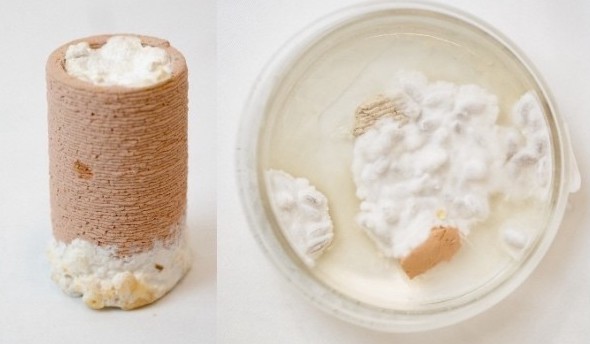
My interest comes from my previous work with mycelium-based material systems, where I explored the ability of fungal growth to bind different materials together. That work made me interested in a larger question:
Can biological attachment be designed at the molecular level, rather than relying only on a living organism to grow across an interface?
In architecture, product design, and construction, materials are commonly joined using mechanical fasteners or synthetic adhesives. Adhesives are effective, but permanent bonding can make multi-material assemblies difficult to repair, separate, reuse, or recycle.
Instead of developing one universal biological glue, I propose a modular protein connector whose two ends can be selected according to the materials being joined.
For the first prototype, I propose a bifunctional protein with the general architecture:
graph LR
A[Cellulose-binding domain] --> B[Flexible linker] --> C[Silica-binding domain]

style A fill:#F7D9D3,stroke:#D98F83,stroke-width:1.5px,color:#4A332F
style B fill:#FBE7E2,stroke:#DFA49A,stroke-width:1.5px,color:#4A332F
style C fill:#F4CFC7,stroke:#D78274,stroke-width:1.5px,color:#4A332F
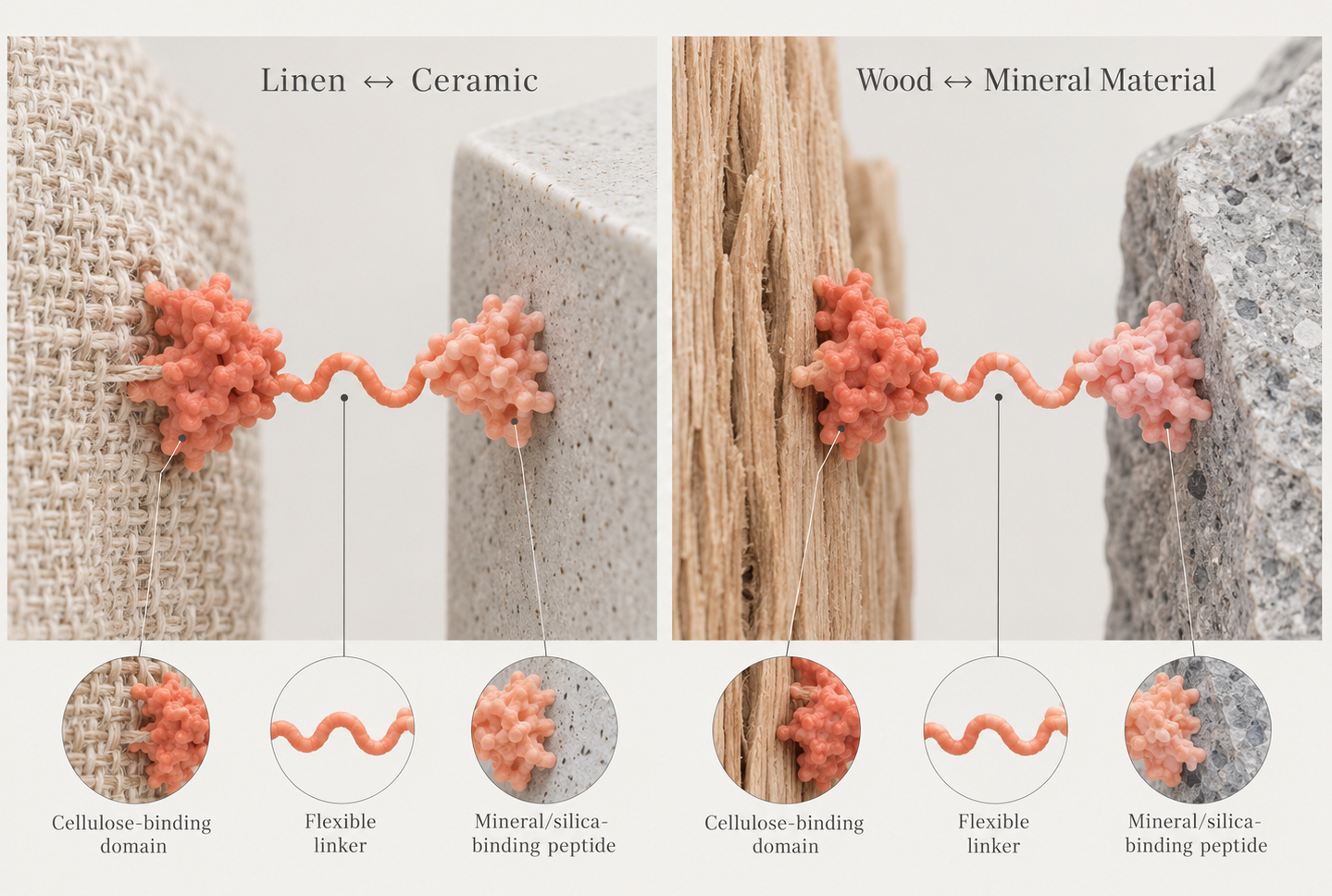
The first domain would recognize cellulose-rich materials such as paper, cotton, linen, hemp, or wood.Cellulose-binding domains, often described as carbohydrate-binding modules, are naturally occurring protein domains that have strong non-covalent affinity for cellulose.The second end of the protein would contain a short silica-binding peptide, giving it affinity toward silica-rich surfaces. A flexible peptide linker would connect the two binding regions and allow them some freedom to orient toward their respective surfaces.
The hypothesis is therefore:
graph LR
A["Cellulose / Paper / Cotton"] -->|binds| B["Cellulose-binding domain"]
B --> C["Flexible linker"]
C --> D["Silica-binding peptide"]
D -->|binds| E["Glass / Silica"]

style A fill:#F7F7F5,stroke:#A8A8A3,stroke-width:1.5px,color:#3F3F3C
style B fill:#F7D9D3,stroke:#D98F83,stroke-width:1.5px,color:#4A332F
style C fill:#FBE7E2,stroke:#DFA49A,stroke-width:1.5px,color:#4A332F
style D fill:#F4CFC7,stroke:#D78274,stroke-width:1.5px,color:#4A332F
style E fill:#F7F7F5,stroke:#A8A8A3,stroke-width:1.5px,color:#3F3F3C
If successful, later versions could be translated into more architecturally relevant interfaces such as:
linen ↔ ceramic or wood ↔ mineral material
The same strategy could potentially work between similar materials as well. A connector containing cellulose-binding functions at both ends could be explored for cellulose-to-cellulose interfaces, while mineral-binding domains could potentially connect two mineral surfaces.
The broader vision is therefore a library of programmable biological connectors, where different material-binding protein domains could be combined depending on which materials need to interface. For this course, I am not trying to demonstrate a structural replacement for epoxy or construction adhesive. My first goal is much smaller and experimentally measurable:
Can I genetically design and manufacture a bifunctional protein that demonstrates measurable interaction with both cellulose and silica-based surfaces?
2. What living system would I use?
I would use an E. coli-based cell-free protein synthesis system (CFPS) as the primary biomanufacturing chassis.
Instead of maintaining an intact genetically engineered organism, cell-free protein synthesis uses the molecular machinery extracted from cells, including ribosomes, enzymes, and other components required for transcription and translation.
The simplified manufacturing process would be:
DNA → transcription → mRNA → translation → Bio-connector protein
The DNA would contain the genetic instructions encoding the complete connector protein.
The cell-free reaction would receive:
DNA template + cell lysate + amino acids + nucleotides + energy system + salts/cofactors
and produce: the bio-connector protein
I am choosing cell-free manufacturing because it allows rapid iteration. If I change the DNA sequence of the connector, I can test a new protein without first establishing an entirely new engineered cell line.
It also creates a direct connection between genetic design and material performance:
change DNA → change protein → test material interaction
Cell-free systems are also well suited to automated, small-volume experiments.
For larger-scale manufacturing in the future, an engineered microbial chassis such as E. coli could potentially produce the protein through fermentation.
Therefore:
Prototype chassis: cell-free protein synthesisFuture scale-up chassis: microbial protein production
Although the primary manufacturing chassis for this project will be an E. coli-based cell-free protein synthesis system, the proteins being manufactured do not need to originate from E. coli.
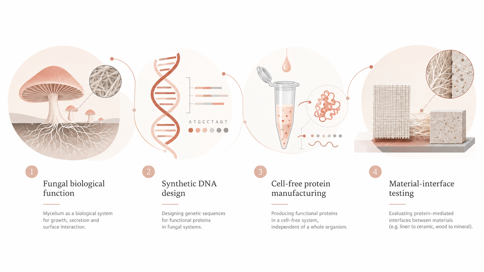
One direction I would like to investigate is the use of fungal-derived surface-active proteins, particularly hydrophobins, as components of the bio-connector system. Hydrophobins are small proteins naturally produced by filamentous fungi that assemble at interfaces and alter surface properties.
Rather than genetically engineering and culturing fungi during the first stage of the project, DNA encoding a selected fungal protein could be introduced into a cell-free expression system and the resulting protein tested directly on different material surfaces.
The project could therefore combine:
In later research, promising protein designs could potentially be transferred back into a fungal chassis, allowing an engineered fungus to produce or secrete the material-binding proteins directly at an interface. This would extend the system from a manufactured protein connector toward a living material capable of growing its own connection.
3. How would I use automation?
I would use automated liquid handling to create a small Design–Build–Test–Learn workflow.
A desktop liquid-handling robot such as an Opentrons system, or a larger automated laboratory platform, could prepare multiple cell-free reactions while precisely controlling reagent volumes. The workflow would be:
Design DNA
↓
Automated cell-free protein production
↓
Apply protein to material samples
↓
Measure binding
↓
Compare designs and conditions
↓
Improve the next design
During early experiments, automation could vary parameters such as protein concentration, pH, salt concentration, linker design, and material type.The first tests could use small cellulose and silica particles or material coupons rather than full architectural components.
For example, multiple wells could contain:
cellulose + connector proteinsilica + connector proteinmaterial + no proteinmaterial + non-specific control protein
After incubation, controlled washing could remove weakly associated protein. A fluorescent reporter or protein-labeling method could initially be used to quantify how much protein remains bound after washing.This would allow me to answer a very basic but essential question:
Does the engineered protein bind preferentially to the material it was designed to recognize?
Once promising conditions are found at small scale, the same protein could be applied to a physical joint between two material coupons.
In the longer term, the same workflow could screen a library of material-binding domains and generate a database connecting:
protein design → material chemistry → binding performance
4. How much needs to be made, and how long would it take?
For this course, I define a meaningful scale as a small material-interface demonstrator supported by replicated laboratory tests.
I would initially make two types of joints:
cellulose ↔ cellulose and cellulose ↔ silica
I would prepare three replicates of each condition:
3 cellulose–cellulose joints + 3 cellulose–silica joints= 6 connector-treated samples
Assume each sample has an overlap area of: 2 cm × 2 cm = 4 cm²
Total treated interface area: 6 × 4 cm² = 24 cm²
For an initial screening assumption, I will estimate approximately: 20 µg of protein per cm²
The exact optimal surface loading is unknown and would need to be experimentally determined.
Protein needed for the physical samples: 24 cm² × 20 µg/cm² = 480 µg
I would also need protein for small-scale binding assays, measurement, repeat experiments, and handling losses.I would therefore set an initial target of approximately: 1 mg of bio-connector protein for one complete prototype campaign.
For an initial calculation, I will assume a usable cell-free yield of: 0.5 mg/mL
The actual yield of this particular fusion protein is currently unknown and would need to be measured.
Required CFPS reaction volume would therefore be approximately:
1 mg ÷ 0.5 mg/mL = 2 mL total cell-free reaction
I would not begin directly with a 2 mL reaction. Initial expression screening could happen at microliter scale. Only after confirming that the protein is successfully produced would the reaction be scaled up.A complete experimental cycle could approximately be:
Protein expression: several hours Protein recovery / concentration / quality check: approximately 1 day Material-binding experiment: approximately 1 day Wash, measurement and physical interface testing: approximately 1 day
Therefore, after the DNA template is available, I estimate a complete design-test cycle could occur in approximately:
2–4 days depending on protein purification and equipment availability.
At larger architectural scales, the manufacturing strategy would likely change.
For example, a surface area of 1 m² is: 10,000 cm²
At the same assumed protein loading of 20 µg/cm²:
10,000 cm² × 20 µg/cm² = 200,000 µg = 200 mg protein
This calculation shows why cell-free synthesis may be ideal for early prototyping while microbial fermentation or another scalable production strategy could eventually become more appropriate for architectural quantities.
5. What I don’t know yet?
The first major unknown is whether both binding domains will still function when combined into one protein.Both cellulose-binding domains and silica-binding peptides have independently demonstrated material affinity. However, combining them into one protein may affect folding, orientation, accessibility, or solubility.The flexible linker may therefore be an important design variable.
The second major unknown is whether molecular surface binding will translate into meaningful mechanical adhesion.
A protein may bind strongly to cellulose or silica at the molecular level but still fail to generate enough collective force to create a useful macroscopic joint. Therefore, I need to distinguish between:
molecular binding and mechanical bonding
The first experimental milestone is proving selective material binding. Mechanical joint strength would be the next level of investigation.The third major unknown is how strongly real material surfaces will affect performance.A clean silica surface and an architectural ceramic surface are not identical. Likewise, purified cellulose differs significantly from wood, linen, or hemp.Variables such as surface chemistry, roughness, porosity, moisture, contamination, and coatings could all influence protein attachment.I would therefore begin with controlled surfaces such as cellulose/paper and glass before progressing toward wood, textile, ceramic, or other construction materials.
Proposed Course-Scale Success Criterion
I would consider the project successful if I can demonstrate that:
A genetically encoded bifunctional protein can be produced using cell-free protein synthesis and shows measurable material-specific binding to both cellulose and silica surfaces compared with appropriate controls.
A successful mechanical joint would be an additional achievement, but it is not required to prove the underlying biological concept.
The longer-term research direction would investigate:
stronger connector variants → similar-material connectors → reversible connectors → architectural material systems → engineered organisms capable of producing connectors locally
The eventual vision is a new type of material interface that is not simply glued together chemically, but whose binding behavior is encoded biologically.
References
Abdelhamid, M. A. A., Motomura, K., Ikeda, T., Ishida, T., Hirota, R., & Kuroda, A. (2014). Affinity purification of recombinant proteins using a novel silica-binding peptide as a fusion tag. Applied Microbiology and Biotechnology, 98, 5677–5684.
Chong, S. (2014). Overview of cell-free protein synthesis: Historic landmarks, commercial systems, and expanding applications. Current Protocols in Molecular Biology, 108, 16.30.1–16.30.11.
Li, S., & Liu, G. (2024). Harnessing cellulose-binding protein domains for the development of functionalized cellulose materials. Bioresources and Bioprocessing, 11, 74.
Silverman, A. D., Karim, A. S., & Jewett, M. C. (2020). Cell-free gene expression: An expanded repertoire of applications. Nature Reviews Genetics, 21, 151–170.
Use of AI
I used ChatGPT (OpenAI, GPT-5.6 Sol) as a learning and research assistant to help me understand molecular biology concepts, explore material-binding proteins and cell-free protein synthesis, evaluate project feasibility, structure the proposal, and develop preliminary scale calculations.
Representative prompts included:
How can I develop a biomanufacturing project that connects biology with architecture and material interfaces while remaining achievable within the course?
Can a genetically encoded protein connector work between similar and dissimilar materials?
Can cell-free protein synthesis be used as the chassis for producing a material-binding fusion protein?
What are existing cellulose-binding and silica-binding protein systems, and how could they be combined into a testable material-interface project?
Based on the HTBAA Homework 1 requirements, develop a realistic biomanufacturing proposal for a genetically encoded bio-connector and clearly identify assumptions and unknowns.
Week 2 HW: DNA Design
Objective
Design a T7-compatible genetic construct for cell-free expression of a bifunctional material-binding protein containing the CBM3a-L cellulose-binding module and the SB7 silica-binding peptide joined by a flexible peptide linker.
Part 1: Expression Target
What molecule am I expressing?
I am expressing an engineered bifunctional fusion protein designed to act as a molecular connector between cellulose-rich and silica-rich material surfaces.
The protein consists of:
CBM3a-L → flexible linker → SB7
CBM3a-L provides cellulose-binding functionality.
The flexible linker separates the two binding modules and allows them to move more independently.
SB7 provides silica-binding functionality.
The broader concept is a modular connector system in which material-binding domains can be recombined depending on the surfaces that need to be joined. This could enable future connector variants for cellulose–cellulose, cellulose–silica, or silica–silica interfaces.
For this homework, I am focusing on the CBM3a-L–linker–SB7 connector for a cellulose-to-silica interface.
What is the sequence and where does it come from?
The fusion protein is composed of three sequence elements.
CBM3a-L CBM3a-L is a cellulose-binding protein domain derived from the CipA scaffoldin of Acetivibrio thermocellus, historically known as Clostridium thermocellum. The sequence used for this project is based on the protein sequence associated with PDB entry 4JO5.
Flexible linker A glycine- and serine-rich flexible linker, (GGGGS)₃, is placed between the two binding modules. The purpose of the linker is to provide spatial separation and flexibility so that the two binding regions are less likely to interfere with one another.
SB7 SB7 is a seven-amino-acid silica-binding peptide with the sequence:
RQSSRGR
It was derived from the silica-binding region of the CotB1 protein from Bacillus cereus.
The complete engineered protein therefore follows the architecture:
CBM3a-L → (GGGGS)₃ → SB7
The exact DNA coding sequence will be designed to encode this fusion protein for expression in the selected cell-free system.
What host or chassis am I using?
The expression chassis is an E. coli-based cell-free protein synthesis (CFPS) system using T7-driven transcription.
Instead of using intact living cells, the cell-free system contains the molecular machinery required for transcription and translation. DNA encoding the bio-connector protein can therefore be added directly to the reaction mixture and used to produce the protein.
This approach allows the protein design to be tested rapidly without first engineering and culturing a complete organism.
Where does the product need to go?
The bio-connector protein is intended to remain soluble in the cell-free reaction mixture after expression. It can then be recovered or used directly in subsequent material-binding experiments.
Because the manufacturing system is cell-free and there is no intact cell membrane that the protein needs to cross, the initial construct does not require a secretion signal peptide.
The expressed protein will eventually be tested on controlled cellulose-rich and silica-rich surfaces before translating the system to more architecturally relevant material interfaces such as linen and ceramic.
Designing the Genetic Parts
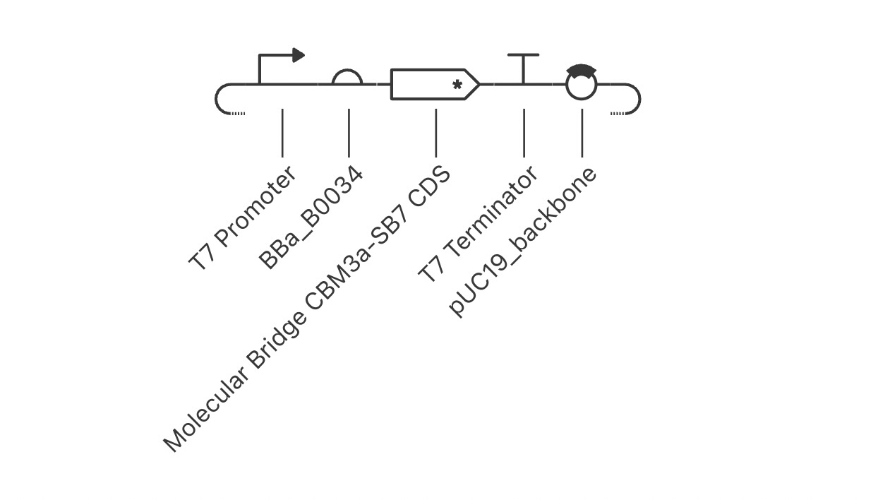
To express the Molecular Bridge protein in an E. coli-based cell-free system, I assembled a T7-driven expression cassette consisting of:
The expression cassette was then placed in a pUC19-derived backbone.
1. T7 Promoter
Part type: Promoter
Sequence: TAATACGACTCACTATAGGG
Length: 20 bp
Source: Existing T7 promoter part in Kernel.
Justification: The T7 promoter was selected because the proposed E. coli-based cell-free protein synthesis system uses T7 RNA polymerase for transcription. The promoter tells T7 RNA polymerase where transcription of the downstream gene should begin.
2. BBa_B0034 Ribosome Binding Site
Part type: Ribosome Binding Site (RBS)
Sequence: AAAGAGGAGAAA
Length: 12 bp
Source: BBa_B0034, imported from the existing Kernel parts library.
Justification: BBa_B0034 is a commonly used bacterial ribosome binding site. It provides a translation-initiation region where the bacterial ribosome can associate with the mRNA before reaching the start codon of the protein-coding sequence.
3. Molecular Bridge CBM3a-SB7 CDS
Part type: CDS (+start +stop)
Length: 642 bp
Protein encoded: 213 amino acids followed by a stop codon.
The protein architecture is:
M → CBM3a-L → (GGGGS)₃ → SB7
The CBM3a-L component is based on the 190-amino-acid protein sequence associated with PDB entry 4JO5. An initiating methionine was added through the ATG start codon.
The DNA coding sequence was designed by reverse-translating the desired protein sequence into DNA codons. It begins with ATG and ends with the stop codon TAA.
Source and design rationale: CBM3a-L is derived from the CipA scaffoldin of Acetivibrio thermocellus and provides the cellulose-binding component of the design. The flexible (GGGGS)₃ linker was selected to provide spatial separation and mobility between the two material-binding regions. SB7 is a seven-amino-acid silica-binding peptide derived from the CotB1 protein of Bacillus cereus.
Justification: This CDS combines a cellulose-binding module and a silica-binding peptide into one engineered fusion protein so that the resulting protein can be tested as a molecular connector between cellulose-rich and silica-rich material surfaces.
Justification: The T7 terminator provides a transcription-stop signal downstream of the Molecular Bridge CDS and defines the end of the T7-driven transcription unit.
5. pUC19-Derived Backbone
Part type: Backbone
Kernel part: pUC19_backbone
Length: 2641 bp
Source: Existing Kernel backbone derived from pUC19 and listed as a copy of BBa_K4411017.
The backbone contains the two primary elements required for propagation in E. coli:
pMB1-derived origin of replication — enables replication of the plasmid in E. coli.
bla / AmpR — provides ampicillin resistance for selection of bacteria carrying the plasmid.
The complete backbone sequence is stored with the Kernel Part.
Justification: The pUC19-derived backbone provides the replication and selection functions required to maintain and amplify the plasmid in E. coli. These functions are useful for producing the DNA template even though the final protein-expression experiment is intended to occur in a cell-free system.
Signal Peptide / Targeting Sequence
No signal peptide was included.
Because the protein will be produced using a cell-free expression system rather than secreted through the membrane of an intact cell, a secretion signal is not required for the initial construct.
The construct is designed to provide a DNA template for T7-driven production of the Molecular Bridge fusion protein in an E. coli-based cell-free protein synthesis system.
Design Hypothesis
If the engineered fusion protein folds correctly and both binding regions remain accessible, the CBM3a-L domain may interact with cellulose-rich surfaces while the SB7 peptide interacts with silica-rich surfaces.The first experiment therefore tests whether two previously characterized material-binding elements can retain their respective functions when combined within a single genetically encoded fusion protein.This does not assume that the protein will function as a structural adhesive. The first goal is to determine whether measurable binding to both material classes can be achieved.
Future Variants
The same modular strategy could later be extended by recombining material-binding domains to create:
CBM3a → linker → CBM3a for cellulose-rich ↔ cellulose-rich interfaces
CBM3a → linker → SB7 for cellulose-rich ↔ silica-rich interfaces
SB7 → linker → SB7 for silica-rich ↔ silica-rich interfaces
This would allow the project to develop from a single fusion protein into a modular family of genetically encoded material connectors.
References
Yaniv, O. et al. (2013). Structure of a family 3a carbohydrate-binding module from the cellulosomal scaffoldin CipA of Clostridium thermocellum with flanking linkers: implications for cellulosome structure. Acta Crystallographica Section F. PDB: 4JO5.
Chen, X., Zaro, J. L., & Shen, W. C. (2013). Fusion protein linkers: property, design and functionality. Advanced Drug Delivery Reviews, 65(10), 1357–1369.
Ikeda, T. et al. (2016). Application of volcanic ash particles for protein affinity purification with a minimized silica-binding tag. The study reports the seven-amino-acid SB7 sequence RQSSRGR.
iGEM Registry of Standard Biological Parts. BBa_B0034 ribosome binding site.
New England Biolabs. pUC19 sequence and map, including the pMB1-derived origin of replication and bla ampicillin-resistance gene.
Thermo Fisher Scientific. T7 promoter sequence and T7 expression-vector documentation.