Hi! I’m currently finishing my Bachelor’s degree in Biophysics and working on my thesis “Metasurfaces for tumoral cells”. I’ve become really interested in materials and how they can be combined with biology to create solutions with a social impact. I’m hoping to build an interdisciplinary career that connects science, technology and biology. I love art and I practice contemporary ballet and also enjoy painting and collecting flowers.
A biofabricated photonic material that can detect changes in the environment through a measurable optical response, ideally a change in color.
2. What living system could make it?
Cellulophaga lytica, because its cells can self-organize into periodic biofilms that produce structural color.
3. How could automation help you make it?
Automation could help control the growth conditions, monitor biofilm formation, and measure optical responses in parallel, allowing systematic optimization of the conditions that produce the desired color.
4. What would it take to make it at a meaningful scale?
I would define a meaningful scale as producing enough biofilm to fabricate a specified number or area of photonic sensors and estimate the required culture area, biomass, production time, and number of parallel cultures.
Include in detail:
(1) What you would like to biomanufacture and why?
I am interested in developing a simple and direct environmental monitoring system that can respond to its surrounding conditions without requiring the environment to be chemically modified. A visible optical response could make environmental changes easier to detect and monitor, while automation could allow the system to continuously monitor changing conditions. This could be useful for environmental monitoring, particularly in the context of air and water pollution and other environmental stressors. In the longer term, I am interested in exploring whether this type of system could be adapted to larger-scale or extreme environments, including potential space applications.
(2) What living system would you use?
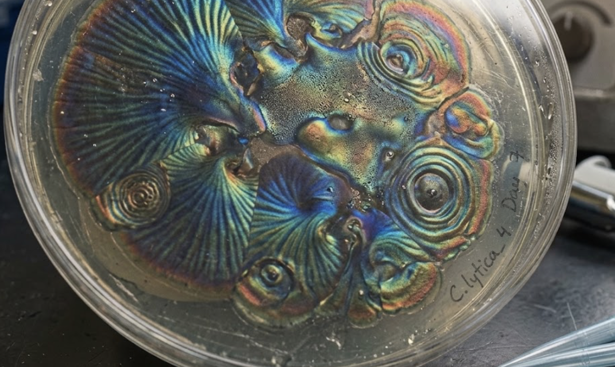
Cellulophaga lytica, a marine bacterium capable of self-organizing into iridescent biofilms with periodic cellular structures that produce structural color. The proposed system assumes that environmental conditions can be used to modulate the biofilm’s optical response.1-3
(3) How would you use automation?
A programmable bioprinter such as the Allevi 3 could be used to automate the deposition of C. lytica in defined patterns and areas. A 2023 study demonstrated that C. lytica could be deposited using an Allevi 3 and maintain the programmed patterns after growth. Automated imaging and optical measurements could then be used to monitor biofilm formation and quantify changes in color or reflected wavelength under different environmental conditions.4
(4) How much would you need to make, and how long would it take?
As an initial prototype, I would define a meaningful scale as a 10 × 10 cm photonic sensing surface. Based on a 2023 study, C. lytica biofilms can be produced on larger surfaces, and an accelerated growth strategy achieved iridescent biofilms in approximately 24 hours. Therefore, I would initially aim to produce a 100 cm² biofilm within approximately 24 hours. This is an engineering estimate based on the reported results and would need to be experimentally validated.4
(5) What don’t you know yet?
• Can the biofilm’s optical response be reliably controlled by environmental conditions such as temperature and hydration?
• Can C. lytica maintain its photonic structure and optical function over time and under changing environmental conditions, with a response that is stable, reversible, and reproducible?
• Can the system maintain reproducible optical properties when scaled to larger areas and adapted to different applications?
AI
I used ChatGPT, Sci -Bot, Notebook and Gemini as a learning and brainstorming tool during the development of this project. I used it to explore possible biomanufacturing systems, clarify scientific concepts, identify technical uncertainties, organize my ideas, and check preliminary calculations. I reviewed and revised the information generated by AI and take responsibility for the final content, assumptions, and decisions presented in this submission.
Representative prompts used:
• “What living systems could produce an adaptive photonic material with a measurable optical response?”
• “What are the advantages and limitations of using Cellulophaga lytica for this concept?”
• “How could automation be applied to the biomanufacturing of Cellulophaga lytica biofilms?”
• “Help me check the back-of-the-envelope calculation for producing a 10 × 10 cm photonic biofilm.”
• “An authentic lab photo of a Petri dish with written marker labels, condensation, and realistic iridescent biofilm patterns.”
References used:
Kientz, B., Vukusic, P., Luke, S., & Rosenfeld, E. (2012). Iridescence of a marine bacterium and classification of prokaryotic structural colors. Applied and Environmental Microbiology, 78(6), 2092–2099. https://doi.org/10.1128/AEM.07339-11
Kientz, B., Luke, S., Vukusic, P., Péteri, R., Beaudry, C., Renault, T., Simon, D., Mignot, T., & Rosenfeld, E. (2016). A unique self-organization of bacterial sub-communities creates iridescence in Cellulophaga lytica colony biofilms. Scientific Reports, 6, 19906. https://doi.org/10.1038/srep19906
Rosenfeld, E., Kientz, B., Ducret, A., Luke, S., Vukusic, P., & Mignot, T. (2012). Effect of abiotic factors on the unique glitter-like iridescence of Cellulophaga lytica. FEMS Microbiology Letters, 333(2), 101–108. https://doi.org/10.1111/j.1574-6968.2012.02614.x
Sullivan, C. J., Brown, K., Hung, C.-S., Tang, J. K.-H., DeSimone, M., Chen, V., Lloyd, P. F., Gupta, M., Juhl, A., Crookes-Goodson, W., Vasudev, M., Dennis, P. B., & Kelley-Loughnane, N. (2023). Iridescent biofilms of Cellulophaga lytica are tunable platforms for scalable, ordered materials. Scientific Reports, 13, 13192. https://doi.org/10.1038/s41598-023-38797-0
Week 2 HW: DNA Design
HTBAA Homework #2: DNA Design
Part 0: Set up in Kernel
I logged in to Kernel and joined the HTBAA 2026 workspace. In the repository HTBAA26_WilliamandMary there is a folder with my name Aranza Cantu Puga, where the folder HW2-Cellulopagha-project and my work done is located.
Part 1: Define your expression target
What molecule are you expressing?
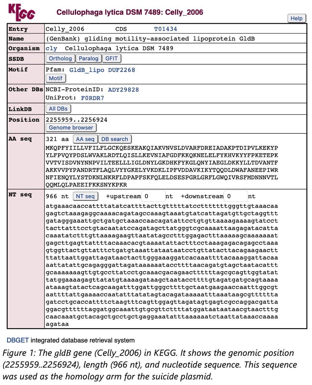
I am not expressing a heterologous protein. Instead, I designed a suicide plasmid to perform a knockout of the native gldB gene in Cellulophaga lyticaDSM 7489. The gldB gene encodes a gliding motility-associated lipoprotein (GldB). Deleting this gene interrupts the cell’s ability to self-organize into periodic biofilms, which are responsible for the structural color (iridescence) of the biofilm.
What is the sequence?
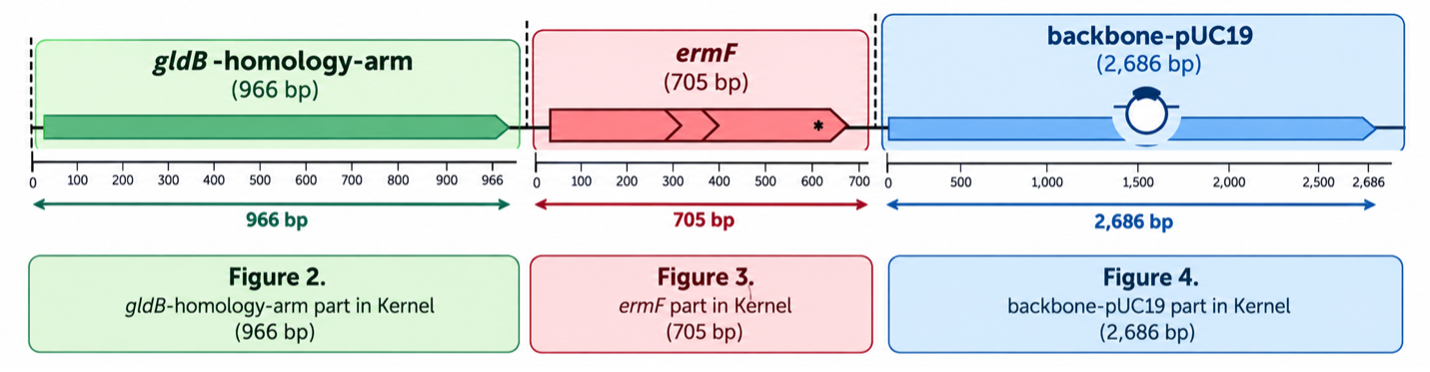
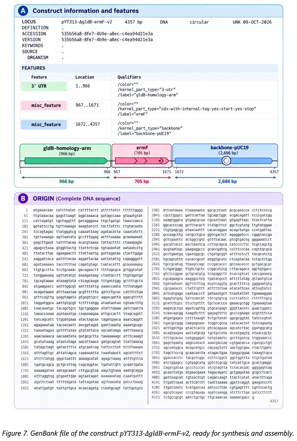
The sequence used as the homology arm is the complete gldB gene (966 bp). It was obtained from the KEGG database (entry: cly:Celly_2006). The selection cassette is the ermF gene (705 bp), which confers resistance to erythromycin and comes from Bacteroides fragilis.
What host or chassis?
The knockout is performed in Cellulophaga lytica DSM 7489 (a marine bacterium). The plasmid is propagated in E. coli DH5α for cloning.
Where does the product need to go?
The cassette needs to integrate into the chromosome of C. lytica via homologous recombination. This is why we use a suicide plasmid (it cannot replicate in C. lytica, so it must integrate or be lost).
Part 2: Design your genetic parts
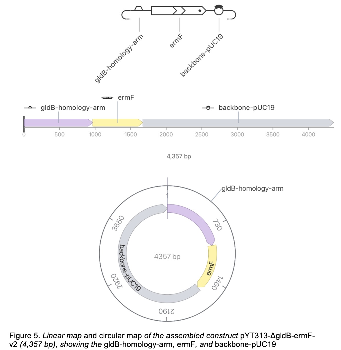
Since my host is a prokaryote (C. lytica for the knockout, E. coli for cloning), I designed a suicide plasmid with the following parts:
Part 3: Assemble your construct
Part 4: Optimize and quality-check
Codon optimization
I did not perform codon optimization because the gldB sequence is used as a homology arm (not as a coding sequence for expression). ermF is a native bacterial resistance gene that is already expressed correctly in C. lytica. Therefore, codon optimization was not necessary for this design.
BLAST / sequence search
I ran a BLAST search of the ermF sequence against the NCBI nucleotide database. The top hits were erythromycin resistance genes from Bacteroides fragilis and related bacteria, with E-values < 1e-100 and 100% query coverage. No significant matches were found against the C. lytica genome, confirming that ermF is heterologous and will not interfere with native genes.
Safety screen
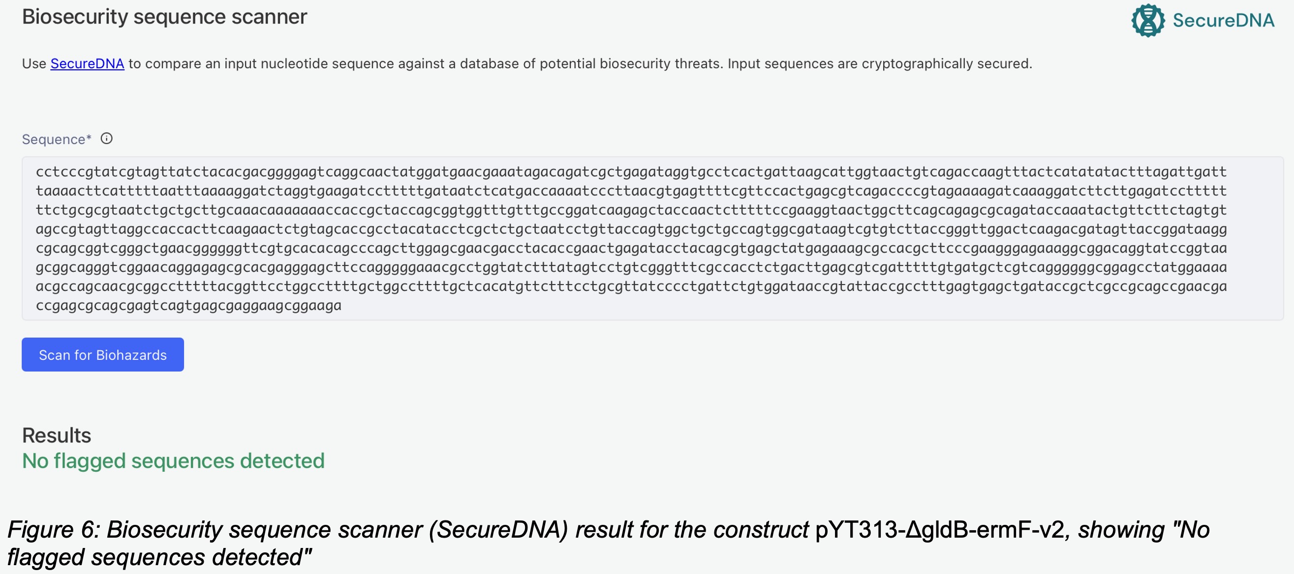
I ran the Biosecurity sequence scanner (SecureDNA) on the construct pYT313-ΔgldB-ermF-v2 in Kernel. The scanner checks for sequences that could be hazardous (e.g., toxin genes, virulence factors). The result was “No flagged sequences detected”, which means the construct is safe. This is expected because the construct contains only a homology arm to a native C. lytica gene and a standard antibiotic resistance marker.
Conclusion:
The construct is suitable for the intended knockout experiment. The gldBhomology arm is specific to C. lytica, and ermF is a standard selection marker with no safety concerns.
Part 5: From design to build
A sequence in software is not yet DNA. Here I explain how my construct (pYT313-ΔgldB-ermF-v2, 4,357 bp) would actually be synthesized and assembled.
How would I build it?
I would build this construct by commercial DNA synthesis of the cassette (gldB-homology-arm + ermF, ~1,671 bp) followed by Gibson Assembly into the pUC19 backbone (2,686 bp). The total construct is 4,357 bp, which is well within the range of commercial synthesis. Gibson Assembly is ideal here because it allows joining multiple fragments in a single isothermal reaction, without needing restriction sites (Gibson et al. 2009). This is simpler and more flexible than traditional restriction cloning.
What does synthesis constrain?
DNA synthesis has error rates and can struggle with sequences that have high GC content, repeats, or secondary structures. My homology arm (gldB, 966 bp) is a native C. lytica sequence, so it may contain some repeats or high-GC regions. To address this, I would order the cassette in two or three smaller fragments and assemble them by Gibson. If errors are detected, I could use protein-mediated error correction (Carr et al. 2004) to remove incorrect sequences before assembly.
Prototype vs. scale
For the prototype, I would order the synthesized cassette and clone it into pUC19 using Gibson Assembly, then transform into E. coli for propagation. This is a standard, cost-effective approach for a single construct. For scale, if I needed many variants of the knockout plasmid (e.g., to test different homology arm lengths), I could use chip-based oligo synthesis (Kosuri et al. 2010; Tian et al. 2004) to generate pools of variants, then assemble them by Gibson. This would be more cost-effective than ordering each construct individually.
References:
Gibson, D. G., et al. (2009). Enzymatic assembly of DNA molecules up to several hundred kilobases. Nature Methods, 6, 343–345.
Carr, P. A., et al. (2004). Protein-mediated error correction for de novo DNA synthesis. Nucleic Acids Research, 32, e162.
Kosuri, S., et al. (2010). Scalable gene synthesis by selective amplification of DNA pools from high-fidelity microchips. Nature Biotechnology, 28, 1295–1299.
Part 6: What don’t you know yet?
The main uncertainties in my current design are:
1. Will the knockout of gldB actually eliminate iridescence?
The main assumption of my project is that gldB is required for the self-organization that produces structural color. However, it is possible that deleting gldB does not completely eliminate iridescence or that other genes compensate for its loss.
How I would find out: I would compare the wild-type and ΔgldB strains visually and with optical measurements (reflectance spectroscopy). If iridescence is not fully lost, I would test additional motility genes (e.g., gldJ, gldK) as alternative targets.
2. Will the suicide plasmid integrate correctly into the C. lytica chromosome?
Homologous recombination is not always efficient, and the plasmid may integrate at the wrong site or not at all. Additionally, C. lytica is a marine bacterium and may be difficult to transform.
How I would find out: I would use colony PCR with primers that flank the gldB locus. A larger PCR product (due to the inserted ermF cassette) would confirm integration. I would also sequence the PCR product to verify the insertion site. If transformation fails, I would optimize the electroporation protocol (voltage, capacitance, recovery time).
3. Will ermF provide reliable selection in C. lytica?
Although ermF is a standard erythromycin resistance marker, its expression level in C. lytica may not be sufficient for selection, or the antibiotic concentration may need optimization.
How I would find out: I would plate transformed cells on increasing concentrations of erythromycin (e.g., 1, 5, 10, 20 µg/mL) and identify the minimum concentration that inhibits wild-type growth but allows transformed cells to grow. If selection is weak, I would test a stronger promoter or a different resistance marker.
Conclusion:
These are the main bets in my design. The knockout of gldB is the central hypothesis and the transformation and selection steps are the main technical risks. Each can be tested with simple experiments (optical measurements, colony PCR, antibiotic titration) before scaling up.
AI
I used ChatGPT (OpenAI) as a learning and brainstorming tool during the development of this project. I used it to explore the biology of Cellulophaga lytica, understand the mechanism of gliding motility, find the gldB gene sequence in KEGG, and organize my ideas for the construct design.
I reviewed and revised all information generated by AI and I take responsibility for the final content, assumptions, and decisions presented in this submission.
Representative prompts used:
“What living systems could produce an adaptive photonic material with a measurable optical response?”
“How does the gldB gene relate to gliding motility and iridescence in Cellulophaga lytica?”
“Help me find the sequence of the gldB gene (Celly_2006) in KEGG.”
“How do I design a suicide plasmid for gene knockout in a marine bacterium?”
“What parts do I need to assemble a knockout construct in Kernel?”
“How do I connect the DNA design from HW2 with the cell-free reaction design for HW3?”
References
Kientz, B., Vukusic, P., Luke, S., & Rosenfeld, E. (2012). Iridescence of a marine bacterium and classification of prokaryotic structural colors. Applied and Environmental Microbiology, 78(6), 2092–2099.
Kientz, B., Luke, S., Vukusic, P., Péteri, R., Beaudry, C., Renault, T., Simon, D., Mignot, T., & Rosenfeld, E. (2016). A unique self-organization of bacterial sub-communities creates iridescence in Cellulophaga lytica colony biofilms. Scientific Reports, 6, 19906.
Sullivan, C. J., et al. (2023). Iridescent biofilms of Cellulophaga lytica are tunable platforms for scalable, ordered materials. Scientific Reports, 13, 13192.
Gibson, D. G., et al. (2009). Enzymatic assembly of DNA molecules up to several hundred kilobases. Nature Methods, 6, 343–345.
Carr, P. A., et al. (2004). Protein-mediated error correction for de novo DNA synthesis. Nucleic Acids Research, 32, e162.
Week 3 HW: Cell-Free Reaction Design
HTBAA Homework #3: Cell-Free Reaction Design
Overview:
In this assignment, I designed cell-free protein synthesis (CFPS) reaction compositions for three target proteins: sfGFP, PETase, and Reteplase. The goal is to optimize the cost per gram of functional protein. I also compared published CFPS formulations and contributed a new reagent to the community inventory.
Part A: Compare cell-free reaction compositions
Why does the concentration of potassium glutamate and magnesium glutamate differ between compositions? Which is more sensitive?
Potassium glutamate (K-Glu) and magnesium glutamate (Mg-Glu) have different roles. K-Glu mainly controls ionic strength and provides glutamate as a compatible solute. Mg-Glu is critical for ribosome function and ATP regeneration. Mg-Glu is more sensitive than K-Glu. Small changes in Mg²⁺ concentration can strongly affect translation, while K⁺ has a broader optimal range. In practice, Mg-Glu is titrated carefully (2–10 mM final), while K-Glu can vary more widely (50–300 mM final).
Explain how a PEP+NTP reaction formulation is different from a Ribose+NMP reaction formulation.
PEP+NTP: Uses phosphoenolpyruvate (PEP) as the energy source and nucleotide triphosphates (NTPs) as building blocks. PEP regenerates ATP through pyruvate kinase. This formulation is fast and high-yielding but expensive because NTPs (especially *CTP, GTP, UTP¨) are costly.
Ribose+NMP: Uses ribose as the energy source and nucleotide monophosphates (NMPs) as building blocks. Ribose is metabolized through the pentose phosphate pathway to regenerate ATP, and NMPs are converted to NTPs by the lysate enzymes. This formulation is cheaper because NMPs are much less expensive than NTPs, but it may be slower and require more optimization.
In short: PEP+NTP = expensive, fast, high yield
**Ribose+NMP ** = cheaper, slower but cost-effective
Choose two cell-free reaction compositions from the literature, compare and contrast the choice of reagents and the concentration of reagents.
Composition 1: PANOx-SP (Jewett & Swartz 2004)
Uses PEP as energy source and NTPs as building blocks
High concentrations of potassium glutamate (175–250 mM) and magnesium glutamate (8–12 mM)
Includes 17 amino acids, folinic acid, NAD, CoA and spermidine
Redox environment: reducing (DTT present)
Best for: soluble proteins without disulfide bonds (like sfGFP)
Composition 2: Cost-optimized formulation (Olsen et al. 2026)
Uses ribose and NMPs instead of PEP and NTPs to reduce cost
Lower concentrations of potassium glutamate (~100–150 mM) and magnesium glutamate (~5–8 mM)
Includes spermidine and improved pH buffering (HEPES-KOH)
Redox environment: oxidizing (GSSG/GSH) for proteins with disulfide bonds
Best for: proteins with disulfide bonds (like Reteplase) and cost optimization
Comparison: PANOx-SP is optimized for high yield but is more expensive. The Olsen formulation is optimized for low cost per gram by replacing expensive NTPs with NMPs and ribose.
Part B: Design reaction compositions for each protein
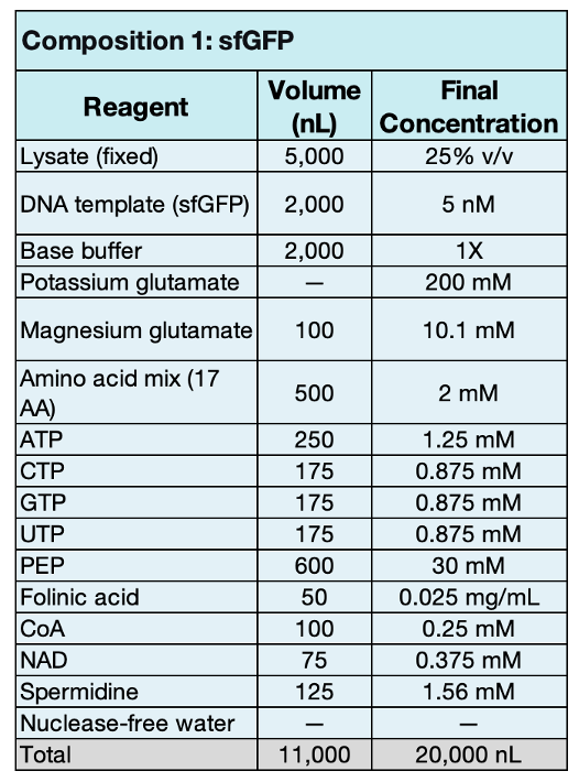
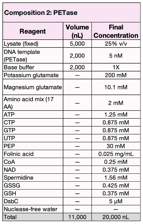
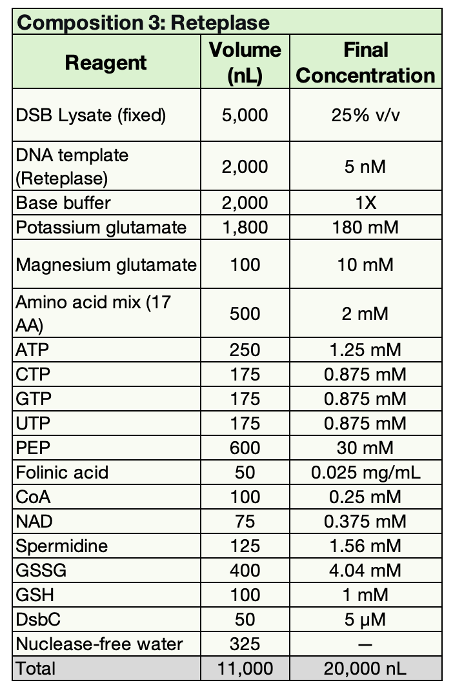
I designed three CFPS compositions for sfGFP, PETase, and Reteplase. Each composition has a total volume of 20,000 nL (20 µL) and follows the fixed components:
Justification: This is a standard formulation for soluble proteins without disulfide bonds. It uses PEP as the energy source and NTPs for transcription. The environment is reducing (no GSSG/GSH), which is compatible with sfGFP. The K-Glu concentration was set to 200 mM, which is within the optimal range of PANOx-SP (175–250 mM).
Justification: PETase has 1 disulfide bond, so it needs a slightly oxidizing environment. The GSSG:GSH ratio is approximately 1.13:1 (0.425 mM : 0.375 mM), which favors disulfide bond formation. DsbC (5 µM) helps correct any mispaired disulfides. The K-Glu concentration was set to 200 mM, which is within the optimal range of PANOx-SP (175–250 mM)
Justification: RReteplase has 9 disulfide bonds and needs a strongly oxidizing environment. DSB lysate is used (no DTT, treated with IAM), and a high GSSG/GSH ratio (4.04 mM : 1 mM) plus DsbC (5 µM) helps correct disulfide pairing.
New Reagent Suggestion: Liposomes (or Nanodiscs)
I suggest adding liposomes or nanodiscs to the CFPS reagent inventory. Membrane proteins (like GldB in C. lytica) are difficult to express in standard CFPS because they are hydrophobic and tend to aggregate. Adding liposomes or nanodiscs provides a lipid environment that mimics the cell membrane, allowing the protein to fold correctly. This is relevant for my photonic biofilm project, where GldB is a membrane lipoprotein.
Part C: Asimov Kernel (optional)
T7-compatible plasmids for cell-free expression
I built three T7-compatible plasmids in Kernel for sfGFP, PETase, and Reteplase. Each plasmid contains a T7 promoter, an RBS, the coding sequence for the target protein, and a T7 terminator in a pUC19 backbone.
T7-sfGFP plasmid:
T7 promoter (drives transcription by T7 RNA polymerase)
RBS-pET (initiates translation)
sfGFP CDS (240 amino acids, 27 kDa)
T7 terminator (ends transcription)
pUC19 backbone (provides ori and AmpR)
Figure 4. T7-sfGFP plasmid map
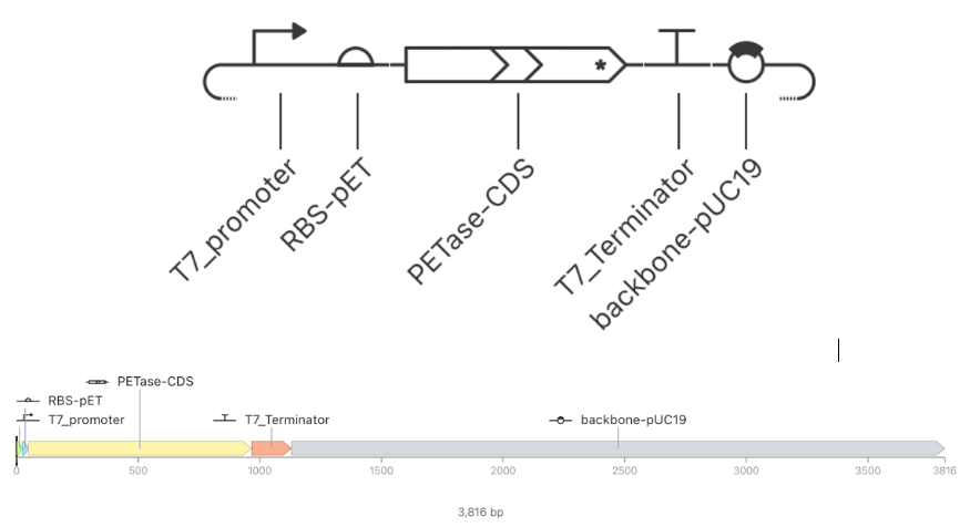
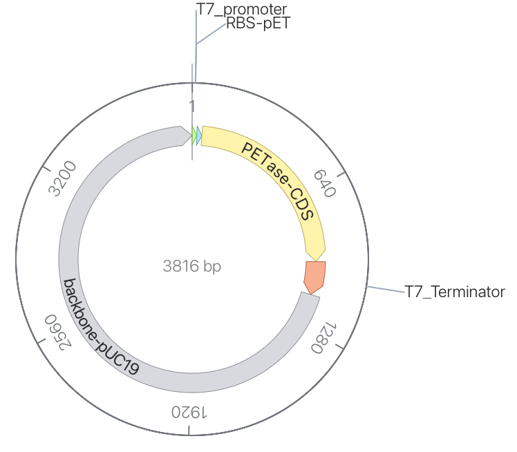
T7-PETase plasmid:
T7 promoter (drives transcription by T7 RNA polymerase)
RBS-pET (initiates translation)
PETase CDS (918 bp, 309 amino acids, 33 kDa)
T7 terminator (ends transcription)
pUC19 backbone (provides ori and AmpR)
Figure 5. T7-PETase plasmid map (3,816 bp). The construct contains a T7 promoter, RBS-pET, PETase CDS, T7 terminator, and pUC19 backbone.
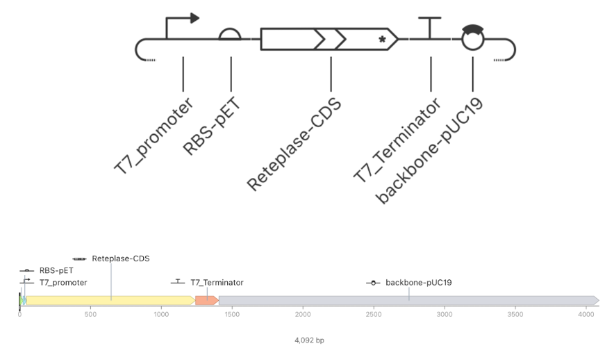
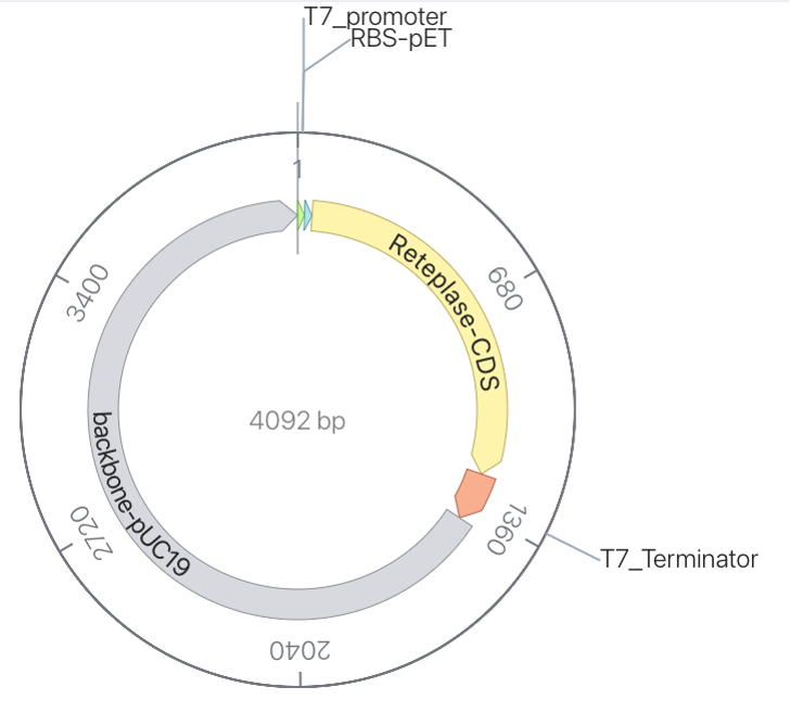
T7-Reteplase plasmid:
T7 promoter (drives transcription by T7 RNA polymerase)
RBS-pET (initiates translation)
Reteplase CDS (1,200 bp, 399 amino acids, 44 kDa)
T7 terminator (ends transcription)
pUC19 backbone (provides ori and AmpR)
Figure 6. T7-Reteplase plasmid map (4,092 bp). The construct contains a T7 promoter, RBS-pET, Reteplase CDS, T7 terminator, and pUC19 backbone.
T7 helper plasmid for Reteplase
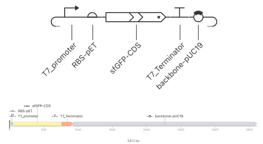
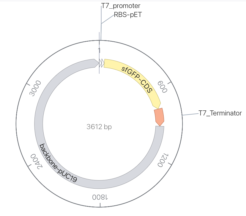
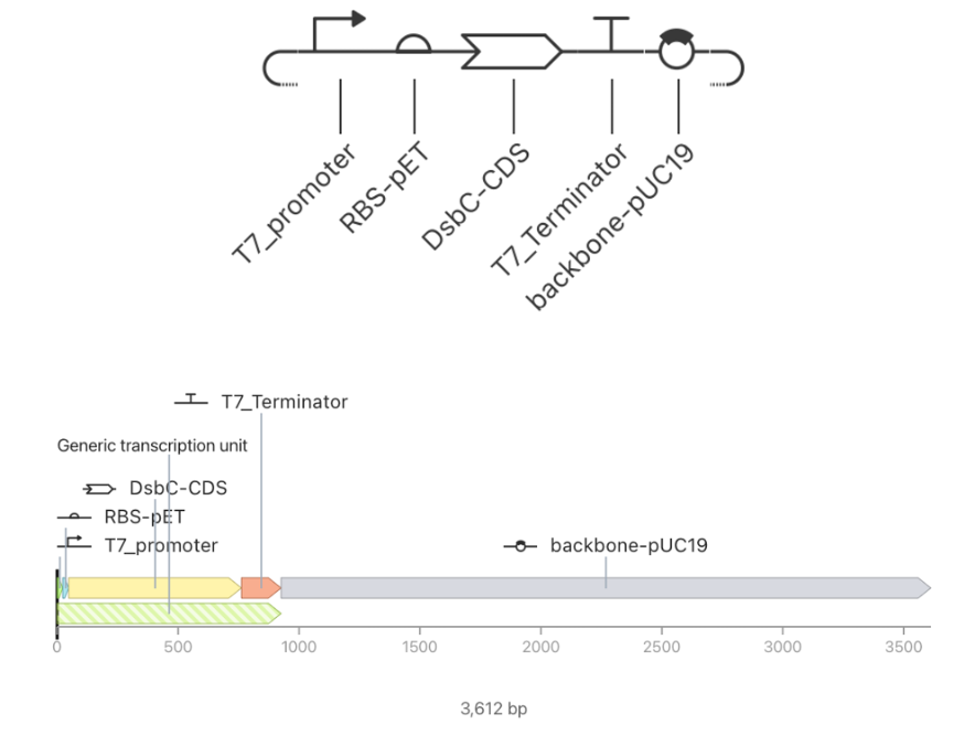
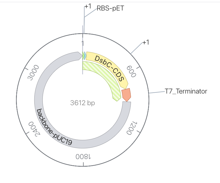
I designed a T7 helper plasmid that expresses DsbC (a disulfide-bond isomerase) to improve the yield of functional Reteplase. DsbC helps correct mispaired disulfide bonds, which is critical for Reteplase’s 9 disulfide bonds. The plasmid contains:
T7 promoter (drives transcription by T7 RNA polymerase)
RBS-pET (initiates translation)
DsbC CDS (741 bp, 247 amino acids)
T7 terminator (ends transcription)
pUC19 backbone (provides ori and AmpR)
The total construct size is 3,612 bp.
Figure 7. T7-DsbC helper plasmid map (3,612 bp). The construct contains a T7 promoter, RBS-pET, DsbC CDS, T7 terminator, and pUC19 backbone.
AI Usage
I used ChatGPT (OpenAI) as a learning assistant to help me understand CFPS concepts, compare published formulations, and structure my design reasoning. I reviewed all AI-generated content, verified it against the literature, and I take full responsibility for the final design decisions.
References
Jewett, M. C., & Swartz, J. R. (2004). Mimicking the Escherichia colicytoplasmic environment activates long-lived and efficient cell-free protein synthesis. Biotechnology and Bioengineering, 86(1), 19–26.
Olsen, M. L., et al. (2026). Design-driven optimization of low-cost reagent formulations for reproducible and high-yielding cell-free gene expression. Nature Communications.
Smith, A. A., et al. (2026). Using a GPT-5-driven autonomous lab to optimize the cost and titer of cell-free protein synthesis. bioRxiv.
Silverman, A. D., Karim, A. S., & Jewett, M. C. (2020). Cell-free gene expression: an expanded repertoire of applications. Nature Reviews Genetics, 21, 151–170.
Rasor, B. J., et al. (2021). Toward sustainable, cell-free biomanufacturing. Current Opinion in Biotechnology, 69, 136–144.