Week 2 HW: DNA Design
HOMEWORK #2
In Homework #1 I proposed biomanufacturing eumelanin in engineered E. coli as the active ingredient for an affordable sunscreen, and I argued from a published benchmark that reached 3.22 g/L of eumelanin from glucose [7]. That was a proposal at the level of strategy. This assignment takes it down to the level of bases.
What follows is a complete expression module: two enzymes, seven parts, one plasmid of 6963 bp. Building it turned up three things that a purely literature-level reading had hidden — the record strain carries more genes than I credited it with, the mutation the paper names does not exist at the residue it names, and the tyrosinase carries a targeting signal I had claimed it did not have. Each of those is documented below where it arises, because they changed the design rather than merely decorating it.
Part 1: Define your expression target
What molecule am I expressing? Not one protein but two enzymes, forming the minimal module of the melanogenesis pathway from central metabolism:
| Enzyme | Role in the pathway | |
|---|---|---|
| 1 | Feedback-resistant DAHP synthase (AroGfbr) | Catalyses the first committed step of the common aromatic pathway, condensing PEP and E4P. Relieving its feedback inhibition is what pushes carbon toward L-tyrosine. |
| 2 | Tyrosinase MutMelA (EC 1.14.18.1) | Hydroxylates L-tyrosine to L-DOPA and oxidises it to dopaquinone. Polymerisation to eumelanin from there is non-enzymatic. |
The tyrosinase: what “MutmelA” actually is
melA comes from Rhizobium etli CFN42, carried on the symbiotic plasmid p42d (protein_id AAM54973.1, plasmid NC_004041) [15].
Chávez-Béjar et al. used a variant they call MutmelA, described as MelA carrying a single nucleotide change replacing Asp535 by Gly [7]. When I retrieved the deposited sequence and translated it, the open reading frame turned out to be 1512 nt encoding 503 amino acids. There is no residue 535.
The most probable explanation, and one that is internally consistent, is that the paper numbers from the start of the cloned fragment rather than from the start codon. The database record carries 318 nt upstream of the ATG, in frame and without stop codons; read from nucleotide 1, that frame gives a 609-residue reading in which position 535 is indeed an aspartate, and it maps to Asp429 of the real protein. The surrounding context is identical in both readings (ADTPVTDPHYVGS), and the change is a single base, GAT→GGT, exactly as described.
That is an inference, not a confirmation. A randomly placed offset would land on an aspartate about 7 % of the time, so the coincidence is meaningful but not proof. I therefore use wild-type melA, which the source itself justifies: the authors state that melanin production begins earlier with the mutant, but that the rate of synthesis is the same with either version [7]. For a first expression module, an unverifiable one-base change that alters only onset timing is not worth carrying.
The tyrosinase carries a Tat signal peptide
Translating the CDS also contradicted something I asserted in my own plan. The protein begins:
This is a textbook twin-arginine (Tat) signal peptide: the arginine pair at positions 5–6, a hydrophobic core, and an AxA cleavage site at A29–F30–A31.
| Signal peptide | residues 1–31 · CDS nt 1–93 |
| Mature protein | residues 32–503 (472 aa) · CDS from nt 94 |
I had written that no signal peptide was included. Cloning the full CDS includes one. Cabrera-Valladares et al. report that the recombinant tyrosinase was expressed intracellularly in E. coli and purified from cell extract [15], which suggests E. coli’s Tat machinery does not efficiently recognise the rhizobial signal — but “I did not include one” and “I included one that appears not to be used” are different claims, and only the second is true.
I keep the native signal peptide, for two reasons. It is what the reference work expressed, so it is the version behind the published titres. And it converts an open question in Part 6 into a testable hypothesis: a functional Tat signal would export folded tyrosinase to the periplasm, which is a concrete candidate answer to where melanin precursors polymerise.
The DAHP synthase
aroGfbr derives from E. coli K-12 aroG, locus b0754 (UniProt P0AB91, protein_id AAC73841.1, RefSeq NP_415275.1). The CDS is 1053 nt encoding 350 aa.
The feedback-resistant allele I selected is D146N. I verified against the deposited sequence that residue 146 is indeed aspartate and that nucleotides 436–438 are GAT, in exact frame with codon 146, so the substitution is a single G→A at nucleotide 436.
This is my choice rather than an inherited one, and worth flagging as such: Chávez-Béjar et al. carried aroGfbr on plasmid pRW300 and never state which mutation it bears [7]. D146N is the best-documented variant for tyrosine overproduction [16]; S180F, P150L and L175D are alternatives I did not pursue.
Host and localisation
Host: E. coli, as proposed in HW1. One host constraint propagates into the whole process design: MelA is rapidly inactivated at 37 °C, so melanin-producing cultures must run at 30–32 °C, a temperature that is not optimal for E. coli growth [7].
Localisation: cytoplasmic expression, with the caveat above that the native CDS carries a Tat signal whose behaviour in E. coli is unresolved. How melanin precursors leave the cell, and whether they polymerise in the periplasm, the medium, or both, is not established in the literature [7]; Part 6 returns to it, because it decides the downstream recovery strategy.
Part 2: Design your genetic parts
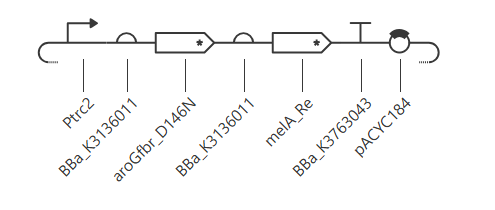
Seven parts. Every sequence below is the one actually registered in Kernel.
1. Ptrc — promoter (91 bp)
IPTG-inducible hybrid trp/lac promoter from pTrc99A [17]; the same promoter that drove the tyrosinase in the reference process, induced at 0.1 mM IPTG [7].
I verified its internal structure rather than trusting the label, and two of the checks mattered:
| Element | Position | Result |
|---|---|---|
−35 TTGACA | 14–19 | ✅ |
| Spacer between −35 and −10 | 20–36 | 17 bp — confirms trc, not tac (which has 16) |
−10 TATAAT | 37–42 | ✅ |
| lac operator (lacO1) | 47–69 | ✅ — IPTG induction is real |
| Shine-Dalgarno | — | absent |
The last row is the consequential one: this promoter part carries no ribosome binding site. The final 22 bp after the operator are 5′ UTR containing a SalI site, not an RBS. Each cistron therefore has to supply its own.
2. RBS1 — ribosome binding site (17 bp)
Built on the BBa_B0034 Shine-Dalgarno core (AAAGAGGAGAAA, the Elowitz RBS, defined as relative efficiency 1.0 in the iGEM Registry) [22], extended with a TACTA spacer.
This substitutes for the native pTrc99A RBS, because the promoter part carries none. The substitution is an improvement rather than a compromise: both cistrons now initiate from characterised parts with published relative strengths, which is exactly what the variant series proposed in Part 6 requires.
B0034 alone would not have worked. Its SD (AGGAG) sits at positions 5–9, leaving only AAA — a 3 nt spacer — before the start codon. The added TACTA brings the spacing to 8 nt, within the 6–8 nt window, and I verified that no spurious ATG is created and that the first ATG in the junction is aroG’s own.
3. aroGfbr (D146N) — CDS (1053 bp)
Derived from Registry part BBa_K1060000, which I confirmed base-for-base to be wild-type E. coli aroG, by a single G→A substitution at nucleotide 436. Codon 146 changes from GAT (Asp) to AAT (Asn), and the protein region reads AAGEFLNMITPQY instead of AAGEFLDMITPQY.
Placed as the first cistron deliberately: flux has to exist before the tyrosinase has anything to consume.
This edit is easy to skip, and skipping it is invisible. My first assembled construct still carried wild-type aroG — Kernel had matched my pasted sequence to BBa_K1060000, which is itself the signal that the substitution had never been applied. Without it, AroG remains phenylalanine-inhibited and the entire flux argument collapses while the plasmid map looks perfectly correct.
4. RBS2 — ribosome binding site (17 bp)
Same construction as RBS1, placed 8 nt upstream of the melA start codon.
This is the primary tuning parameter of the design, for the reason given in Part 6: the downstream cistron carries the catalytic enzyme, and the known bottleneck is tyrosinase capacity rather than tyrosine supply.
5. melA — CDS (1512 bp)
Wild-type tyrosinase from R. etli CFN42 (AAM54973.1), for the reasons set out in Part 1. Requires Cu²⁺ as cofactor — which is what makes cofactor-level control of the production phase possible. Residues 1–31 are the Tat signal peptide.
One practical note for anyone reproducing this: the ENA record for AAM54973.1 returns 1830 nt, including 318 nt upstream of the start codon. The sequence above is trimmed to the CDS. Pasting the raw record would register 318 nt of non-coding sequence as part of a CDS.
6. Terminator — BBa_K3763043 (28 bp)
I checked that it is a real intrinsic terminator rather than a label: AGGCCATCC is the exact reverse complement of GGATGGCCT, so the sequence folds into a stem-loop, and it ends in a T-rich tail. Both features of a Rho-independent terminator are present.
7. pACYC184 — backbone (4245 bp)
GenBank X06403. Sequence not reproduced here for length; import the GenBank record rather than pasting raw sequence, so the origin and resistance genes arrive annotated.
p15A origin, low-to-medium copy number, chloramphenicol marker. Used as the tyrosinase backbone in the reference work, and its p15A origin is compatible with ColE1 plasmids, so tyrC and pheACM can be added later on a second plasmid without redesigning this one — which is precisely the upgrade path from this module to the record strain. I verified after assembly that both the tetracycline and chloramphenicol resistance genes are present and intact.
Architecture
A single Ptrc driving a bicistronic operon, rather than two independent cassettes. The original authors set this precedent themselves, transcribing tyrC and pheACM as an operon from one trc promoter [7].
The reason one promoter suffices is the most interesting design point here: the two-phase production strategy is not transcriptional control, it is cofactor control. Both genes are induced together with IPTG, but tyrosinase is synthesised as an inactive apoenzyme because Cu²⁺ is absent from the medium. Adding CuSO₄ (20 µg/mL) at mid-exponential phase activates the enzyme already present and permits de novo synthesis of active enzyme [7].
This matters because tyrosinase activity during growth depletes intracellular L-tyrosine and cripples the cell: strain W3110MG failed to grow at all in minimal medium lacking supplemented tyrosine when copper was present from the start [7]. Decoupling growth from production therefore requires no second inducible promoter — only withholding a metal ion.
Part 3: Assemble your construct
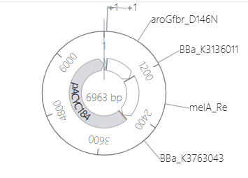
| Feature | Coordinates | Length |
|---|---|---|
| Ptrc | 1–91 | 91 bp |
| RBS1 | 92–108 | 17 bp |
| aroGfbr CDS | 109–1161 | 1053 bp |
| RBS2 | 1162–1178 | 17 bp |
| melA CDS | 1179–2690 | 1512 bp |
| Terminator | 2691–2718 | 28 bp |
| pACYC184 | 2719–6963 | 4245 bp |
| Total | 6963 bp |
Verification performed on the assembled sequence. I checked these computationally rather than by eye:
- Reading frame intact across both CDSs, each with its own start and stop codon — they are independent cistrons, not a translational fusion. aroG: 1053 nt → 350 aa, stop
TAA. melA: 1512 nt → 503 aa, stopTAA. - No accidental internal stop codons in either cistron.
- Shine-Dalgarno to start-codon spacing of 8 nt on both ribosome binding sites.
- No premature terminator between the cistrons.
- Residue 146 of AroG reads N, confirming the feedback-resistance substitution survived assembly.
- Backbone length exactly 4245 bp with both resistance genes intact, confirming the vector imported without truncation.
The insert (everything but the backbone) is 2718 bp, or about 2.7 kb.
Part 4: Optimize and quality-check
Codon optimization
The two coding sequences call for opposite decisions, and that contrast is itself the useful result.
aroGfbr is already an E. coli gene expressed in E. coli, so optimization has essentially nothing to act on. Re-coding it would also make it harder to distinguish from the chromosomal copy in the sequence search below. I left it native. Measured GC: 52.1 %.
melA is the gene that genuinely needs it. R. etli is a GC-rich alphaproteobacterium, and the measured GC content of the native CDS is 59.5 % — already at the top of the 40–60 % window, and higher locally.
Constraints applied: GC held within 40–60 %, avoidance of long GC runs and of predicted secondary structure near the ribosome binding site, and removal of restriction sites that clash with the assembly junctions. On that last point I had to correct my own plan: I had intended to remove BamHI, SspI and EcoRV sites, but neither CDS contains any. The sites that actually exist are PstI at nt 124 and 1426 of melA, and SalI at nt 826 of melA — the latter mattering because the Ptrc part carries a SalI site of its own at position 76.
Sequence search (BLAST)
Expected and observed: aroGfbr returns a near-identical match to native E. coli aroG differing at a single codon, which independently confirms the D146N substitution landed where intended. After codon optimization, melA diverges substantially from the R. etli gene at the nucleotide level while the translated protein remains identical — so the meaningful check here is protein-level rather than nucleotide-level identity. The CDS is a re-coded natural gene, not a novel sequence.
Safety screen
I expected a clean report, and the expectation is worth justifying rather than assuming. Neither enzyme is toxin-related: tyrosinase is a pigment-forming monophenol oxidase, and DAHP synthase is a primary-metabolism enzyme present in E. coli’s own genome. R. etli is a nitrogen-fixing plant symbiont, not a pathogen. The most plausible flag would be a generic hit on the antibiotic resistance markers in the backbone, which is expected of any cloning vector and is not a hazard of the design.
Simulation
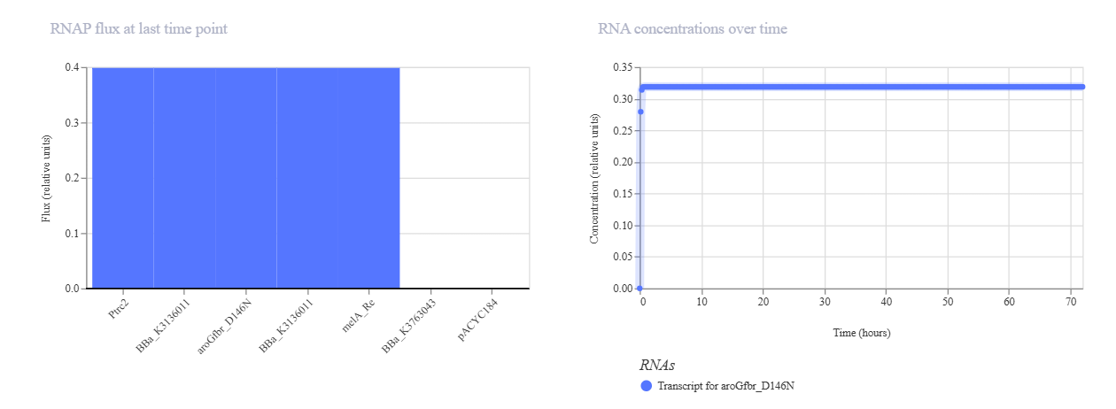
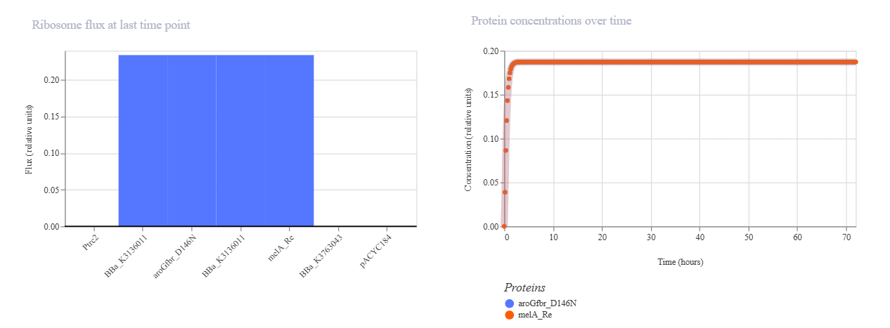
Worth stating what a simulation of this construct can and cannot settle. A genetic-circuit simulator models transcription and translation from promoter and RBS strengths, so it can speak to the relative expression of the two cistrons — which is exactly the Part 6 question about RBS2. It does not model allosteric feedback inhibition, aromatic amino acid flux, copper-dependent apoenzyme activation, or non-enzymatic polymerisation. It would therefore show no difference between aroGfbr and wild-type aroG, which is precisely why the sequence-level verification in Part 3 was the check that mattered.
Part 5: From design to build
How would I build it? The insert is ~2.7 kb with two coding sequences plus regulatory parts — a natural fit for Gibson assembly from three or four synthetic fragments carrying 20–40 bp overlaps, assembled into linearised pACYC184 in a single isothermal reaction [19]. This is a real improvement over the route the original authors had to take, which chained SspI, BamHI, BstZ17I and EcoRV digestions across several subcloning steps to reach the equivalent plasmid [7]. Collapsing that serial restriction-and-ligation process into one reaction is precisely what the method was developed for.
What does synthesis constrain? The difficult fragment is melA, for the same measured reason it needed codon optimization: 59.5 % GC, originating from a GC-rich genome, and GC-rich structured sequences are where de novo synthesis errors concentrate. Error correction is therefore not optional here. Protein-mediated mismatch error correction, using mismatch-binding proteins to remove erroneous duplexes from synthesized pools, substantially reduces error frequency and is what makes it realistic to order a difficult fragment as synthetic DNA rather than screening clone by clone for a correct one [20]. Codon optimization helps twice over, since breaking up GC runs makes the fragment easier to synthesize as well as easier to translate.
Prototype vs. scale. These diverge sharply, mirroring the two-scale automation argument from HW1. To build one construct and test whether it works, column-synthesized fragments plus Gibson assembly is cheapest and fastest. But the automation plan I proposed in HW1 is a combinatorial screen across RBS strength, induction timing and CuSO₄ concentration on the OT-2, and that is a library, not a construct. Producing hundreds of RBS and promoter variants by individual column synthesis is economically absurd; synthesizing them as oligo pools on high-fidelity microchips and recovering subsets by selective amplification is what brings per-sequence cost down to where a combinatorial screen is affordable [21]. So: one construct by Gibson, the variant library by chip-synthesized pools.
Part 6: What don’t you know yet?
1. Is the ribosome binding site on the second cistron strong enough? In a bicistronic operon the downstream gene is typically expressed at a lower level, and here the downstream gene is the catalytic one. The reference data indicate this is the wrong direction in which to err: strain VH33MGTR accumulated roughly 4 g/L of L-tyrosine alongside its 3.22 g/L of melanin, meaning tyrosine synthesis was outrunning tyrosinase capacity, and the authors identify increasing tyrosinase activity as the route to improvement [7]. An underpowered RBS2 would deepen a bottleneck that already exists.
How I would find out: build a variant series on the same B0034 SD core, varying only the spacer length, and screen in 96-well plates reading absorbance at 400 nm (one OD400 unit corresponds to 0.066 g/L eumelanin [7]). Select for the highest ratio of melanin to residual extracellular tyrosine. Swapping gene order is a variant worth including.
| Variant | Sequence | Spacer |
|---|---|---|
| v2 | AAAGAGGAGAAATA | 5 nt |
| v3 | AAAGAGGAGAAATAC | 6 nt |
| v4 | AAAGAGGAGAAATACT | 7 nt |
| v5 | AAAGAGGAGAAATACTA | 8 nt (current) |
| v6 | AAAGAGGAGAAATACTAG | 9 nt |
| v7 | AAAGAGGAGAAATACTAGA | 10 nt |
I verified for all six that no spurious ATG is introduced and that the first start codon in each junction remains melA’s own.
2. Will MelA fold and stay active in E. coli at production temperature? It is a copper-dependent enzyme from a mesophilic alphaproteobacterium, already known to inactivate rapidly at 37 °C [7]. Expression level and folding are separate questions from catalytic activity, and a 59.5 % GC heterologous CDS is a plausible candidate for misfolding. How I would find out: a Cu-supplemented plate assay for visible pigment as the functional readout, paired with soluble-versus-insoluble fractionation on SDS-PAGE to check whether the protein partitions into inclusion bodies. If it does, the design variants to try are a weaker RBS2, lower cultivation temperature, or a solubility tag.
3. Does the Tat signal peptide actually export the tyrosinase, and does that determine where the melanin ends up? This question did not exist in my plan until I translated the CDS and found the twin-arginine signal described in Part 1. Whether precursors polymerise in the periplasm, the extracellular medium, or both is unresolved in the literature [7], and a functional Tat signal is a concrete mechanistic candidate: Tat exports folded proteins, so a periplasmic tyrosinase would make the periplasm the site of conversion. Against that, Cabrera-Valladares et al. purified the recombinant enzyme from cell extract [15], suggesting the rhizobial signal is poorly recognised by E. coli.
This is not an academic question for this project. It determines whether recovery is acid precipitation from clarified supernatant or something requiring cell lysis, and it connects directly to the secretion-route question I raised in HW1 [9] — including the LPS problem, since anything recovered as membrane vesicles carries endotoxin on its surface by construction.
How I would find out: fractionate a production culture into supernatant, periplasmic wash and cell pellet, and quantify A400 in each across a time course. Run it in parallel against a signal-peptide-deleted variant (CDS starting at nt 94 with an added ATG) — if the fractionation profiles are identical, the signal is not being used, and that is a clean answer either way.
References
[7] Chávez-Béjar, M. I., Balderas-Hernandez, V. E., Gutiérrez-Alejandre, A., Martinez, A., Bolívar, F., & Gosset, G. (2013). Metabolic engineering of Escherichia coli to optimize melanin synthesis from glucose. Microbial Cell Factories, 12, 108. https://doi.org/10.1186/1475-2859-12-108
[9] Kong, X., Zhang, J., Li, Y., Liu, Y., Zhang, Y., Ye, C., & Wang, H. (2024). Genetically modified E. coli secreting melanin (E.melanin) activates the astrocytic PSAP-GPR37L1 pathway and mitigates the pathogenesis of Parkinson’s disease. Journal of Nanobiotechnology, 22, 690. https://doi.org/10.1186/s12951-024-02955-x
[15] Cabrera-Valladares, N., Martinez, A., Piñero, S., Lagunas-Muñoz, V. H., Tinoco, R., de Anda, R., Vázquez-Duhalt, R., Bolívar, F., & Gosset, G. (2006). Expression of the melA gene from Rhizobium etli CFN42 in Escherichia coli and characterization of the encoded tyrosinase. Enzyme and Microbial Technology, 38(6), 772–779. https://doi.org/10.1016/j.enzmictec.2005.08.004
[16] Lütke-Eversloh, T., & Stephanopoulos, G. (2007). L-tyrosine production by deregulated strains of Escherichia coli. Applied Microbiology and Biotechnology, 75, 103–110. https://doi.org/10.1007/s00253-006-0792-9
[17] Amann, E., Ochs, B., & Abel, K. J. (1988). Tightly regulated tac promoter vectors useful for the expression of unfused and fused proteins in Escherichia coli. Gene, 69(2), 301–315. https://doi.org/10.1016/0378-1119(88)90440-4
[18] Rose, R. E. (1988). The nucleotide sequence of pACYC184. Nucleic Acids Research, 16(1), 355. https://doi.org/10.1093/nar/16.1.355
[19] Gibson, D. G., Young, L., Chuang, R.-Y., Venter, J. C., Hutchison, C. A., & Smith, H. O. (2009). Enzymatic assembly of DNA molecules up to several hundred kilobases. Nature Methods, 6, 343–345. https://doi.org/10.1038/nmeth.1318
[20] Carr, P. A., Park, J. S., Lee, Y.-J., Yu, T., Zhang, S., & Jacobson, J. M. (2004). Protein-mediated error correction for de novo DNA synthesis. Nucleic Acids Research, 32(20), e162. https://doi.org/10.1093/nar/gnh160
[21] Kosuri, S., Eroshenko, N., LeProust, E. M., Super, M., Way, J., Li, J. B., & Church, G. M. (2010). Scalable gene synthesis by selective amplification of DNA pools from high-fidelity microchips. Nature Biotechnology, 28, 1295–1299. https://doi.org/10.1038/nbt.1716