I’m expressing alpha-1-antitrypsin, a glycoprotein encoded by the SERPINA1 mRNA (GenBank accession K01396.1; NCBI Gene ID 526). I used the mRNA-derived CDS rather than the genomic SERPINA1 sequence because it is already spliced, contains no introns, and can be cloned directly as a continuous open reading frame into an expression vector. This protein requires post-translational modifications, so I chose CHO cells as the chassis. Moreover, this host is easier to scale up than HEK 293 cells. The protein has to be secreted, and the sequence already includes the native signal peptide, which directs the protein to the secretory pathway and is cleaved to release the mature protein, so no additional signal peptide is needed. The sequence is the following:
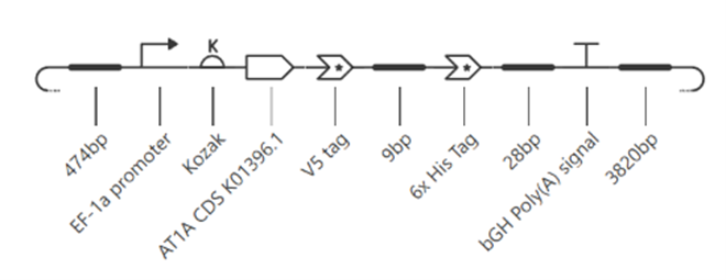
The chart below details the genetic parts needed for the construct. The backbone already contains the majority of the necessary components for the expression cassette, including the promoter and the His-tag.
Part type
Name
Where it came from?
Justification
Promoter
Promotor EF-1α
SnapGene/Backbone
A strong human constitutive promoter that tends to be stable in long-term culture. [1]
5’ UTR_Kozak
Kozak
Kozak, M [2]
Kozak consensus sequence
Signal peptide
Native signal peptide from A1AT
NCBI
Directs A1AT to the secretory pathway for direct recovery from the medium.
CDS
K01396.1
NCBI
This is the CDS that encodes A1AT and includes the signal peptide.
Tag
6xHisTag
SnapGene/Backbone
This tag will help during the purification process.
Transcription Stop
BGH PolyA
SnapGene/Backbone
It ensures efficient polyadenylation and the stability of mRNA in the cytoplasm.
Vector
pTracer™-EF/V5-His A
SnapGene
High-level expression vector, designed for visual detection of transformed E. coli cells and transfected mammalian cells.
Part 3: Assemble your construct
The construct uses the pTracer™-EF/V5-His A vector as the backbone. This vector already provides the elements required for expression in mammalian cells: the EF-1α promoter (a strong constitutive promoter that functions in CHO cells), a C-terminal V5/6xHis tag, and a polyadenylation signal. It also carries the ampicillin resistance gene (AmpR) for selection in E. coli and the bleomycin/Zeocin resistance gene (BleoR) for selection in mammalian cells. Therefore, only a few elements had to be added: a Kozak sequence and the α1-antitrypsin coding sequence (GenBank K01396.1), including its native signal peptide, which directs the protein to the secretory pathway. The insert is cloned into the multiple cloning site between the XbaI and Acc65I sites, which cut the vector once and allow directional cloning.
Codon optimization was performed for Cricetulus griseus (CHO) to match the host’s codon usage bias, which can improve translation efficiency and mRNA stability. Although human and CHO codon preferences are similar, they are not identical, and optimization also allowed me to remove unwanted restriction sites and avoid regions of extreme GC content that complicate synthesis.
The codon-optimized sequence of the CDS of A1AT is the following:
After optimizing the sequence, a BLAST against the NCBI nucleotide collection was run. All top hits were human SERPINA1 (alpha-1-antitrypsin) transcripts and cDNA clones, led by Homo sapiens serpin family A member 1 (SERPINA1), transcript variant 2, mRNA (NM_001002236.3), with 99% query coverage and 84.56% identity. This confirms that the CDS encodes alpha-1-antitrypsin.
The final genetic construct was evaluated using an automated biosecurity safety screening tool to detect potential biosecurity threats. The screening returned zero flagged sequences, confirming the complete absence of controlled pathogens, toxins, select agents, or dangerous viral elements. As a result, the construct is composed exclusively of benign mammalian genetic parts and a safe codon-optimized human therapeutic gene, rendering it fully compliant with biosecurity protocols, eligible for commercial DNA synthesis, and safe for standard BSL-1 laboratory containment.
Part 5: From design to build
As it was mentioned before, most of the part are already in the backbone vector, so for the physical construction, Gibson Assembly will be used. It is an isothermal single-step enzymatic method operating at 50 °C that utilizes a 5’ T5 exonuclease to generate single-stranded overhangs, a Phusion DNA polymerase to fill internal gaps, and a Taq DNA ligase to covalently seal nicks between overlapping double-stranded DNA fragments [3]. To mitigate synthesis-derived errors such as microdeletions and mismatches inherent to oligonucleotide precursor chemical synthesis, MutS protein-mediated error correction will be employed, wherein the Thermus aquaticus MutS mismatch-binding protein selectively binds to flawed heteroduplexes to physically resolve error-free DNA fractions, reducing mutation frequency by over 15-fold down to ~1 error per 10,000 bp [4].
To test the construct at a prototype level, enzymatic digestion, agarose electrophoresis, and in silico pattern comparison effectively verify overall plasmid integrity. Then Sanger sequencing allows determination of the presence of point mutations. On the other hand, large-scale bioprocess optimization requiring parallel screening of genetic parts demands high-throughput synthesis technologies. For scaled-up screening, programmable DNA microchips enable the massively parallel synthesis of thousands of custom oligonucleotides in microfluidic arrays at low cost, coupled with MutS mismatch filtration to purge errors from high-density pools.
Part 6: What don’t you know yet?
Glycosylation Profile: I don’t know if the glycosylation profile is similar to the endogenous A1AT. CHO cells do not possess the same glycosylation machinery as human cells, and glycan composition has been shown to influence the immunogenicity and stability of therapeutic proteins. A suitable approach would be to analyze the glycosylation pattern by HPLC using fluorescently labeled glycans, allowing comparison of the glycan profile of recombinant and endogenous A1AT. If needed, α2,6-sialyltransferase can be co-expressed to improve the glycosylation profile [5].
Signal Peptide Cleavage: It is unknown whether the native hA1AT signal peptide will drive 100% efficient ER/Golgi translocation and extracellular secretion in CHO cells. To find this out, quantifying extracellular versus intracellular protein fractions by ELISA and Western blot is a good option.
Long-Term Clonal Productivity and Epigenetic Stability in Bioreactors: Although the human EF-1α promoter is resistant to long-term DNA methylation, extended passaging and fed-batch bioreactor conditions could cause clonal drift. So, it would be necessary to conduct a 12-week cell line stability study in Erlenmeyer shake flasks with and without selection pressure, tracking cell-specific productivity and titer via ELISA.
References
[1] X. Wang et al., “The EF‐1α promoter maintains high‐level transgene expression from episomal vectors in transfected CHO‐K1 cells,” J. Cell. Mol. Med., vol. 21, no. 11, pp. 3044–3054, Nov. 2017, doi: 10.1111/jcmm.13216.
[2] M. Kozak, “An analysis of 5’-noncoding sequences from 699 vertebrate messenger RNAs,” Nucleic Acids Res., vol. 15, no. 20, pp. 8125–8148, 1987, doi: 10.1093/nar/15.20.8125.
[3] D. G. Gibson, L. Young, R. Y. Chuang, J. C. Venter, C. A. Hutchison, and H. O. Smith, “Enzymatic assembly of DNA molecules up to several hundred kilobases,” Nat. Methods, vol. 6, no. 5, pp. 343–345, 2009, doi: 10.1038/nmeth.1318.
[4] P. A. Carr, J. S. Park, Y. J. Lee, T. Yu, S. Zhang, and J. M. Jacobson, “Protein-mediated error correction for de novo DNA synthesis,” Nucleic Acids Res., vol. 32, no. 20, 2004, doi: 10.1093/nar/gnh160.
[5] M. E. Lalonde, I. Koyuturk, D. Brochu, J. Jabbour, M. Gilbert, and Y. Durocher, “Production of α2,6-sialylated and non-fucosylated recombinant alpha-1-antitrypsin in CHO cells,” J. Biotechnol., vol. 307, pp. 87–97, Jan. 2020, doi: 10.1016/j.jbiotec.2019.10.021.